Fig. 50.1
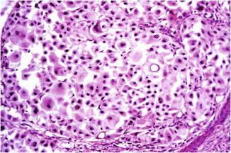
Balloon cell melanoma. Enlarged polygonal cells with vacuolated (clear) cytoplasm and central hyperchromatic nucleus in at least 50 % of cells

Fig. 50.2
Balloon cell melanoma. Nuclear pleomorphism, intranuclear cytoplasmic pseudoinclusions, atypia, and mitoses help to distinguish melanoma from balloon cell nevus
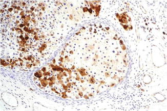
The cells mainly show the immunohistochemical features of conventional melanoma cells (S-100, HMB45, melan-A expression) (Fig. 50.3). Although it is generally believed that balloon melanoma cells represent a degenerative change, the immunohistochemical and electron microscopic findings suggest that the balloon tumor cells are most likely metabolically active melanocytic cells. In particular, electron microscopy shows abundant cytoplasmic membrane-bound vacuoles, few or no melanosomes, and only infrequent abnormal premelanosomes.


Fig. 50.3
Balloon cell melanoma. HMB45 positivity helps to rule out other clear cell tumors
Balloon cells are usually sparse in the primary melanoma but have a potential of constituting the entire lesion in metastasis. In few cases, the metastasis of melanoma is composed of balloon cells, whereas the primary melanoma does not exhibit any balloon cells and predominantly consists of spindle-shaped and epithelioid cells.
A peculiarity of BCMM is the general lack of melanin, in contrast to conventional melanoma and balloon nevus cells.
Differential Diagnosis
Clinically, BCMM could mimic other types of malignant melanoma, basal cell carcinoma, squamous cell carcinoma, fibromatous lesions, and cutaneous adnexal tumors.
Histological differential diagnosis are clear cell tumors such as clear cell sarcoma (malignant melanoma of soft tissue), clear cell carcinoma (including basal cell carcinoma, adnexal tumors such as malignant clear cell acrospiroma and clear cell syringoma, and metastases from tumors of the kidney, breast, large bowel, lung, salivary glands and thyroid), malignant granular (clear) cell tumor, hibernoma, xanthoma, sebaceous neoplasms, seminoma, perivascular epithelioid cell tumors (PECOMAS), and atypical fibroxanthoma.
Stay updated, free articles. Join our Telegram channel

Full access? Get Clinical Tree


