| Deep tear trough, skeletonization of the inferior orbital rim, malar depression |
| Grade tear trough, skeletonization of inferior rim, cheek or malar hollowness, lower lid fat prominence, lower lid laxity, lower lid excess skin, festoons, “V” deformity |
| History of ventral abdominal or umbilical hernia (potential donor sites for autologous fat) |
| Component | Concentration | Amount |
|---|---|---|
| Normal saline | 0.9% | 450 ml |
| Lidocaine (plain) | 1% | 50 ml |
| Epinephrine | 1 : 1000 (1 mg/ml) | 0.5 ml |
| Sodium bicarbonate | 8.4% | 5 ml |
Introduction
The majority of our aesthetic oculofacial surgical procedures involve the excising and lifting of redundant and descended tissues. In recent years, the role of volume loss in the aging process has received more focus and there is an increasing awareness of how restoring volume in the face can address this process. Adding autologous volume enhancement to our surgical armamentarium can be a tremendously useful adjunct.
Autologous fat transfer is not a new concept but advances by Dr. Sidney Coleman have made the procedure more effective and safer. Instead of injecting harvested fat as a bolus where it will likely not survive owing to insufficient blood supply, the fat is placed as multiple droplets. This maximizes uptake of the transfer and is less likely to produce palpable nodules.
The donor site for autologous fat should be discussed with the patient before surgery. The patient should be counseled that the donor site will not result in a therapeutic contouring of adipose beds as seen with traditional lipoaspiration. Suitable locations include the abdomen and outer thigh. In women, the outer thigh usually has a good supply of fat, whereas in men the abdomen may be a more fruitful donor site. A history of abdominal hernia should direct harvesting from the outer thigh. Care is taken with abdominal harvesting to avoid peritoneal perforation. Tumescent anesthesia solution is given at the donor site as described in Table 47.3 . The total amount of lidocaine given is far below the toxic dose, which is greater than 50 mg/kg of lidocaine. Triamcinolone, which is often included in standard tumescent solutions, is intentionally omitted for fat transfer as this may result in adipose cell death.
After harvesting, blood, tumescent anesthetic, lysed adipocytes and free fatty acids must be separated from the fat pearls to be grafted. There are several methods for separation and these are all performed under aseptic and relative anaerobic conditions including passive sedimentation, filtration, and centrifugation. Our preferred technique is passive sedimentation followed by removal of the supranatant and infranatant by gently rolling the fat over a non-adherent dressing (Telfa pad). The fat is emulsified and transferred to 1 ml syringes for transfer.
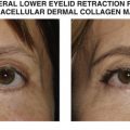
While autologous fat transfer can be performed as a stand-alone procedure, large quantities of fat are required as well as a prolonged recovery and potentially less predictable uptake of the tissue. However, when performed as a surgical adjunct to aesthetic surgery, this can be a powerful tool to address the limitations of lifting and tightening alone. In particular, autologous fat transfer can be used to volumize the inferior orbital rim, malar depressions and to blend the eyelid–cheek junction during lower blepharoplasty. Patients are increasingly aware of the benefits of autologous fat transfer and are readily accepting of this technique.
Fat transfer, although autologous, is not without complications. The most feared complication is death resulting from fat emboli causing ischemic stroke. Blindness can result from central retinal artery occlusion similar to hyaluronic acid gel fillers. Fat emboli can also cause regional necrosis of the face, depending on the arterial supply that is interrupted. More common complications of fat transfer are related to the plane of the injection. Fat transfer more superficially can be unforgiving with the thin periocular skin leading to lumps, bulges, depressions, and persistent edema at the injection site. Management is conservative, consisting of local steroid injection and possibly 5-fluorouracil. Surgical excision, if contemplated, should be delayed by at least 6 months.
Related posts:


Stay updated, free articles. Join our Telegram channel

Full access? Get Clinical Tree


