Fig. 10.1
A case of enchondromatosis with a simple leg length discrepancy. Ipsilateral femoral and tibial shortening were present. We performed simultaneous ipsilateral femoral and tibial lengthening using unilateral external fixators

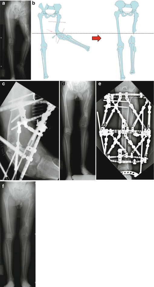
Fig. 10.2
A case of severe left lower limb deformity due to Ollier’s disease. Radiograph of anteroposterior view of lower limbs before surgery (a); operative planning for the first surgery (b); in the first step, osteotomy and gradual deformity correction were performed on the femur with Taylor spatial frame (c); after removal of the Taylor spatial frame (d); in the second step, lengthening of the tibia and ankle mobilization were performed with Taylor spatial frame (e); after completion of the treatment (f)
10.2.2 Fibrous Dysplasia
Fibrous dysplasia is an anomaly characterized by widening of the affected bone with cortical thinning and the presence of fibro-osseous tissue in the interior of the bone. Polyostotic fibrous dysplasia that is associated with autonomous endocrine hyperfunction and café au lait spots is known as the McCune-Albright syndrome.
Pathological fractures and deformities are common. Shepherd’s crook deformity is a well-known progressive varus deformity with shortening of the femur. Valgus osteotomy, plating, and hip nailing are common surgical procedures for femoral lesions.
However, conventional treatment is curettage of the lesions and bone grafting, but the authors do not recommend this treatment. Curettage of fibro-osseous lesions causes massive bleeding and grafted bone is gradually absorbed. Curettage of the lesions and bone grafting might lead to frequent recurrence of dysplastic bone.
Surgeons who treat patients with fibrous dysplasia need to consider the importance of the deformity correction but not of the fibrous dysplasia region itself. It is important to correct the deformity and to obtain a normal mechanical axis to prevent pathological fracture and deformity progression. Extension of fibrous dysplasia into the interior of the bone becomes quiescent with the cessation of growth.
Our recommended treatment consists of osteotomy and gradual deformity correction using an Ilizarov ring fixator or a Taylor spatial frame and mechanical axis realignment of the proximal part of the femur without internal fixation. Even at the site of fibrous dysplasia, bone formation is similar to normal bone.
For severe deformities at the femoral neck, curettage and cortical fibula bone grafts should be performed to prevent further deformity (Fig. 10.3).


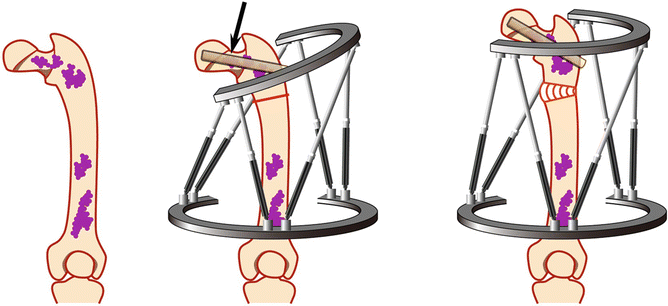
Fig. 10.3
A case of fibrous dysplasia with a cystic bone tumor lesion, pathological fracture, and varus deformity of the proximal femur (Shepherd’s crook deformity). We performed tumor curettage, fibula bone graft, and deformity correction. Black arrow indicates grafted fibula
10.2.3 Osteofibrous Dysplasia (Ossifying Fibroma)
Osteofibrous dysplasia is either the benign counterpart of a neoplastic process that produces an adamantinoma or the result of spontaneous regression of an adamantinoma. The lesion is most often found in the shaft of the tibia. This tumor actively grows during childhood and adolescence and then becomes quiescent with the cessation of growth.
Conventional treatment involves curettage and bone graft, but local recurrence is frequent, and the cortex thins and expands with the bowing deformity. Curettage and bone grafting are likely to induce severe deformity and leg length discrepancy, especially in children under the age of 5 years, in addition to a high incidence of recurrence.
With sufficient informed consent from the patient or persons in parental authority, we recommend en bloc tumor resection and reconstruction with bone transport because this treatment avoids complications and recurrence (Fig. 10.4) (Karita et al. 2004; Tsuchiya et al. 2004).


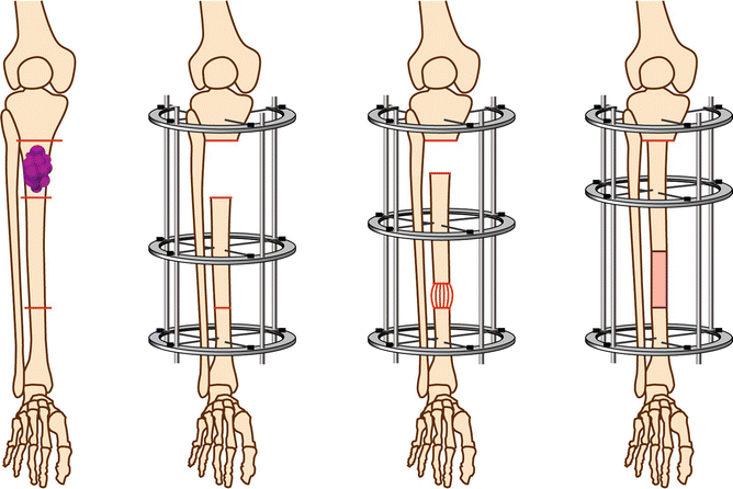
Fig. 10.4
A case of osteofibrous dysplasia with thinned and expanded tibial cortex with a bowing deformity. We performed bone transport after en bloc tumor resection to reconstruct the defect
10.2.4 Exostosis (Osteochondroma)
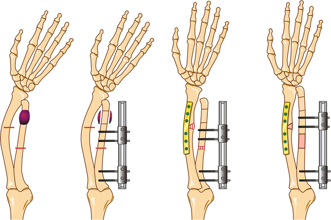
Multiple exostoses are a hereditary disorder of enchondral bone growth and result in asymmetrical retardation of longitudinal bone growth. The most common deformity of the forearm is a combination of relative shortening of the ulna, bowing of either or both forearm bones, increased ulnar tilt of the distal epiphysis of the radius, ulnar deviation of the hand, progressive ulnarward translocation of the carpus, and dislocation of the proximal radial head. Our treatment involves three steps: Step 1 is tumor resection, step 2 is osteotomy of the radius and ulna followed by a radial correction using internal fixation or an unilateral fixator, and step 3 is ulnar lengthening (Fig. 10.5).


Fig. 10.5
A case of exostosis with shortening of the ulna, ulnar tilt of the distal epiphysis of the radius, and ulnar deviation of the hand. Radial head is dislocated. We performed ulnar tumor resection, radial deformity correction and internal fixation, and ulnar lengthening using a unilateral fixator. In proportion to ulnar lengthening, dislocation of the radial head was completely reduced. The ulna is overlengthened several millimeters to prevent recurrence of the deformity
10.3 Treatment of Malignant Bone Tumors
Tumor surgery for malignant bone tumors has evolved from amputation to limb-saving and joint-saving surgery (Tsuchiya et al. 1999b). Reconstruction can result in some difficulties such as an extensive defect, loss of healthy soft tissue, and the need for chemotherapy in some cases.
Biological reconstruction after tumor resection is extremely beneficial for the patients. Once the resected area is completely reconstructed from natural tissue, the patients do not need to worry about infection or breakage for their entire lifetime, unlike with a mega-prosthesis. The distraction osteogenesis technique for biological reconstruction is safe and provides the best qualified living bone. However, the main disadvantage of this technique is the mental burden and complications resulting from long-term external fixation.
Epiphyseal preservation and reconstruction using the distraction osteogenesis technique can provide excellent outcomes in selected cases resulting in sturdy reconstruction and reproduction of the native limb. The Ilizarov fixator or the Taylor spatial frame is used mainly for juxta-articular reconstruction, and a unilateral fixator is used for diaphyseal reconstruction or arthrodesis.
Limb lengthening is beneficial for growing children who suffer from leg shortening as a result of tumor excision or irradiation of the epiphyseal plate. Arthrodesis using distraction osteogenesis is also effective for the treatment of infected tumor prostheses. Furthermore, distraction osteogenesis for late limb length discrepancy and failure after limb-saving surgery appears to be similar to that of benign conditions because there is little influence of chemotherapy or irradiation and soft tissue is also repaired.
It is easier to manage benign and low-grade tumors because expertise and experience are needed for the treatment of high-grade tumors. In the latter case, accurate timing of antibiotic administration, change of dressing, and adjustment of lengthening rate are essential to achieve successful reconstruction.
10.4 Reconstruction After Excision of Malignant Tumor
10.4.1 Selection of Patients
The most important consideration for the use of this procedure is the indication. Patients with a low-grade tumor can be safely treated by distraction osteogenesis because they have no risk induced by chemotherapy and healthy soft tissues are well preserved, which leads to good bone regeneration. Both diaphyseal and metaphyseal defects are the most suitable to be reconstructed by distraction osteogenesis. Distraction osteogenesis enables joint preservation and provides excellent limb function. Patients with less than a 15 cm defect are good candidates for this procedure. However, patients with a > 15 cm defect can be treated in combination with an intramedullary nailing. The intramedullary nailing shortens the treatment period.
Stay updated, free articles. Join our Telegram channel

Full access? Get Clinical Tree


