19 Alternative flaps for breast reconstruction
Synopsis
Many women today have undergone procedures on their abdomen, such as liposuction and abdominoplasty, that preclude the TRAM/DIEP breast reconstructive option.
The skilled microsurgeon needs to be able to utilize other flaps for women in this category.
Alternative flaps may include transverse upper gracilis free flap (TUG); superior gluteal artery free flap (SGAP); inferior gluteal myocutaneous flap (IGAP); deep femoral artery perforator (DFAP), and lumbar artery perforator flap (LAP).
Introduction
The modern era of autogenous breast reconstruction began with the TRAM flap, popularized by Dr Carl Hartrampf in the early 1980s. This was the procedure of choice until the 1990s, when the goal of muscle preservation became more apparent. Perforator flap breast reconstruction in the early 1990s by the authors’ group at Louisiana State University Medical Center was the next significant advance in breast reconstruction. By injecting fresh abdominoplasty specimens, it was determined that the skin and fat could be transferred without sacrifice of the rectus abdominus muscle. This led to the first DIEP flap for breast reconstruction by Allen in 1992.1 The inception of free tissue transfer allowed an infinite range of possibilities to appropriately match donor and recipient sites.2
Patient preference and body habitus has played a role in determining where autologous tissue is taken. Some women do not like the long abdominal scar and prefer not to have surgery on their abdomen. Women’s body shape plays a role in determining where to harvest tissue from. Many women can be categorized into two main body shapes: pear and apple. The apple shaped women predominantly have fat in their abdomen and pear shapes have more fat in their thighs and buttock.
The surgeon needs to take all this into consideration before determining the appropriate donor site that meets the needs of the patient. Now, with several options, the plastic surgeon has more to choose from in his/her armamentarium.
This chapter will review several alternative flaps for breast reconstruction with donor sites, including the medial thigh and the buttock region. The choices of medial thigh flaps discussed include the transverse upper gracilis, the medial circumflex femoral artery both the musculocutaneous and septocutaneous variations, and the superficial femoral artery perforator flap. In terms of the buttock donor site, the superior and inferior gluteal artery perforator flaps with the septocutaneous variations will be discussed. The lumbar artery perforator and the deep femoral artery perforator flap will also be considered.
History
Transverse upper gracilis free flap (TUG)
Orticochea in 1972 was the first to describe immediate transfer of a large amount of skin without using the ever popular delay maneuvre.3 The premise of the skin survival was thought to be through the vasculature supplying the underlying muscle. This was the concept for the TUG flap. McGraw et al. improved upon this theory by conceptualizing the idea of a perforator vessel otherwise known as musculocutaneous perforators.4 The gracilis free flap became widely accepted and used, however the skin paddle overlying the muscle, when included, became precarious for survival. Yousif et al. in 1992 mapped out the gracilis muscle perforators and devised a transverse skin orientation using the upper third of the muscle with predictable reliability of the skin paddle.5,6
Hallock in 2003, reported modifications of the TUG flap by identifying the perforator off of the medial circumflex femoral artery. He used this flap predominantly for lower leg reconstruction for trauma patients.7 Also in the same year, the TUG flap based on the medial circumflex femoral artery was first described for breast reconstruction. Arnez et al. performed the free TUG flap on seven patients for breast reconstruction, five flaps were successful and two flaps were lost.8 Schoeller and Wechselberger in 2004 described 12 TUG flaps for breast reconstruction and all were successful.9 This paper has led to the current popularity of the TUG flap for breast reconstruction.
Peek et al. in 2005 further delineated the anatomy of the blood supply to the gracilis muscle and surrounding skin. His group described the variations of the TUG perforator flap for breast reconstruction. In 2009, he dissected 24 fresh cadavers defining the perforators into either a musculocutaneous or septocutaneous course.10 Finally, the superficial femoral artery perforator was first described by Beak in 1983 and Song et al. in 1984.11,12 This flap was first used by our group for breast reconstruction in 2008, where three flaps were performed for breast reconstruction.
Superior/inferior gluteal artery perforator free flap (SGAP/IGAP)
The superior gluteal myocutaneous free flap by Fujino was first described in 1975 for breast reconstruction. The myocutaneous superior gluteal artery free flap for breast reconstruction was popularized by Dr Bill Shaw.14–18 However, a short vascular pedicle frequently required vein grafting, increasing the difficulty and complications of this technique. The first breast reconstruction with an inferior gluteal myocutaneous flap was performed in 1978 by LeQuang.19
Basic science/disease process
Patients with invasive ductal/lobular or DCIS/LCIS are most commonly seen in our practice. Detail about these diseases is outside the remit of this chapter and therefore the reader should review specific breast cancer texts to ascertain more information about these processes.
Diagnosis/patient presentation
Patients usually present to our practice with either active breast cancer or after they have completed treatment. The immediate reconstruction population is usually referred by their breast surgeon for surgical options for breast reconstruction at the same time as unilateral or bilateral mastectomy. The breast surgeons affiliated with our practice are performing more and more nipple sparing procedures, depending on the cancer diagnosis, location, size and lymph node involvement. These procedures allow us to bury the flap, and, ultimately, they have the best aesthetic appearance.
The delayed reconstruction population can either have an implant that has a capsular contracture, which can be painful or asymmetric, or the patient wishes to have them removed for personal reasons. In these patients, more skin is required to make up the original tissue loss versus the buried flap in the immediate reconstruction group. The alternative breast flaps from the buttock/thigh will give most women the volume and skin they need, but one must consider adjunctive procedures if the volume match is not identical.
Transverse upper gracilis free flap (TUG)
Patient selection
As described previously, there are many factors the reconstructive surgeon must take into consideration when finding the ideal donor site for breast reconstruction. Certain questions need to be asked. Is this patient undergoing immediate or delayed reconstruction? Has the patient received radiation or are they going to receive postoperative radiation? Is a skin sparing or nipple sparing mastectomy going to be used? Do they want to be the same size, smaller or larger? The answers to these questions help the surgeon gauge if more skin is needed, the type of volume acquired, and types of incisions to be used.
A thorough history is obtained. It is important that there is a discussion with the breast surgeon to determine the oncological procedures needed as these may impact on the type or timing of breast reconstruction. Finally, consideration must be given to the patient’s body habitus, as well as to their preference.
Anatomy
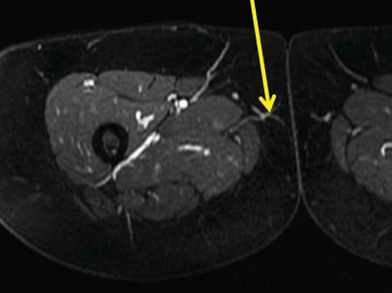
The gracilis muscle is a narrow muscle extending from the pubic tubercle to the medial upper surface of the tibia. Its actions include thigh adduction and flexion, as well as leg flexion and medial rotation. It is categorized as a type II muscle by Mathes and Nahai with one dominant and one minor pedicle.13 The motor innervation is by the obturator nerve arising from the lumbar plexus L3–L4 with its anterior branch supplying the gracilis muscle. The blood supply is from the medial circumflex femoral artery from the profunda femoris artery in which the dominant pedicle enters the medial aspect of the gracilis 6–10 cm inferior to the pubic tubercle. This vasculature then divides into a musculocutaneous and sometimes a septocutaneous branch. This division usually takes place in the upper third of the muscle. The venae comitantes follow the perforators and enter into the femoral vein. The saphenous vein also augments drainage to this area.
Treatment/surgical technique
Many of our patients undergo a preoperative CTA or MRA of the medial thigh to determine the perforator location and size. This study also allows us to differentiate between musculocutaneous versus septocutaneous perforators. It can also help determine the presence of a superficial femoral artery perforator.
The patient is seen the day before surgery and the markings are placed at this time. Identifying the medial edge of the adductor longus and measuring 8–10 cm inferior from the pubic tubercle, a mark is placed on the skin. The hand held Doppler is used to isolate the arterial signal of the medial circumflex femoral artery and or perforator through the skin. The skin island measures roughly 7 cm in width and 20–26 cm in length. The markings start lateral to the adductor longus muscle and course to the midline of the posterior thigh, centered over the key perforator or medial circumflex femoral artery.
Using a 10-blade, the superior limits of the incision marking are incised first, starting laterally and extending medially to posterior. Identification and preservation of the saphenous vein is done, sparing superficial lymph nodes. The incision is continued to the inferior marking to identify the inferior extent of the saphenous vein. A suprafascial dissection begins lateral to medial, identifying the adductor muscle. The deep fascia of the adductor longus is incised vertically. A septocutaneous perforator will be first visualized here. If this is not present, then a musculocutaneous perforator identification commences, first dissecting from an anterior direction and then switching to a posterior direction over the gracilis muscle. In some instances, the perforators are too small and then the flap is converted to a TUG flap with a small section of muscle taken. If the perforator is observed, it is dissected through the muscle until it enters the medial circumflex femoral vessels. The pedicle is followed to its origin from the profunda femoris artery and vein. The pedicle is then clipped at its origin. The flap is weighed and brought to the recipient site. Anastomoses are to the internal mammary vessels or their perforators. The harvested flap is long, narrow, and curved. Suturing the proximal and distal ends of the flap together results in a conical shape with a large base and pointed top. The flap coning creates an excellent breast shape. The donor site is closed in layers over a suction drain.
Hints and tips
• Preoperative imaging will shorten your operative time by one-third.
• Always locate the adductor when the patient comes in for preoperative markings. It will make the dissection faster in the operating room due to the consistency of the anatomy in this flap.
• Once the vessels are identified, dissect under the adductor from its posterior border, then switch to the anterior border to finish the dissection to the superficial femoral artery/vein.
• Keep the saphenous vein intact to keep the percentage of lymphedema minimal.
• Use a knife when dissecting the superior portion of the flap so as not to stun the lymph nodes and again to minimize lymphedema.
Complications
The complications of the TUG flap are similar to those of a medial thigh lift. We have seen the tendency for the drains to stay in for 2–7 weeks. With the prolonged drainage, the infection rate is around 20%. This is treated with adequate drainage and antibiotics. The patient may develop transient lower leg edema shortly after the procedure. We encourage a compressive garment over the donor site to be worn for 6 weeks, to compress the dead space and expedite healing.
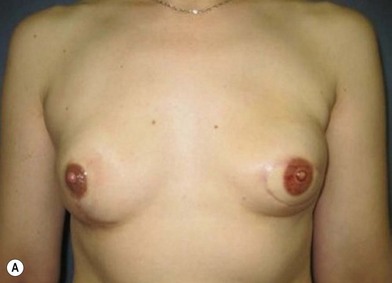
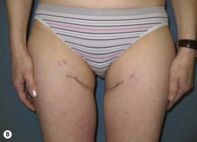
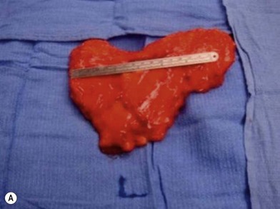
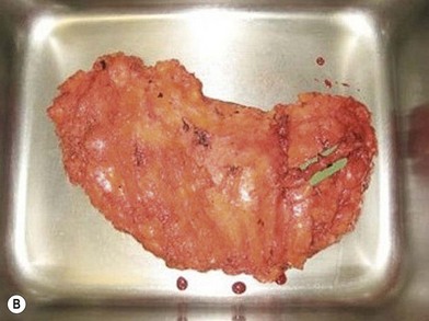
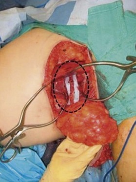
Distortion of the external genitalia or inferior migration of the scar has not occurred in our 71 cases. Preoperative placement of the upper incision near the inguinal crease helps hide the scar. Aesthetically, the upper thigh can be scooped out compared with the lower part. There is a natural narrowing of the thigh superiorly, so this is usually not an issue. If this becomes an issue, liposuction at a second stage can improve contour (Figs 19.1–19.5).

Fig. 19.1 MRA of transverse upper gracilis (TUG) free flap. The yellow arrow demonstrates the gracilis perforator from the gracilis branch of the medial circumflex femoral artery.

Stay updated, free articles. Join our Telegram channel

Full access? Get Clinical Tree

