Fig. 15.1
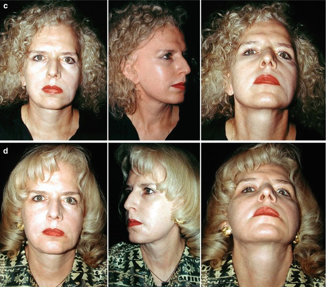
(a) A 25-year-old female patient presenting with sequelae of left-sided facial paralysis. (b) Two years after suspension of the left hemiface with flaps without dermal epithelium, selective myectomies in the right melolabial sulcus; removal of the canine tooth, zygoma, risorius, triangularis, and quadratus bottom; segmental rhytidoplasty and lipo-injection of 40 mL in the upper lip of the left side, left cheek, and zygomatic region; and lipo-injection of 10 mL subsequently every 6 months on four occasions were performed. The aesthetics of the patient improvement were very noticeable, and there was a high degree of regeneration in the motor muscle innervation. (c) Eight years later. (d) Twenty years later
A 25-year-old female patient presented with sequelae of left-sided facial paralysis (Fig. 15.1). This patient was treated with a combined procedure carried out in 1986, which included: (1) suspension of the left hemiface with flaps without dermal epithelium; (2) selective myectomies in the right melolabial sulcus, removing the following muscles – canine tooth, zygoma, risorius, triangularis, and quadratus bottom; (3) segmental rhytidoplasty; and (4) lipo-injection of 40 mL in the following areas: upper lip of the left side, left cheek, and zygomatic region and subsequently lipo-injection of 10 mL every 6 months on four occasions. The aesthetics of the patient’s improvement were very noticeable, and there was a high degree of regeneration in the motor muscle innervation, seeing the patient 2 years later (Fig. 15.1), 8 years later (Figs. 15.5 and 15.6), and 20 years later (Fig. 15.1) [3].
A 33-year-old female patient presented sequelae of facial paralysis in the right side of her face and aesthetic and functional deformity on the lips of the right side (Fig. 15.2). This patient was treated with only four sessions of lipo-injection introducing every 6 months fat grafts of 6 mL on the upper lip and 4 mL lower lip. There were excellent results both aesthetic and functional (Fig. 15.2).
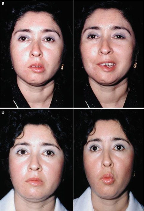

Fig. 15.2
(a) A 33-year-old female patient presented sequelae of facial paralysis in the right side of her face and aesthetic and functional deformity on the lips of the right side. (b) Postoperative condition after treatment with only four sessions of lipo-injection introducing every 6 months 6 mL on the upper lip and 4 mL lower lip of fat grafts. There are excellent results both aesthetic and functional
15.3.2 Parry-Romberg Disease (Fig. 15.3)
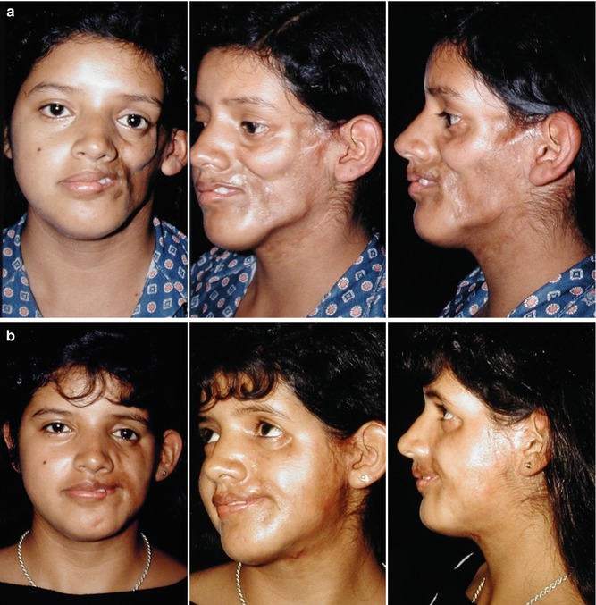
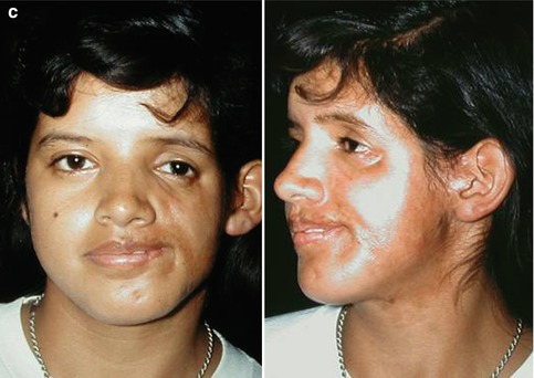
Fig. 15.3
(a) A 27-year-old female patient presented sequels of Parry-Romberg disease with depressions in the cheek, mandibular region, and left chin. (b) A combined treatment using a galeal flap, cartilage grafts in the first surgical session, free grafting of the dermis fat, and four sessions every 6 months of lipo-injection and fragmented fascia. (c) A favorable result is observed 4 years after the start of treatment and 6 months after the latest infiltration of fat and fascia
A 27-year-old female patient presented sequels of Parry-Romberg disease with depressions in the cheek, mandibular region, and left chin (Fig. 15.3). In this patient, a combined treatment plan using a galeal flap, cartilage grafts in the first surgical session, free grafting of the dermis fat, and four sessions every 6 months of lipo-injection and fragmented fascia was performed (Fig. 15.3). Four years after the start of treatment and 6 months after the latest infiltration of fat and fascia, a favorable aesthetic result is observed (Fig. 15.3).
A 24-year-old female patient presented sequels of Parry-Romberg with severe deformities (Fig. 15.4). In a first surgical session, cartilage grafts, galeal flap with fascia and muscle, and the first session of lipo-injection of 40 mL of fat were used. Three times every 6 months, 25, 15, and 5 mL of fat grafts were applied. The patient was improving slowly and after the last lipo-injection, there were very favorable aesthetic results (Fig. 15.4).
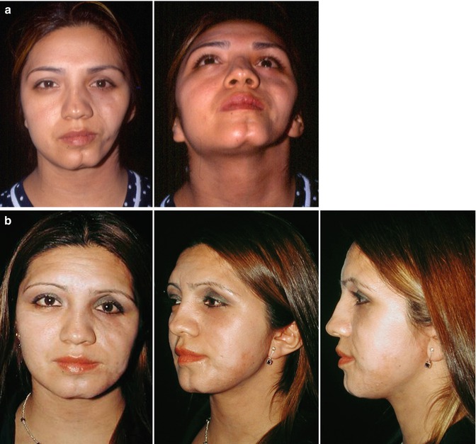

Fig. 15.4
(a) A 24-year-old female presented sequelae of Parry-Romberg with severe deformities. (b) There were very favorable aesthetic results following a first surgical session of cartilage grafts, galeal flap with fascia and muscle, and the first session of lipo-injection of 40 mL of fat. Three times every 6 months, 25, 15, and 5 mL of fat grafts were applied. The patient was improving slowly and after the last lipo-injections
15.3.3 Facial Scars (Fig. 15.5)
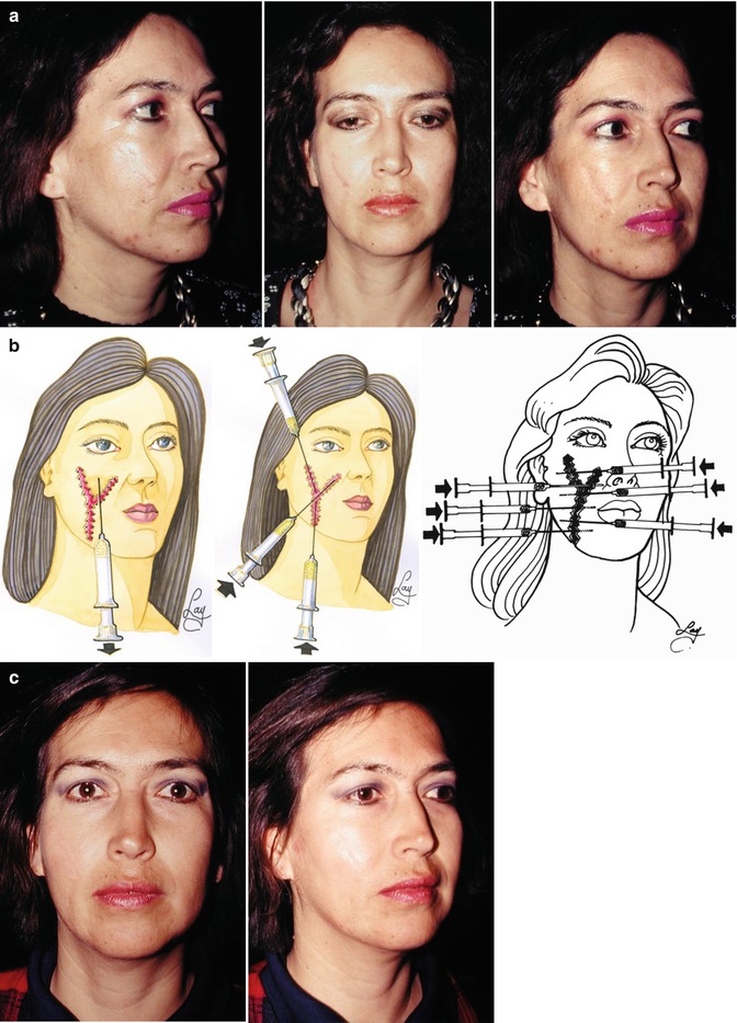
Fig. 15.5
(a) A 30-year-old female patient had an accident with facial trauma of the right cheek that caused skin scars, bumps in neighboring areas, and depressions in the affected area. (b) A combined treatment by removing the scar edges in a W fashion avoiding the straight sutures was used. Also a suture was placed across the small flaps of the edges in W liposuction of the bulky area located along the zygoma and lipofilling the sub-subcutaneous musculoaponeurotic system (sub-SMAS) in the depressed areas. (c) One year later an excellent aesthetic improvement was seen, both in the scar and also in the outline of the right cheek
Stay updated, free articles. Join our Telegram channel

Full access? Get Clinical Tree


