Fig. 63.1
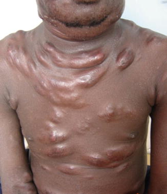
ATLL, acute form: disseminated papules on both legs, nodules on the trunk and extremities (Courtesy of Dr. Dr Moussa Diallo, Dept. of Dermatology, University Hospital Dakar, Senegal)

Fig. 63.2
ATLL, acute form: multiple tumors on the trunk (Courtesy of Dr. Dr Moussa Diallo, Dept. of Dermatology, University Hospital Dakar, Senegal)
Pathology
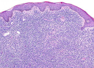
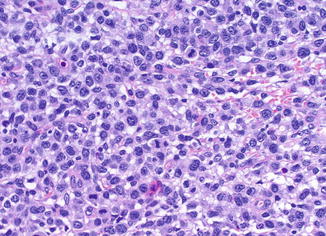
In the acute and lymphomatous form, a superficial or diffuse dermal infiltrate of medium-sized to large pleomorphic cells with or without epidermotropism is found (Figs. 63.3 and 63.4). The findings resemble MF. In chronic or smoldering forms, often a subtle perivascular infiltrate with only a few atypical cells is present. The tumor cells exhibit a CD2+ CD4+ CD5+ CD8- CD25+ betaF1+ phenotype and may express CD30 but usually show downregulation of CD3 and lack CD7. In addition, expression of FOXP3 is typically found in ATLL. Monoclonal rearrangement of T-cell receptor genes and clonal integration of HTLV-1 into the host cell genome can be detected. In the peripheral blood, circulating tumor cells with convoluted nuclei are referred to as flower cells.


Fig. 63.3
ATLL, lymphomatous form: dense dermal infiltrates of atypical large lymphoid cells

Fig. 63.4
ATLL, lymphomatous form: large tumor cells with pleomorphic nuclei and abundant cytoplasm
Differential Diagnosis
Due to overlapping clinical, histological, and immunophenotypic features, the most important differential diagnoses are MF, Sézary syndrome, cutaneous anaplastic large cell lymphoma, and cutaneous peripheral T-cell lymphoma, unspecified. They differ from ATLL by the absence of HTLV-1 infection.
Stay updated, free articles. Join our Telegram channel

Full access? Get Clinical Tree


