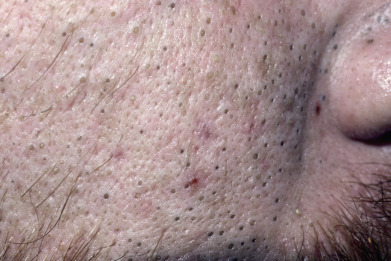
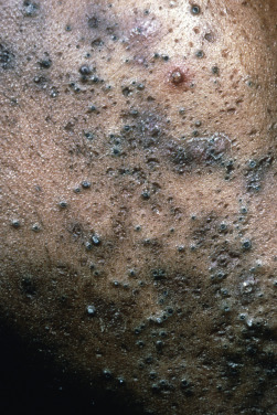
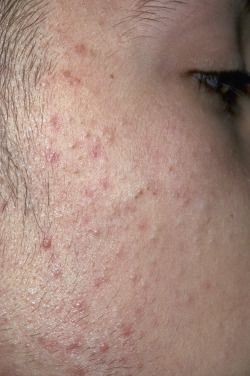
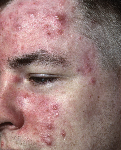
Typical acne is so commonplace that its comedones, papules, and pustules are immediately recognizable. There exist, however, a wide spectrum of acne lesions, acneiform eruptions, and rosacea that are exhibited here to assist the deciphering medical provider.
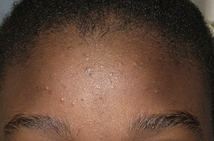
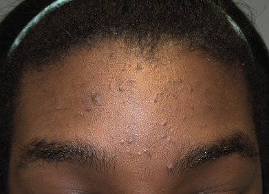
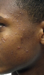
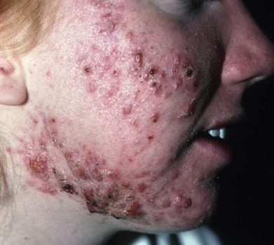
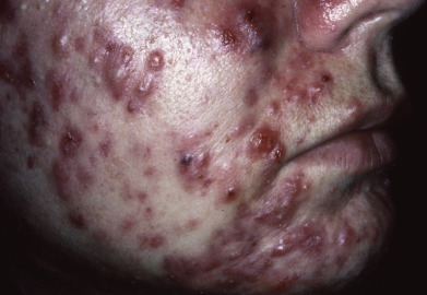
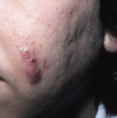
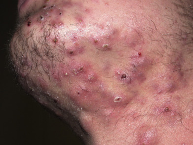
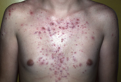
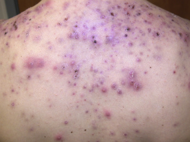
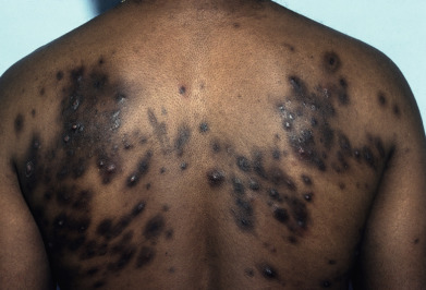
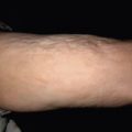
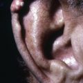
The earliest lesions of acne are open and closed comedones that are often first present on the forehead of adolescent patients. Papules, pustules, nodules, cysts, and scarring can also be seen in the typical acne-prone anatomic regions of the face, upper back, chest, shoulders, and upper arms. The most severe form of acne, acne fulminans, is characterized by joint pains, painful nodulocystic lesions, and even fevers. Other acneiform eruptions include the scarring follicular pustules of acne keloidalis nuchae, the more monomorphous pustular eruption of corticosteroid-induced acne, and the pustules and nodules of gram-negative folliculitis.
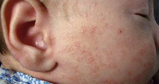
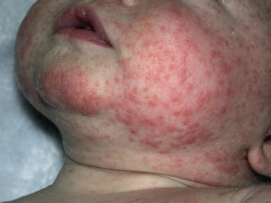
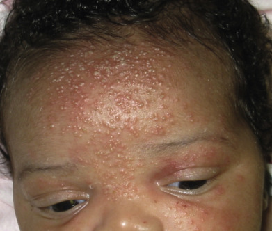
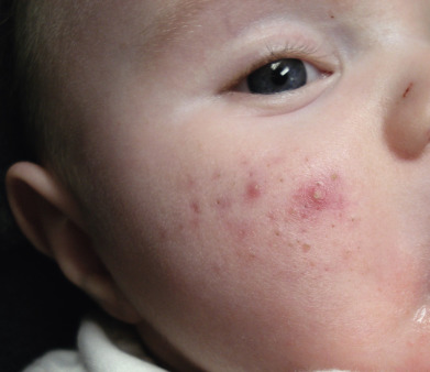
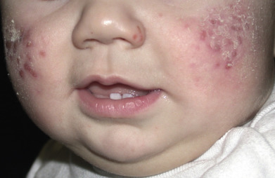
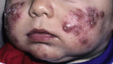
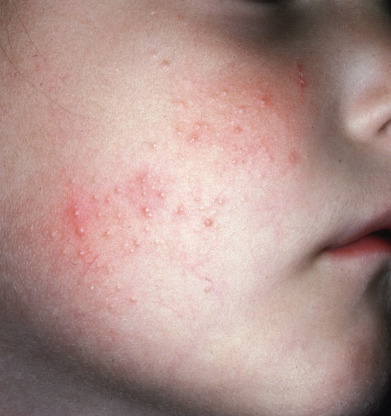
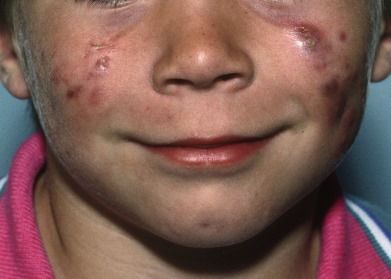
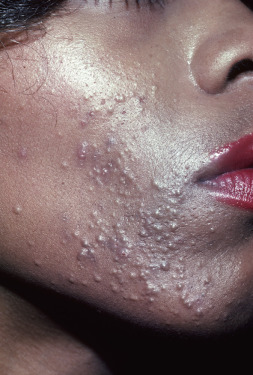
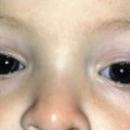
In neonates, the transient micropustular eruption on the head and neck that has been historically known as neonatal acne has now been renamed neonatal cephalic pustulosis due to its lack of comedones, nodules, or scarring. Infantile acne, in contrast, occurs mostly on the cheeks of infants and toddlers and is a true subset of acne with its comedones, papules, pustules, nodules, and potential scarring. Finally, periorificial dermatitis is a distinctive acneiform eruption notable for papules and pustules located around the mouth, nose, and eyes of children and younger adults.
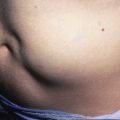
Hidradenitis suppurativa is also included in this chapter with its chronic inflammatory abscesses, nodules, and sinus tract formation in the axillae, inframammary folds, inguinal folds, and gluteal cleft.
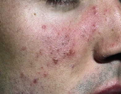
Rosacea is also highlighted here to allow for direct comparison with acne. Flushing, erythema, and fine telangiectasias seen on the convex surfaces of the face define the erythrotelangiectatic subtype of rosacea, whereas the papules, pustules, and in some cases nodules are present in the papulopustular and glandular forms.
Most conditions in this chapter will depend on clinical findings to arrive at a diagnosis, but in some cases a skin biopsy or bacterial culture will be useful. This portion of the atlas features examples of important clinical findings to recognize in patients with acne and the many acneiform eruptions.