Models
Type
Rejection type
Guinea pig-rat
Discordant
Hyperacute rejection (HAR)
Hamster-rat
Concordant
Acute cellular xenograft rejection (ACXR)
Mouse-rat
Concordant
Acute cellular xenograft rejection (ACXR)
Composite Tissue Xenopreservation Model
A composite tissue xenotransplantation model is described between rats and mice, which are concordant species. The inferior epigastric artery and vein-based groin flap are used as a composite tissue flap. This flap includes skin, fat, lymphatic tissue and vessels [13]. The donor group is Sprague-Dawley rats weighing between 350 and 450 g. The recipient/carrier group is mice weighing about 50 g.
Surgical Procedure
All surgeries were performed under general anesthesia and sterile conditions. Two transplantations were performed. The first operation was the xenotransplantation of the rat groin flap to the mouse and the second operation was the transfer of the xenopreserved groin flap to the rat. The experimental protocol is summarized in Fig. 33.1.
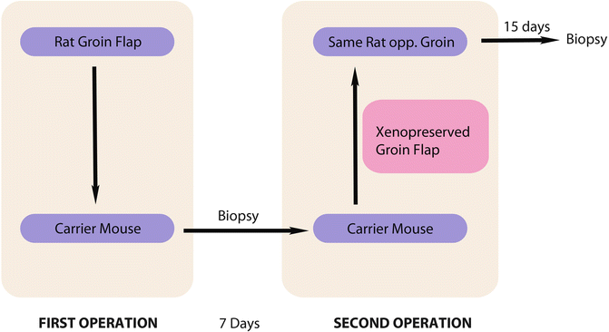

Fig. 33.1
Experimental protocol
First Operation
In the donor rat, under ketamine anesthesia, the groin region was shaved, prepped and draped. A groin flap based on inferior epigastric vessels with 1 × 1 cm dimensions was harvested. Following the harvest of the flap, pedicle dissection was continued, including the femoral artery and vein (Fig. 33.2).
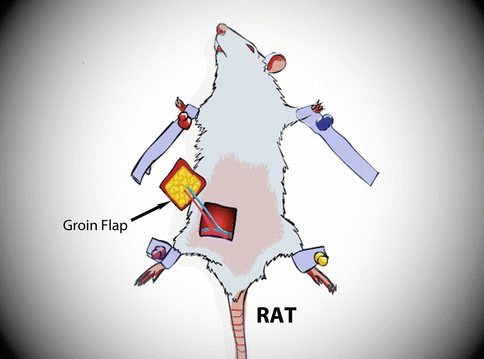

Fig. 33.2
The groin flap, based on femoral vessels, is harvested from rat
In the recipient/carrier mouse, a midsagittal incision was performed and the submandibular gland was excised. The external jugular vein was prepared as the recipient vein. Later, the sternocleidomastoid muscle was excised and the common carotid artery was exposed. The common carotid artery was prepared as the recipient artery.
When both donor and recipients were ready, the xenotransplantation was performed. The femoral pedicle of the groin flap was cut, and the flap was transferred to the neck region of the mouse. Vascular anastomoses were performed with interrupted 11/0 etilon (Ethycon, Johnson & Johnson, NJ, USA) sutures and both anastomoses were in end-to end fashion (Fig. 33.3). The flap was inset with 4/0 polyglactin sutures. The defect at the donor rat was closed primarily (Fig. 33.4).
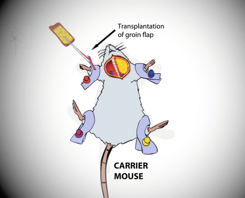
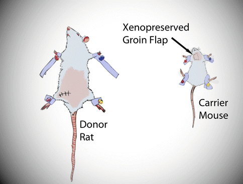

Fig. 33.3
The groin flap is transferred to the neck of the carrier mouse

Fig. 33.4
Appearance of the donor rat and carrier mouse at the end of the first operation
To prevent acute cellular xenograft rejection ACXR, 20 mg/kg/day cyclosporine A (CsA) treatment was given to the recipient/carrier mouse. Xenotransplanted groin flaps were evaluated daily for 7 days and infection and vascular problems were monitored.
Second Operation
At the end of 7 days, a second operation was performed. The xenotransplanted groin flap, which was now xenopreserved groin flap, was returned back to the donor rat (Fig. 33.5). The xenopreserved groin flap was re-harvested from the neck of the recipient/carrier mouse, based on the femoral vessels. After harvest, biopsies were taken from skin, subcutaneous tissues and femoral vessels. The opposite groin region of the donor rat was prepped and draped. Femoral vessels at this site were prepared for anastomoses. The xenopreserved groin flap was returned back to the opposite groin of the donor rat. End-to end arterial and veinal anastomoses were performed with interrupted 10/0 etilon (Ethycon, Johnson&Johnson, NJ, USA) sutures and flap inset was completed.
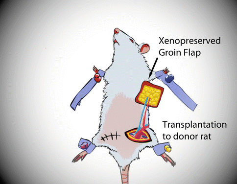

Fig. 33.5
Second operation: transfer of xenopreserved groin flap to the donor rat
Following the second operation, the xenopreserved flap was evaluated daily in terms of flap viability and infection. No immunosuppressive drugs were administered after second operation. At the 15th postoperative day, the rats were sacrificed and biopsies were obtained from skin, subcutaneous tissue and vessels. All biopsies were evaluated histologically.
Results of Composite Tissue Xenopreservation
Macroscopic Evaluation
Stay updated, free articles. Join our Telegram channel

Full access? Get Clinical Tree


