Microsurgery continues to evolve, integrating advanced imaging, robotic techniques, perfusion assessment, artificial intelligence, anastomotic devices, and enhanced recovery after surgery pathways. High-resolution ultrasound and augmented reality can improve flap design and operative efficiency, while robotic microsurgery expands reconstructive possibilities and reduces donor-site morbidity. Emerging wireless monitoring systems enable continuous postoperative flap surveillance, improving salvage rates. Sutureless anastomotic technologies promise shorter procedures with the potential for less user variability. Enhanced recovery after surgery protocols improve patient outcomes and have increased the feasibility of outpatient free flap breast reconstruction. These technological advancements can reduce complications and improve reconstructive outcomes and patient safety.
Key points
-
•
High-resolution ultrasound and smartphone-based thermography have been increasingly used as preoperative tools for lymphatic and perforator mapping.
-
•
Virtual surgical planning has enhanced intraoperative accuracy and efficiency. Combining virtual surgical planning with augmented reality can enhance perioperative planning and intraoperative efficiency.
-
•
Robotic microsurgery continues to expand its indications beyond traditional flap harvest to include microsurgical and supermicrosurgical anastomoses.
-
•
Wireless, wearable, and implantable monitoring technologies are emerging as postoperative flap surveillance methods, providing continuous, objective data on flap perfusion, potentially increasing salvage rates.
-
•
Artificial intelligence can optimize clinical decision-making through improved preoperative planning and enhance surgical education through objective, artificial intelligence-driven feedback.
Abbreviations
| 3D | 3 dimensional |
| ERAS | enhanced recovery after surgery |
| ICGA | indocyanine green angiography |
| POD | postoperative day |
| UHF-US | ultrahigh-frequency ultrasound |
Introduction
Microsurgery remains one of plastic surgery’s most dynamic and rapidly evolving subspecialties, continually integrating novel technologies and multidisciplinary approaches to enhance patient care. Recent advancements in imaging technologies, robotic surgery, augmented reality, and artificial intelligence have helped to redefine surgical efficiency while significantly broadening the scope of techniques available to reconstructive surgeons. Innovations in perforator mapping, virtual surgical planning, and real-time perfusion assessment allow for increasingly precise and individualized reconstructions for various defects. Simultaneously, developments in robotic-assisted microsurgery, sutureless anastomotic devices, and advanced flap monitoring are helping to reshape and redefine the future landscape of reconstructive surgery. Combining these emerging fields of research and technology with rapid advancements in machine learning demonstrates the potential for significant enhancements in surgical planning, execution, and training for microsurgical reconstruction. This article summarizes important recent advancements in microsurgery, highlighting innovations that have already improved clinical outcomes and exploring technologies likely to redefine the specialty in the coming years.
Imaging and microsurgery
Perforator Mapping and Flap Design
The use of high-resolution ultrasound has become an essential tool for preoperative planning. Ultrasound significantly simplifies flap planning by precisely identifying and characterizing perforator anatomy, including vessel caliber, flow, and spatial relationships in real time. ,,, These advancements have been especially beneficial in extremity reconstruction, where ultrasound-guided perforator mapping can reduce operative time and improve flap survival by aiding precise flap design and minimizing unnecessary dissection. Ultrasound-based perforator mapping also facilitates the design of propeller and perforator flaps, particularly in the lower extremity, and has repopularized anatomically variable flaps like the superficial circumflex iliac artery perforator flap. , The advent of ultrahigh-frequency ultrasound (UHF-US) has further expanded capabilities by enabling the visualization of lymphatic vessels for lymphovenous bypass, and fine perforator arborization beneath the skin, allowing for the safe elevation of ultrathin flaps. , Compared to high-frequency ultrasound (10–20 MHz), which provides greater penetration, UHF-US (30–70 MHz) offers superior resolution for superficial structures, including small lymphatics and subdermal perforator branches. , Notably, modern ultrasound platforms no longer require bulky consoles; handheld wireless systems like Clarius and Butterfly now provide high-resolution imaging in a portable, user-friendly format. These systems offer reconstructive surgeons a noninvasive and accessible approach to microsurgical planning.
Thermography, particularly smartphone-based infrared imaging, has emerged as a cost-effective adjunct to traditional and newer imaging modalities. Recent systematic reviews highlight the accuracy of thermography and its growing role in reconstructive surgery, reporting sensitivities for perforator mapping that approach 90% for detecting viable perforators. The portability and real-time feedback of smartphone-based thermography have significant advantages, particularly in rural or resource-depleted settings where access to more advanced imaging, such as computed tomographic angiography, is limited. As the resolution and processing power of thermographic devices continue to improve, thermography has the potential to become an increasingly valuable and cost-effective adjunct to traditional perforator mapping methods; however, it lacks the high-resolution capability of high-frequency ultrasound and UHF-US.
Virtual Surgical Planning and Augmented Reality
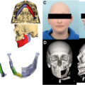
Three-dimensional (3D) printing, particularly through stereolithography, has expanded virtual surgical planning capabilities by allowing precise anatomic models, customized cutting guides, patient-specific implants, and nonlinear fixation plates to be fabricated. Integrating virtual surgical planning into microsurgery has significantly improved preoperative planning and intraoperative efficiency, especially in complex reconstructions of the head, neck, and extremities. , Using computed tomography data, surgeons can visualize a patient’s anatomy in 3 dimensions, allowing them to virtually plan their flap and create customized intraoperative cutting guides and patient-specific implants. , Such technologies have been particularly transformative in microsurgical reconstructions of the head and neck, where accurate restoration of bone and soft tissue directly influences functional and esthetic outcomes and has significantly advanced our ability to replace “like with like.”
Virtual planning in mandibular reconstruction improves the accuracy of bone alignment, reduces ischemia time, and shortens operative durations. The precision of virtual surgical planning-guided osteotomies has enabled the immediate placement of dental implants during fibula flap reconstruction, facilitating osseointegration before adjuvant radiation therapy and improving long-term prosthodontic outcomes. This strategy underpins the “Jaw in a Day” concept, which has been propelled forward by the accuracy and customization made possible through virtual surgical planning. Moreover, 3D-printed fixation plates can be designed with nonlinear configurations not achievable with traditional hand-bent linear plates, offering improved adaptability to patient-specific anatomy.
Recent developments have expanded virtual surgical planning capabilities beyond bone reconstruction to complex soft-tissue reconstructions. Advanced navigation systems integrating augmented reality and virtual plans facilitate accurate flap inset, vessel alignment, and avoidance of critical structures during surgery. ,, Recent studies report high spatial fidelity between augmented reality projections of the anterolateral thigh and deep inferior epigastric artery perforators and actual intraoperative anatomic findings, underscoring the potential of augmented reality to enhance surgical accuracy during flap harvest and inset. ,, Additionally, combining virtual surgical planning with augmented reality offers surgeons real-time intraoperative navigation, significantly enhancing precision, reducing complication risks, and streamlining surgical decision-making. , As computer software and three-dimensional technology continue to advance, virtual surgical planning combined with augmented reality can become an essential component of preoperative and intraoperative planning in reconstructive microsurgery to improve surgical efficiency and patient outcomes.
Robotic microsurgery
Flap Harvest: Updates on Use and Expanded Indications
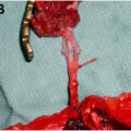
Robotic surgery has been increasingly integrated into microsurgical practice, particularly for flap harvest in autologous breast reconstruction. Recent systematic reviews and clinical studies demonstrate that robotic-assisted harvest of deep inferior epigastric perforator (DIEP) and latissimus dorsi flaps provides outcomes comparable to traditional open approaches, with added benefits such as reduced donor-site morbidity, smaller incisions, and improved donor site cosmesis. ,,, Although initial robotic surgeries reported longer operative times, recent data indicate this duration decreases as surgeons accumulate experience. A recent systematic review of robotic DIEP flap reconstructions reported high overall success rates with minimal complications, suggesting the robotic approach as a promising alternative for abdominal-based breast reconstruction. ,, Similarly, robotic-assisted latissimus dorsi harvest has been validated as a safe and effective technique in autologous breast reconstruction, offering improved patient satisfaction regarding esthetics and reduced donor-site morbidity. Notably, robotic DIEP harvest allows for significantly smaller fascial incisions, but requires identification of a short intramuscular perforator course to be feasible, making preoperative imaging essential. In contrast, robotic latissimus flaps are less suited when a skin paddle is needed. However, they eliminate the need for a sizable donor-site incision when the flap is harvested for muscle-only applications.
Beyond breast reconstruction, robot-assisted surgery has expanded into harvesting omentum and gracilis muscle flaps. , Robotic omentum flap harvest provides minimally invasive access and excellent visualization for vascular dissection while minimizing postoperative pain and intra-abdominal complications, and potentially reducing the risk of abdominal wall morbidity (eg, hernia development) after surgery. ,, Robotic techniques have also been used for free gracilis muscle flap harvest with significantly smaller donor-site incisions than conventional approaches, resulting in improved donor-site esthetics and decreased donor-site morbidity. The evolving applications for robotic flap harvest highlight the potential utility of robotic surgery when prioritizing minimally invasive techniques to mitigate donor site morbidity. Before robotic surgery is introduced on a large scale in reconstructive microsurgery, further evidence demonstrating cost-effectiveness within health care systems and additional surgeon training and proficiency are needed.
Microsurgical Anastomoses
Recent innovations in robotic surgery have expanded the indications for robotic-assisted microsurgery beyond flap harvest and now include microsurgical and supermicrosurgical anastomoses for lymphatic procedures. , Dedicated robotic microsurgical platforms, such as the Symani Surgical System and MUSA (MicroSure, the Netherlands), have demonstrated feasibility and safety in performing microvascular and supermicrosurgical anastomoses in clinical and preclinical studies. , Malzone and colleagues recently reported a randomized preclinical trial where robotic systems completed microvascular arterial and venous anastomoses with high patency rates, comparable to traditional techniques, highlighting the potential of robotic assistance in microsurgery for challenging anatomic sites or extremely small vessels (<0.8 mm). Additionally, robotic supermicrosurgery has been applied in treating breast cancer-related lymphedema to successfully perform lymphaticovenous anastomoses with outcomes equivalent to nonrobotic techniques, demonstrating feasibility of the robot’s use in lymphatic reconstruction.
Robotic-assisted microsurgical anastomoses offer potential advantages, including enhanced precision through tremor filtration, improved ergonomics, and the ability to perform procedures in anatomically challenging sites. , These systems can scale down surgeon hand movements by up to 20:1, allowing for refined instrument control by eliminating physiologic tremor. This level of precision is particularly advantageous in confined or difficult-to-access regions such as intraperitoneal, intraoral, or deep pelvic spaces, where traditional instrumentation may be limited. , Early clinical experiences demonstrate that these technologies provide consistent outcomes and increased accuracy compared nonrobotic methods and may potentially broaden the technical availability of microsurgery to surgeons. ,,, However, there are inherent disadvantages to the robotic platforms. This includes significant costs required to acquire and maintain robotic surgical platforms, which limit widespread adoption in health care systems. Furthermore, while robotic platforms show promise for supermicrosurgical procedures, most microsurgeons are not routinely performing anastomoses on vessels that fall within the range of 0.3 to 0.8 mm, where the utilization of a robot would be most beneficial. Cost-effectiveness analyses are necessary before widespread implementation can be justified, particularly within cost-sensitive health care systems, highlighting the need for continued development to balance efficiency, outcomes, and long-term financial sustainability.
Perfusion assessment and flap monitoring
Updates in Flap Perfusion Assessment
Near-infrared fluorescence imaging with indocyanine green angiography (ICGA) has become an essential tool during microsurgical reconstruction, enabling intraoperative visualization of tissue perfusion and vascular patency. ICGA provides surgeons with immediate data on flap perfusion, allowing real-time decisions that can decrease postoperative complications such as fat necrosis and total flap loss. In DIEP flap breast reconstruction, recent meta-analyses demonstrated that intraoperative use of ICGA significantly reduces the rate of postoperative fat necrosis and reoperation compared to clinical judgment alone. , Smit and colleagues confirmed fluorescence imaging with ICGA as the most accurate intraoperative tool to assess flap perfusion, noting increased flap survival when adjustments were made based on this assessment. Similarly, Singaravelu and colleagues found that the systematic application of ICGA significantly improved clinical outcomes and was cost-effective in breast and head and neck reconstruction, substantially reducing reoperation rates and postoperative complications.
Limitations to ICGA include a reliance on subjective interpretation and the requirement for intravenous dye injection. This has led to interest in emerging technologies such as laser speckle contrast and hyperspectral imaging, which are currently being explored for their advantages over traditional fluorescence-based methods. For example, laser speckle contrast imaging enables rapid, noncontact visualization of microvascular blood flow, providing dynamic, high-resolution perfusion maps without the need for injectable contrast agents. , Recent studies suggest laser speckle contrast imaging offers reliable perfusion assessment comparable to ICGA, with potential advantages including repeated measurements and real-time flow visualization throughout surgery. , Although these technologies remain investigational, their introduction highlights a broader trend toward minimally invasive, real-time perfusion assessments during reconstructive surgery. As these technologies continue to develop, microsurgeons will have increasingly powerful tools to assess flap perfusion and viability during surgery, improving patient outcomes.
Updates on Wireless, Implantable, and Nonimplantable Systems for Flap Monitoring
Postoperative flap monitoring has increasingly emphasized wireless and implantable devices designed to detect flap compromise at the earliest stage. Current monitoring techniques, such as clinical assessment, handheld Doppler probes, and wired near-infrared spectroscopy devices like ViOptix, have limitations. These include intermittent rather than continuous assessment, restricted mobility due to tethered cables, and reliance on subjective interpretation. ViOptix utilizes near-infrared spectroscopy to continuously measure tissue oxygen saturation (StO 2 ), whereas the Spectros T-Stat device employs visible light spectroscopy to assess hemoglobin oxygenation at the capillary level. Both technologies have demonstrated the ability to detect flap compromise earlier than clinical examination, with some studies reporting that they can identify perfusion deficits several hours before clinical examination. , Recent clinical trials of wearable wireless multisensor devices have demonstrated improved sensitivity and reliability compared to traditional monitoring. Tomioka and colleagues validated a wireless wearable system combining thermal, oxygenation, and perfusion sensors, enabling earlier detection of flap compromise and rapid surgical intervention, enhancing salvage rates compared to standard clinical monitoring alone. Similarly, Guo and colleagues reported promising results with wireless implantable optical probes, highlighting their ability to continuously monitor flap oxygen saturation, providing immediate alerts of vascular compromise.
Emerging noninvasive optical methods, such as diffuse reflectance spectroscopy, also show promise. Moreno-Oyervides and colleagues recently introduced a portable optical instrument based on this technology, designed to offer continuous, real-time flap monitoring with high sensitivity to changes in hemoglobin concentration. This allowed for the reliable detection of flap compromise and the ability to distinguish between arterial and venous occlusions in animal models. These early innovations represent a promising area of investigation toward reliable and minimally invasive surveillance methods that could improve flap monitoring and increase salvage rates in the setting of arterial or venous compromise.
Artificial intelligence in microsurgery
Artificial intelligence can transform clinical decision-making in microsurgery through enhanced preoperative planning and intraoperative assessments. Recent studies have demonstrated that machine learning algorithms can streamline preoperative planning by identifying optimal perforators on computed tomography angiography, reducing planning time and enhancing surgical predictability. , Intraoperatively, artificial intelligence-driven tools such as augmented reality overlays and computer-vision systems offer real-time anatomic visualization, potentially decreasing complications by helping surgeons through complex flap dissection and inset. Deep learning has also been applied intraoperatively to analyze perfusion imaging, automating the quantification of flap viability and assisting surgeons in objectively interpreting complex imaging data during flap inset. , The ability of these technologies to provide real-time, data-driven feedback highlights a promising adjunct to assist surgeons with preoperative and intraoperative decision-making.
In surgical education, artificial intelligence-driven technologies such as virtual reality simulators and automated skills assessment platforms have shown promise for improving resident training. Recent evidence supports the integration of artificial intelligence-driven performance metrics into microsurgical simulators, allowing trainees to receive immediate, objective feedback on technical skills such as instrument handling, motion efficiency, and anastomotic precision. , Morris and colleagues reviewed using artificial intelligence-enhanced training modules in surgical education, demonstrating improved technical skill acquisition through artificial intelligence-guided feedback, personalized adaptive learning, and objective skill assessment. Such artificial intelligence-based platforms can identify specific areas for improvement and accelerate the acquisition of microsurgical skills among trainees through real-time feedback. , As these educational technologies become more accessible, they can enhance resident training in microsurgical techniques by providing consistent, objective, and personalized educational experiences. ,,
Innovation with anastomotic devices
Recent advances in microsurgery have emphasized sutureless anastomotic techniques designed to enhance surgical efficiency, reduce ischemia time, and decrease technical complexity. Mechanical coupling devices traditionally used for venous anastomoses are being increasingly explored for arterial applications. Gerling and colleagues reported successful preclinical results with the Vaso-Lock device, a mechanical sutureless coupling system for arterial anastomoses, demonstrating robust patency and shorter operative times compared to hand-sewn techniques in a swine femoral artery model. Additionally, systematic reviews by Ribaudo and colleagues and Maellela and colleagues discussed multiple innovative sutureless methods, including mechanical couplers, adhesives, and laser-assisted techniques, highlighting their benefits in reducing vessel trauma, procedural time, and improving reliability compared to hand-sewn techniques. However, the authors noted the need for additional clinical validation, device refinement, and cost-effectiveness evaluation before widespread adoption.
Venous coupling devices, notably the GEM microvascular coupler, are widely used due to their proven reliability, rapid application, and consistently high patency rates, particularly in breast and head and neck reconstructive surgery. These couplers significantly reduce operative duration and intraoperative variability, improving outcomes irrespective of surgeon experience level. Nevertheless, their limitations include potential difficulties in coupling thick-walled or calcified vessels, compliance issues with significant vessel mismatch that could predispose to arterial or venous thrombosis, and device-associated costs. Ongoing research and development continue to refine coupling device designs, aiming for broader clinical applicability with arterial anastomoses, reduced complications, and improved economic viability. As these devices and methods become increasingly refined, sutureless techniques could become routine in not only venous but also arterial anastomoses to enhance intraoperative efficiency. ,,
Related posts:

Stay updated, free articles. Join our Telegram channel

Full access? Get Clinical Tree


