Indications and approaches for pediatric free tissue transfer have expanded in recent years, and most flaps historically described in the adult population are now routinely used in children. Those embarking on pediatric microsurgical operations must understand expected growth and development and should incorporate these factors into the reconstructive plan. Virtual surgical planning is revolutionizing preoperative planning and intraoperative execution and should be considered standard-of-care in complex cases. New technologies including microsurgical robotics and artificial intelligence will continue to refine and improve pediatric microsurgical outcomes in the years to come.
Key points
-
•
Pediatric microsurgery is safe and effective, and can obviate the need for multi-stage procedures such as tissue expansion and pedicled flaps.
-
•
Soft tissue and bony reconstruction in the pediatric population needs to account for stage of development, and flaps should be planned according to expected and desired growth.
-
•
Virtual surgical planning allows for more predictable reconstruction with shorter operative times and specification of hardware tailored to the pediatric skeleton.
-
•
Indications for pediatric microsurgery continue to expand, and advancements in robotic and artificial intelligence technologies may further change the paradigm of care.
Abbreviations
| DVT | deep vein thrombosis |
| FP | Facial paralysis |
| FVFG | free vascularized fibula grafts |
| LMWH | low molecular weight heparin |
| SIEA | superficial inferior epigastric artery |
Introduction
After Jacobson and Suarez pioneered the field of microsurgery in 1960 with the use of the operating microscope, Malt and McKhann performed a successful mid-humeral replantation on a 12 year old boy. ,, Despite early doubts about the ability to anastomose pediatric-sized vessels, pediatric free tissue transfer has grown to display equal to or higher success rates than adult microsurgery, and indications have and continue to expand. ,
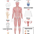
We herein provide an overview of microsurgical reconstruction of the geographic-specific pediatric defect, namely craniofacial/head and neck, upper extremity, and lower extremity. Fig. 1 summarizes the various etiologies and some example microsurgical options for specific defects.
Indications for pediatric microsurgery.
( Illustration by D. Low, MD [Philadelphia, PA].)
General principles of pediatric microsurgery
Preoperative Considerations
The Food and Drug Administration announced in 2017 that based on earlier animal studies, repeated or prolonged sedation in children under 3 year old can have a negative impact on neurocognitive development and advised that young patients should receive these medications only when necessary and under caution for elective procedures. , Since then, ongoing clinical studies suggest multiple anesthetic exposures have a higher risk for cognitive and behavioral problems. Microvascular procedures are often lengthy, yet alternative methods of reconstruction can also be equally time-consuming or require multiple stages. Therefore, in young children, the choice of reconstruction should consider cumulative sedation time. In microsurgery, strategies to expedite operative time and decrease anesthesia exposure, including 2 team approaches to free flaps, should be considered. ,,,
Flap preferences have gradually shifted from muscle flaps to free perforator flaps for soft tissue defects. , Muscles exert biomechanical forces on bone which impact the balance of the skeletal structure. This idea led to the concern that the absence of a muscle at the donor site for a free flap may cause skeletal structure and growth abnormalities, but this concern was recently disproved. , Compared to muscle flaps, fasciocutaneous flaps may offer relatively less tissue, and pediatric compared to adult skin may have greater skin recoil, requiring a larger skin paddle than expected. , Donor site primary closure may not always be possible ; tissue expansion and delayed closure may avoid poor scarring with donor site skin grafts. , Ultimately, both muscle and fasciocutaneous flaps are considered safe and effective in children.
Intraoperative Considerations
Though early case reports cited vasospasm and small vessel size as risks for failure in pediatric microsurgery, a review including 504 cases reported only one case of flap failure reportedly caused by vasoconstriction. ,,,,, Even transient vasospasm is not encountered more frequently than in adults ,, ; careful dissection, ,, minimal vessel adventitia removal, peripheral nerve blocks, and maintaining warmth have all been suggested to reduce risk. Vasospasm intraoperatively is typically managed with topical papaverine and 2% lidocaine. ,,,
It was once thought that the smaller size of children’s blood vessels could cause failure due to poor perfusion of the flap. Although pediatric vessels are smaller, they are not proportionally smaller. The vessel-to-flap-volume ratio is often more favorable than in adults. ,,, However, some studies cite higher flap failure rates in children under 5 year old, likely due to the technical difficulty of suturing extremely small vessels. In these younger patients, procedural optimization involves microscope magnification (16–20x), , multiple arterial anastomoses if the pedicle is less than 0.5 mm, , and involvement of experienced microsurgeons. A bolus of heparin at the time of anastomosis, if given, is typically dosed at 50 to 100 U/kg up to 5000 U intravenously. ,, Structural differences in pediatric anatomy have been described. Serletti and colleagues described the vessels of children under 10 year old as thin with a “gelatinous” consistency. Parry and colleagues speculated that the muscular layer of the vessel walls in young children may not be developed, and though this hypothesis has been widely cited, histologic evidence is lacking.
Postoperative Considerations
Postoperative anticoagulation and aspirin practices vary. Children are less likely to develop thromboses compared to adults due to higher circulating levels of anti-coagulative proteins, lower pro-thrombotic precursors, and less reactive vascular endothelium due to lack of chronic disease. ,, Some surgeons prescribe neither aspirin nor anticoagulation routinely except in the setting of trauma or complications. ,,,, Others routinely use heparin, low molecular weight heparin (LMWH), or aspirin in variable doses and durations. ,,,,,, When used, the typical heparin dose is 100 to 150 U/kg/day, , the typical LMWH dose is 1 mg/kg/day subcutaneously, , and the reported aspirin doses are 1 to 5 mg/kg/day, 75 mg/day, or 81 mg/day. Reye syndrome is a rare but potentially fatal illness with encephalopathy and liver failure associated with aspirin use in children, particularly during recovery from viral illness. Although no complications have been reported after pediatric free tissue transfer, aspirin is generally not recommended in children under 12 years due to this risk. Clinicians should monitor for recent viral illness and recognize early signs (vomiting, lethargy, confusion, hyperventilation, tachycardia) and late signs (decorticate rigidity, seizures, pupil hyporeactivity, respiratory failure).
A recent review of 73 pediatric free flaps found no benefit to routine anticoagulation or aspirin in pediatric microsurgery beyond deep vein thrombosis (DVT) chemoprophylaxis, which is not even always indicated for children. The current recommendation for DVT chemoprophylaxis in pediatric trauma patients is LMWH at 0.5 mg/kg/dose subcutaneously every 12 hours for all adolescents over 15 year old and all post-pubertal children under 15 year old with an Injury Severity Score of greater than 25 ; no monitoring of Anti-Xa levels is needed. There are no specific guidelines for children whose recipient bed has been irradiated.
Young children may be uncooperative postoperatively, so construct protection through splinting, casting, or even external fixators may be necessary in addition to postoperative sedation. Postoperative pain and anxiety can trigger vasospasm, so peripheral nerve blocks and sedatives may be beneficial for flap perfusion in addition to comfort and compliance.
Children who sustain traumatic injuries are at greater risk for anxiety, depression, substance abuse, and other mental health disorders later in life. Family engagement, child life specialists, and skilled pediatric rehabilitation specialists can help improve compliance , and provide psychological support. Despite the possible cooperation challenges, children tend to recover quickly and adapt well to residual disability. ,,
Craniofacial/head and neck
There is a wide range of indications for pediatric microsurgical reconstruction of the cranium, face, and neck. Reasons include congenital and progressive causes of both bony and soft tissue asymmetry, recalcitrant cleft palate, facial paralysis (FP), and both benign and malignant tumors. Ultimately, reconstruction in children may involve any combination of soft-tissue filling for volume enhancement, subunit restoration, bony replacement with occlusal restoration, and neuromuscular transfer for functional repair.
Congenital Facial Asymmetry, Progressive Atrophy, and Cleft Palate
Pediatric congenital facial asymmetry covers a range of disorders, including craniofacial microsomia, Treacher Collins syndrome, and orbitofacial clefting, secondary to deformities of the first two pharyngeal arches with resultant deficiencies in the midface and mandible. The hypoplasia and asymmetry of bone, muscle, subcutaneous tissue, and skin of the mid-face and lower face, provides a natural role for microsurgical reconstruction.
Bony microsurgical reconstruction of the mandible in pediatric hemifacial microsomia is well described with success rates over 90%. Free flaps are not first line, as the advancement of distraction osteogenesis has obviated their need in cases of small-to-moderate defects of Pruzansky grade I or II with preservation of the ramus or condyle. Nonvascularized iliac and costochondral bone grafts have been used in these patients with variable outcomes secondary to bone resorption and unpredictable impacts on growth. However, for patients with Pruzansky grade III mandibles lacking sufficient bone stock for distractor placement and an unstable or nonexistent condyle, free flap reconstruction has evolved as the most appropriate mode of reconstruction. In particular, iliac, scapular, and fibular flaps have been described. ,, Preoperative virtual surgical planning (VSP) and prefabricated cutting jigs for precise congenital mandibular reconstruction can facilitate further orthognathic procedures. Cleveland and colleagues show a 100% flap success rate in Pruzansky Grade III mandibles undergoing preplanned free vascularized fibula grafts (FVFG) transfer and all patients who required dental implants obtained them successfully. Due to the long and segmental blood supply, the FVFG allows for multi-segment osteotomies more readily than parascapular and iliac flaps, though iliac bone has been noted to have a more natural mandible-resembling curvature and height. This can make it easier to place osseointegrated implants in tooth-bearing areas. FVFG can be harvested without long-term orthopedic implications with only 1 case of an unspecified abnormal gait in 87 patients in one study. The same study reported hardware removal in 25% of patients, though 13 of 22 cases (59%) were elective removal with only 1 case (4.5%) due to long-term plate exposure, with the remaining were noted as unknown reasons. The researchers suggested hardware may be maintained long-term in pediatric reconstruction cases with minimal long-term complications or growth/skeletal impact, in comparison to those in the adult literature that report up to 20% hardware exposure rates. ,
Soft tissue reconstruction is more variable than bony mandibular reconstruction. In addition to the aforementioned congenital etiologies, it includes Parry-Romberg syndrome, a disorder involving unilateral atrophy of the skin and subcutaneous tissue and less commonly of the facial skeleton. As Inigo and colleagues note in their experience with 118 cases of hemifacial microsomia and Romberg’s disease, soft tissue free flaps can offer the ideal reconstructive modality as reconstructions (1) must be malleable, (2) must undergo minimal change in size and position, and (3) should mimic surrounding tissue. Saadeh and colleagues reviewed 93 patients who underwent 112 superficial inferior epigastric artery (SIEA), groin, and circumflex scapular flaps for facial asymmetry and had improved subjective symmetry, improved esthetics, and no flap losses. They also note the effect on local skin and soft tissue texture, tone, and color and recommend free tissue transfer as early as 6 years of age. Various flaps have been described for soft tissue asymmetry including the omental, scapular/parascapular, latissimus dorsi muscle, groin, serratus anterior muscle, SIEA, anterior lateral thigh, and others. ,,,,,
Primary and revision cleft palate repairs primarily utilize local flaps. Meta-analysis data indicate palatal fistula occurs in 6.4% to 8.6%. ,, Large fistulas or those encased in significant local scar may require free tissue transfer. MacLeod et al. first described the free radial forearm for closure of large palatal fistulae in 1983. Schwabegger and colleagues recommend free tissue transfer with greater than 3 failed closure attempts, significant scar burden, unsuitability for mucosal or buccal flaps, and fistula size more than 1 cm 2. A systematic review of 65 patients, aged 3 to 55 years, and a median fistula size of 8 cm 2, found the most common flap choice was radial forearm flap (N = 37), noting its hairless skin paddle, long pedicle, thinness, and folding ability. The authors suggested utilizing the inherent flap skin for oral or nasal lining rather than prelaminating, anastomosing to the facial vessels with rare use of vein grafts, and repairing subsequent small dehiscence with flap re-advancement. The most complex technical aspect of palatal free flaps is choosing the pedicle pathway, with some advocating a tunnel through the cheek and others utilizing a submucosal or subcutaneous tunnel or even a tunnel through the retromolar trigone or preexisting alveolar cleft. Gur and colleagues recommend incising the mucosa rather than tunneling and utilizing a skin tail on the radial forearm along the pedicle to subsequently close the defect. This provides for good surgical exposure of the pedicle and easier control of unexpected bleeding.
Dynamic Facial Reanimation
FP in children under 15 years of age has an incidence of 21.1 cases per 100,000 per year. Compared to the adult population in which Bell’s or idiopathic causes predominate, pediatric FP is more commonly congenital or acquired (ie, perinatal trauma). Syndromes associated with FP include Moebius syndrome, Goldenhar-Gorlin syndrome, CHARGE (coloboma, heart defects, atresia choanae, retardation of growth, genital or urinary abnormalities and ear anomalies), hemifacial microsomia, and others. Free muscle transfer is the gold standard treatment of dynamic smile reconstruction, with pectoralis minor, latissimus dorsi, serratus, and gracilis all described. , The free gracilis, first described by Harii in 1976, is favored secondary to reliable anatomy, low donor site morbidity, easy customization, and a natural 2 team approach ( Fig. 2 ). ,,, Most modern surgeons harvest a segmented gracilis based on fascicular territory to reduce donor site morbidity and utilize the facial vessels as recipient vessels due to ease of access and caliber relative to the superficial temporal vessels. Donor nerve selection is perhaps the biggest decision point in free muscle transfer for pediatric patients. Cross-facial nerve with grafting, first described in 1980, remains the donor nerve of choice due to its spontaneous and more natural appearing effect on commissure excursion, but the masseteric nerve is used in bilateral cases or when looking to increase the power of commissure excursion with dual-innervation. ,,, Axonal density is the primary determinant of the degree of facial movement.
Free segmented gracilis for facial reanimation demonstrating anastomosis to the facial artery and vein as recipient vessels and a cross-facial sural nerve graft coaptation to the obturator nerve. An implantable Doppler is utilized.
( Illustration by D. Low, MD [Philadelphia, PA].)
Extensive physiotherapy is needed for neural training. Delaying facial reanimation until school age (5–6 years) allows time for children to become mindful and more active participants in operative care and therapy, as well as providing for larger vessel size. Ultimately, timing of intervention is tailored to the specific child and family, balancing the tradeoff of better rehabilitation cooperation in older children versus better neuroplasticity in younger children.
Head and Neck Malignancies
Head and neck malignancies account for 12% of all pediatric cancers with an incidence of 1.49 cases per 1,000,000 person-years. Pediatric tumors tend to be benign and mesenchymal in origin than in adults. Neural tumors and soft tissue sarcomas affect infants and toddlers while lymphomas and thyroid malignancies affect older children. Ameloblastoma, a benign tumor of ectodermal tooth enamel or dental lamina, is the most common mandibular growth in children. Free flaps for tumor-related head and neck reconstruction are well established.
Parham and colleagues describe a useful “subunit” approach based largely off of variations in development and skeletal maturity, segmenting into scalp/cranium/skull base, orbit, maxilla, and mandible. Growth and development are generally least concerning when it comes to cranial and orbital reconstruction, as skeletal maturity in these regions approaches that of adulthood by age 6 to 8 years. The mid-facial and lower facial skeleton, however, sees significant growth in puberty.
Given the timing of resection is dictated by oncologic urgency, ideal timing of bony versus soft tissue reconstruction is debated, as some studies have suggested younger age (<9 years) to be a risk factor while others found more complications in the older age group. , Demonte and colleagues performed 9 soft tissue free flaps for large skull base defects and found adequate vessel size, no need for vein grafts, and no significant adverse effect on craniofacial development. Though success rates for osseous and soft tissue flaps generally do not differ, researchers advocated for an individualized approach utilizing soft tissue flaps and waiting for skeletal maturity before performing bony flaps. At our institution, we regularly utilize VSP to plan oncologic resection and fibula osteotomy design, custom cutting guides, and custom hardware ( Fig. 3 A–D ).
A 15 year old girl with Ewing sarcoma of left mandible reconstructed with 2 segment osteocutaneous free fibula. ( A ) Preoperative 3D rendering of computed tomography scan (CT) max/face with overlayed resection plan ( red ). ( B ) Preplanned 2 segment fibula with custom hardware. ( C ) Postoperative front and lateral clinical views. ( D ) Postoperative 3D CT demonstrating appropriate alignment of fibula and custom hardware.
( Images courtesy of C. Ligh, MD [Philadelphia, PA].)
The same principles apply to bony reconstruction when comparing malignancy and congenital pathologies, and there still remains some debate whether osseous flaps continue to grow with the patient. General consensus is that condylar preservation in the case of mandibular reconstruction allows for growth while radiotherapy inhibits. , Hardware generally has minimal long-term exposure risk and some advocate it can be left in place with growth of the facial skeleton. Dental rehabilitation and proper occlusion likely promote mandible growth and facial symmetry. ,,
Clinics care points
-
•
Muscle and fasciocutaneous flaps are considered safe and effective in the pediatric population; though, consideration for prolonged or multiple anesthetic exposures should be noted during surgical planning and discussed with families.
-
•
Microscope magnification should be considered in younger children, due to size of vessels.
-
•
There is no consensus on post-operative anticoagulation and aspirin practices, though in patients under 12 years should closely monitor for Reye syndrome
-
•
Post-operative management (construct protection and pain management/sedation) is crucial in the pediatric population as compliance is variable and pain and anxiety have been shown to trigger vasospasm.
-
•
Virtual surgical planning (VSP) should be heavily considered for bony reconstruction in head and neck cases, as they can improve patient outcomes.
Related posts:

Stay updated, free articles. Join our Telegram channel

Full access? Get Clinical Tree


