Indications and approaches for pediatric free tissue transfer have expanded in recent years, and most flaps historically described in the adult population are now routinely used in children. Those embarking on pediatric microsurgical operations must understand expected growth and development and should incorporate these factors into the reconstructive plan. Virtual surgical planning is revolutionizing preoperative planning and intraoperative execution and should be considered standard-of-care in complex cases. New technologies including microsurgical robotics and artificial intelligence will continue to refine and improve pediatric microsurgical outcomes in the years to come.
Key points
-
•
Pediatric microsurgery is safe and effective, and can obviate the need for multi-stage procedures such as tissue expansion and pedicled flaps.
-
•
Soft tissue and bony reconstruction in the pediatric population needs to account for stage of development, and flaps should be planned according to expected and desired growth.
-
•
Virtual surgical planning allows for more predictable reconstruction with shorter operative times and specification of hardware tailored to the pediatric skeleton.
-
•
Indications for pediatric microsurgery continue to expand, and advancements in robotic and artificial intelligence technologies may further change the paradigm of care.
Abbreviations
| AI | artificial intelligence |
| FVFG | free vascularized fibular graft |
| FVET | Free vascularized epiphyseal transfer |
| LLB | Limb-length discrepancy |
| PLP | phantom limb pain |
| RLP | residual limb pain |
| VCA | Vascularized composite allotransplantation |
| VSP | Virtual surgical planning |
Hand and upper extremity
Microsurgical needs in the hand and the upper extremity can be largely divided into congenital and acquired indications. Congenital anomalies include absent digits, radial longitudinal deficiency, and pseudoarthrosis. Acquired indications include trauma or oncologic resections and may include replantation or revascularization, bony reconstruction, free functional muscle transfer, and vascularized composite allotransplantation.
Congenital Anomalies: Absent Digits
Absent digits may be due to adactyly, symbrachydactyly, transverse failure of formation, cleft hand, or constriction ring/amniotic band syndrome. ,,,,, While children adapt well to some absent digits and others do well with pollicization, some scenarios benefit from toe-to-hand transfers: (1) absent thumb distal to carpometacarpal (CMC) joint; (2) absent thumb, index, middle, and ring fingers, but with an intact small finger; (3) intact thumb but otherwise absent fingers; and (4) complete adactyly of all digits. The goal is to restore at least one finger and thumb for opposition to allow pinch and grip.
In a study of 15 children who underwent 20 toe-to-hand transfers, the average age at surgery was 4.9 years and patients or their parents were surveyed at a mean follow-up of 7.6 years. The patients had subjective functional scores comparable to “normal” children and reported essentially no morbidity with minimal difficulty walking at 2 weeks postoperatively. However, in another series of 16 cleft hand patients with 20 toe transfers at a surgical mean age of 6 years, grip and pinch strength never exceeded 30% of normal values. Revisions included tenolysis, bony fusion or revision fixation for nonunion or malunion, and skin graft revision. Donor site morbidity is usually minimal, and most patients are asymptomatic without problematic clinical or radiologic findings at up to 17.4 year follow-up.
Congenital Anomalies: Radial Longitudinal Deficiency
The Vilkki procedure is the classic treatment for congenital radial club hand reconstruction when centralization is unsuccessful. The second metatarsophalangeal joint with a viable physis is transferred to the native ulna in a Y configuration to create a one-bone forearm and avoid severe radial deviation. A 30 year study on 34 congenital radial club hand reconstruction demonstrated that the procedure resulted in better wrist mobility without restricting ulnar growth until at least age of 11 years. Patients should undergo active hand therapy and centralization with wrist distraction as young as possible, and the best age for the free flap is age 3 to 6 years, as wrist tightness worsens after age of 3 years. A modification on the classic Vilkki procedure in patients with type 3 radial longitudinal deficiency (diminutive radius) uses the toe metatarsal to reconstruct the radius for a 2 bone forearm. The toe tendon can reconstruct the interosseous membrane to decrease the risk of longitudinal instability. In their patient with 2.5 year follow-up, active wrist flexion to 70° and active wrist extension to neutral was achieved with maintenance of passive pronosupination. At 7 year old, the patient still had open physes and was without donor site complications.
Congenital Anomalies: Pseudoarthrosis
Pseudoarthrosis is a rare condition associated with neurofibromatosis with congenital dysplasia or progressive deformity of the arm either congenitally or after minor trauma resulting in poor bony healing. , Progressive bowing of the forearm can lead to slow deterioration of arm function. Treatment options include open reduction internal fixation (ORIF) with or without autologous bone grafting, external fixation, vascularized bone graft, and radioulnar fusion into a one-bone forearm. In a systematic review of 47 studies of 84 cases, Siebelt, and colleagues recommend first-line reconstruction with free vascularized fibular graft (FVFG) given an 87% union rate with good function.
Acquired Anomalies: Replantation and Revascularization
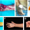
Success rate of digit replantation in children is cited at 63% to 97%. While the smaller caliber of all the structural components makes it technically challenging, the likelihood of spasm is thought to be no different from that in adults. Indications for replantation in children is broader given better functional outcomes even in those amputated proximal to the flexor digitorum superficialis (FDS) insertion and in index or small finger replantations ( Fig. 1 A–G ). Children have better nerve regeneration both peripherally and central neuroplasticity to avoid motor dysfunction, chronic pain, and pain syndromes.
A 14 year old boy sustained a right ring finger subtotal amputation and a small finger total amputation with a wood splitter ( A ). The small finger was repaired as a composite graft. The ring finger was replanted with bony fixation of the oblique fracture with crossed K-wires ( B ). Both flexor tendons were repaired. One digital artery was repaired with a dorsal foot vein graft; a dorsal vein was repaired; both digital nerves were primarily repaired ( C ). By 7 months postoperatively, the patient had excellent range of motion ( D–F ), demonstrated good bony healing ( G ), and returned to all activities including football and wrestling.
(Figures courtesy of Ines C. Lin, MD (Philadelphia, PA).)
There are, however, notable challenges, especially in polytrauma, including reduced blood volume and greater surface area, making the pediatric patient more prone to clinically significant blood and insensible losses. Postoperative leeching can also cause anemia if unattended. For bony fixation, every effort should be made to protect the physis to allow for future growth. Postoperatively, compliance is an issue, and long-term splinting or casting is necessary to avoid disruption of fine repairs. Children can escape from even extensive postoperative dressings, so extreme joint flexion in cases of direct repair should be avoided.
Acquired Anomalies: Bony Reconstruction
Unique considerations in pediatric bony reconstruction include growth potential and preservation of the joint surface for long-term pain-free joint usage. The proximal fibula epiphysis with the physis can be taken based on the epiphyseal branch of the anterior tibial artery. A strip of the biceps femoris tendon can also be taken for soft tissue reconstruction. The donor site is evaluated for knee lateral ligament stability and common peroneal nerve injury. Peroneal palsy is cited at 50%, although most are transient.
An international multi-institutional review on free vascularized epiphyseal transfer (FVET) for proximal humerus osteosarcoma reconstruction identified 27 patients. The median age at surgery was 7 years and the median defect length was 12.5 cm. Complications included flap failure (7%), fracture (41%), avascular necrosis of the fibula head (4%), infection (4%), and hardware failure (4%). Peroneal nerve palsy was seen in 59%, of which 11% were permanent. At the recipient site, longitudinal growth was seen in 89%. The biceps femoris tendon and lateral collateral ligament were used for shoulder joint capsule or rotator cuff stability.
Acquired Anomalies: Free Functional Muscle Transfer
In cases of compartment syndrome, brachial plexus injury, or central nervous issues (ie, cerebral palsy, traumatic brain injury), free functional muscle transfer may help restore function. The gracilis, semitendinosus, pectoralis major, and latissimus dorsi are all described donor muscles. ,
Zuker and colleagues reported 6 pediatric cases of Volkmann ischemic contracture treated with free functional gracilis for digital flexor and extensor reconstruction at an average age of 6.1 years. While there were notable differences compared to the normal arm, there were dramatic improvements in overall active range of motion at all hand joints as well as grip and pinch strengths. One child required tenolysis and another developed wrist flexion contracture from imbalance with their native weakened wrist extensors. Wrist fusion may avoid the latter complication, but this would be recommended only after full bony growth.
Hattori and colleagues described 3 cases of pediatric complete brachial plexus avulsion injury treated with double free muscle transfer. The first gracilis supplied by the spinal accessory nerve was used to restore elbow flexion and finger extension while the second muscle, performed 3 months later, was supplied by the fifth and sixth intercostal nerves to restore finger flexion. These transfers were also supplemented by transfer of other intercostal nerves to the triceps for elbow extension and to sensory nerve branches. At 3 year follow-up, the outcomes were better than in adults. There was some imbalance between elbow flexion and extension (nerve transfer to native triceps) that caused elbow contracture, but the muscles grew in proportion to skeletal growth and the contractures did not worsen over time. Motivating and explaining the postoperative rehabilitation protocol in detail to both the patients and to supportive parents is paramount.
Acquired Anomalies: Vascularized Composite Allotransplantation
Vascularized composite allotransplantation (VCA) is a life-enhancing, but not life-saving procedure that can even decrease life expectancy. In addition to progressive, direct end-organ damage from immunosuppressive medications, patients are at increased risk for opportunistic infections and de novo malignancy, which is related to the cumulative time of immunosuppression. This is a serious consideration in the pediatric patient who will hopefully live decades after their transplant. , Despite these downsides, benefits in the pediatric population include greater neuroplasticity and improved psychosocial and emotional development especially among those with acquired amputations.
The first bilateral pediatric hand-forearm VCA took place in 2015 in a patient who was already immunosuppressed from kidney transplantation. The donor was matched with the appropriate size, skin color, and growth status. Post-operatively, the patient successfully engaged in a variety of physical exercises and musical instrumentation with both arms. With each annual visit, the bone age of the allotransplant increased appropriately. Four-year follow-up demonstrated appropriate growth of bilateral both bones similar to normal values in healthy children ( Fig. 2 A, B ).
An 8 year old boy underwent bilateral upper extremity vascularized composite allotransplantation at the level of the forearm. Radiographic imaging demonstrated growth from the time of transplantation and bony fixation ( A ) to postoperative year 7 ( B ). The patient continues to use his bilateral transplanted arms and is now driving 9 years postoperatively.
( Images courtesy of L.S. Levin, MD [Philadelphia, PA].)
Lower extremity
Microsurgical reconstruction of the lower extremity is indicated in limb salvage after trauma, oncologic resection, or congenital conditions.
Limb Salvage and Amputation in Children
When considering reconstructive options, both limb salvage and amputation have specific challenges specific to pediatrics. Limb-length discrepancy (LLD) requiring orthopedic revision is common after trauma or resections involving growth plates, so skeletally immature patients must be followed closely by an orthopedics team. Amputation also presents challenges specific to pediatric patients, particularly due to bony overgrowth relative to soft tissue, which can occur in up to 40% with complications including pain, pressure ulcers, skin perforation, and resulting avoidance of prosthetic use.
Factors influencing LLD prediction include age at injury, physis involvement, and remaining growth potential of native or transplanted physes. Rotationplasty, a non-microsurgical technique, is performed after removal of the knee joint as an alternative to amputation; the distal tibia is rotated 180°, with the ankle joint then able to act as the knee joint within a prosthesis. It is performed for patients under 5 year old who are expected to have large LLDs (>4–5 cm) after injury/resection and preferred to avoid multiple future orthopedic procedures for LLD. ,, Amputation is indicated for large deficits when the sciatic nerve or its branches are not intact. , In children, amputation is performed at a weight-bearing level (eg, Syme or Boyd amputation) when possible. Syme and Boyd amputations are disarticulations through the ankle that preserve the distal epiphyseal plate and reduce the chance of bony overgrowth. Children are able to stand on the residual limb at this level without prostheses.
Reconstruction for Soft Tissue Deficit
Approximately 75% of pediatric lower extremity free tissue transfer is performed for the treatment of Gustilo-Anderson grade 3B injuries due to high-energy mechanisms including motor vehicle accidents, lawnmower injuries, and crush injuries. ,, Children are less likely to sustain lower extremity injuries requiring free tissue transfer compared to adults, likely due to the use of car seats and greater bone plasticity which can withstand the force better than in adults.
Lower extremity tumors in children are most often of bony origin, but resection of a large amount of soft tissue may be required for clearance, and radiation changes may necessitate the transfer of healthy vascular tissue. The reconstructive ladder for soft tissue reconstruction and principles of lower extremity microsurgical reconstruction are the same in children as in adults. Both muscle and fasciocutaneous flaps generally grow with the child and are commonly used. ,,, In dorsal foot deformities, the flap can be measured with the foot in full plantarflexion and with a wavy distal margin over the metatarsal phalangeal joints to prevent the development of overriding toes as the foot grows.
Postoperatively, dangle protocols vary, though authors do not report that pediatric dangle protocols differ from those in adults. ,, Bony or ligamentous damage from the injury can result in progressive equinus, varus, or valgus deformities, and physeal damage can arrest growth in the affected limb. ,
Reconstruction for Bony Deficit
Indications for lower extremity use of the free vascularized fibular graft (FVFG) in pediatric patients include segmental bone loss, growth potential, and joint reconstruction. The fibula remodels and hypertrophies under the biomechanical forces of the recipient site, , and its vascularized nature improves timing to bony healing and reduces the risk of infection. It can be used alone for segmental defects greater than 6 cm as in adults, and variations of the FVFG are used for pediatric-specific indications. In 88% of pediatric patients undergoing lower extremity reconstruction with FVFG, full weight-bearing is achieved with return to normal activities. However, McCullough et al reported that 44.8% of free fibula patients had a delayed complication including fracture and LLD. LLD has been reported in up to 69% of children. ,
Children often return to activity quickly, increasing fracture risk. Therefore, attempts have been made to strengthen this construct, including double-barreled free fibulas for proximal tibia or distal femur and the Capanna technique. The Capanna technique uses a longer free fibula within the medullary canal of an allograft for larger intercalary bony reconstruction , to improve bone-healing time and fracture healing outcomes compared to allograft alone. Allografts have ingrowth from surrounding soft tissues, but this only extends approximately 60% into the depth of the cortex. A free fibula flap within the allograft augments this and gradually incorporates into the allograft. While this technique may not decrease the rate of fracture, it does shorten time to bone healing, reduce infection rate, improves functional scores, and most fractures that occur in the allograft do not require revision surgery in contrast to allograft alone.
FVET of the proximal fibula has been performed in children who require reconstruction with bone that will grow, including joint reconstruction. , The anterior tibial branch is the most reliable pedicle for this procedure and it results in the most growth (0.44–1.72 cm per year) compared to dual pedicle or peroneal artery, , which is also associated with premature epiphyseal closure. Growth also varies by recipient site: less in the femur (∼0.5 cm/year) than in the upper extremity or the mandible. Hip reconstruction with FVET has had few mixed reports.
Complications shared by all variations of FVFG include late fracture, nonunion, hardware failure, infection, peroneal nerve palsy, , donor site valgus deformity, and LLD. Nonunion and infection are more common in children undergoing chemotherapy. ,
Related posts:

Stay updated, free articles. Join our Telegram channel

Full access? Get Clinical Tree


