Clinical Features.
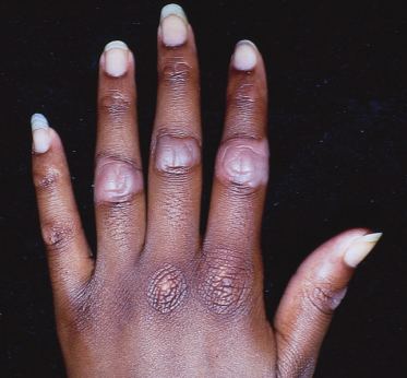
The onset of knuckle pads is generally inconspicuous, occurring over a period of months or years. Idiopathic knuckle pads of childhood are most often reported in adolescence; onset may be earlier for familial forms. Knuckle pads are most commonly described over the dorsa of the PIP joints, but can occur over the dorsal aspects of any joints of the hands and feet [2]. The lesions are flat or convex with a slightly rough surface. They may have prominent pigment change in people with darker type IV or V skin (Fig. 96.2). A possibly related idiopathic variant has been described that involves lateral, rather than dorsal aspects of the PIP joints, dubbed ‘pachydermodactyly’ [15]. People with the familial form may also have thickened plaques over the knees and elbows (Fig. 96.3) [3]. Knuckle pads are usually asymptomatic but can be disfiguring.
Fig. 96.2 This 14-year-old girl has idiopathic knuckle pads of childhood. Note the prominent hypopigmentation typically seen in dark-skinned patients.

Prognosis.
There is no uniformly successful treatment for knuckle pads. Lesions usually persist for several years, or remain indefinitely, but spontaneous resolution has been reported [3].
Differential Diagnosis.
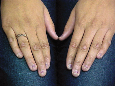
Knuckle pads should be distinguished from other cutaneous disorders, especially those with more ominous associations [3]. A medical history, physical examination and skin biopsy are usually sufficient to make the correct diagnosis. In children, the differential diagnosis includes callosities, hypertrophic scars, deep granuloma annulare, rheumatoid nodules, Gottron’s papules (Fig. 96.4
Stay updated, free articles. Join our Telegram channel

Full access? Get Clinical Tree


