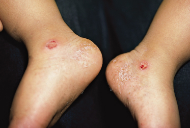
Fig. 88.2 Infantile acropustulosis. Erosions on the ankle are the result of vigorous rubbing to alleviate pruritus.
Courtesy of Dr Neil S. Prose.
Laboratory studies are not helpful in making the diagnosis. Cultures of the lesions for bacteria, fungi [3] and viruses [7] are negative. Blood count, erythrocyte sedimentation rate, stool evaluation for ova and parasites [3], Venereal Disease Research Laboratory test (VDRL), rapid plasmin reagin test (RPR) [20], Gram stain, purified protein derivative (PPD) [20], total complement activity, hepatitis B surface antigen and antibody [3], Tzanck preparation [16], T helper and T suppressor cell number and ratio [14,20] and chest radiograph [20] are all normal.
Elevated levels of circulating eosinophils have been reported in some patients with IA. With the exception of one case of profound hypereosinophilia accompanied by an elevated white blood cell count (WBC) without identifiable cause [20], there seems to be no correlation between exacerbations of IA and blood eosinophilia [5,8,12]. In addition, there is a lack of correlation between blood eosinophilia and the type of cellular infiltrate (neutrophilic versus eosinophilic) contained in the pustules and dermis. Markedly elevated levels of serum immunoglobulin E (IgE) during the active phase of the disease have been reported in two cases [4,14]. One patient had neither a personal nor a family history of atopy and the results of a radio-allergosorbent test (RAST) were negative.
The diagnosis of IA rests primarily upon the characteristic clinical findings. It is self-limited, with complete resolution by 3 years in almost all cases. It is speculated that the earlier the disease has its onset, the longer it may persist [1].
Differential Diagnosis.
The clinical picture of recurrent attacks of palmar and plantar pruritic pustules differentiates IA from most other pustular eruptions seen in infancy. The following should be ruled out prior to making the diagnosis of IA.
Scabies infestation is generally foremost on the list of differential diagnoses for IA. The morphology and distribution of the lesions may be similar to those of IA, as may be the chronicity and intensity of the associated pruritus. On histological examination of scabies, particularly in the area of a mite, there is spongiosis in the underlying spinous layer, often with resultant vesicle formation. Scabies is usually diagnosed from skin scrapings, and most patients with IA are treated unsuccessfully for scabies. Some feel that scabies should be the only differential diagnosis considered for IA [8].
Impetigo may have a similar histological picture, showing a subcorneal bulla containing numerous neutrophils, but the stratum malpighii underlying the bulla discloses spongiosis with neutrophils migrating through it. IA lesions may become secondarily infected from scratching; however, in its absence, Gram stains and cultures are negative, and the response to antibiotics should allow differentiation.
Pompholyx or dyshidrotic eczema is extremely rare in infants and the histological picture of subcorneal pustules is not present. Prominent spongiosis with fluid-filled spaces with few inflammatory cells in the superficial epidermis is observed [2].
Erythema toxicum neonatorum commonly affects newborns, usually after 12 h, almost always by 48 h after birth, and, occasionally, up to several weeks of age. Presenting as small erythematous papules and pustules on blotchy erythematous bases, it is usually widespread, favours the trunk and back more than the extremities, and is asymptomatic. It usually resolves spontaneously without recurrence or sequelae in 2–5 days.
Transient neonatal pustular melanosis
Related posts:


Stay updated, free articles. Join our Telegram channel

Full access? Get Clinical Tree


