References
1 Van Praag MC, Van Rooij RW, Folkers E et al. Diagnosis and treatment of pustular disorders in the neonate. Pediatr Dermatol 1997;14:131–43.
2 Sahn EE. Vesiculopustular diseases of neonates and infants. Curr Opin Pediatr 1994;6:442–6.
3 Moisson YF, Wallach D. Les dermatoses pustuleuses de la periode neonatale. Ann Pediatr (Paris) 1992;39:397–406.
4 Epps RE, Pittelkow MR, Su WP. TORCH syndrome. Semin Dermatol 1995;14:179–86.
5 Remington JS, Klein JO, Wilson BW, Nizet V, eds. Infectious Diseases in the Fetus and Newborn Infant, 7th edn. Philadelphia: W.B. Saunders, 2010.
6 Cherry JD. Cutaneous manifestations of systemic infections. In: Feigin RD, Cherry JD, Demmler-Harrison GJ, Kaplan S, eds. Feigin & Cherry’s Textbook of Pediatric Infectious Diseases, 6th edn. Philadelphia: Saunders Elsevier, 2009:755–80.
7 Groark SP, Jampel RM. Violaceous papules and macules in a newborn: dermal erythropoiesis associated with congenital cytomegalovirus infection. Arch Dermatol 1989;125:116–17.
8 Penouil MH, Bressieux JM, Mehaut S et al. Hematopoiese cutanee neonatale revelant une rubeole congenitale. Ann Dermatol Vénéréol 1996;123:334–6.
9 Brough AJ, Jones MD, Page RH et al. Dermal erythropoiesis in neonatal infants: a manifestation of intrauterine viral disease. Pediatrics 1967;40:627–35.
10 Bowden JB, Hebert AA, Rapini RP. Dermal hematopoiesis in neonates: report of five cases. J Am Acad Dermatol 1989;20:1104–10.
11 Gottesfeld E, Silverman RA, Coccia PF et al. Transient blueberry muffin appearance of a newborn with congenital monoblastic leukemia. J Am Acad Dermatol 1989;21:347–51.
12 Mehta V, Balachandran C, Lonikar V. Blueberry muffin baby: a pictoral differential diagnosis. Dermatol Online J 2008;14:8.
13 Uhara H, Shiohara M, Baba A et al. Transient myeloproliferative disorder with vesiculopustular eruption: early smear is useful for quick diagnosis. J Am Acad Dermatol 2009;60:869–71.
14 Hoeger PH, Veelken N, Foeldvari I et al. Neonatal onset of rash in Still’s disease. J Pediatr 2000;137:128–31.
15 Prieur AM. A recently recognised chronic inflammatory disease of early onset characterised by the triad of rash, central nervous involvement and arthropathy. Clin Exp Rheumatol 2001;19:103–6.
16 Stein SL, Paller AS, Haut PR et al. Langerhans cell histiocytosis presenting in the neonatal period. Arch Pediatr Adolesc Med 2001;155:778–83.
17 Chen SH, Chopra K, Evans TY et al. Herpes gestationis in a mother and child. J Am Acad Dermatol 1999;40:847–9.
18 Campo-Voegeli A, Muñoz F, Mascaro JM et al. Neonatal pemphigus vulgaris with extensive mucocutaneous lesions from a mother with oral pemphigus vulgaris. Br J Dermatol 2002;147:801–5.
Viral Infections
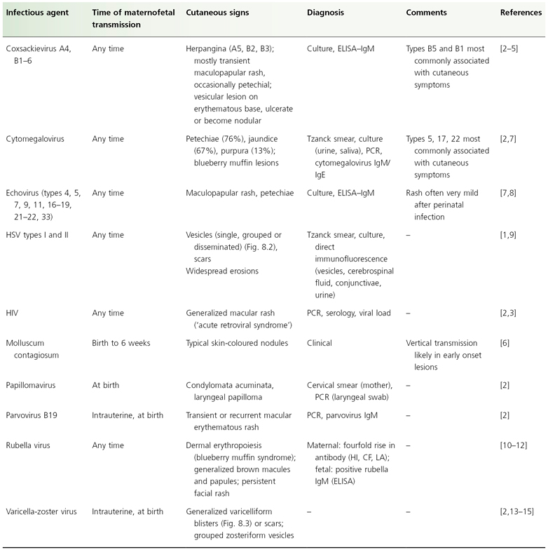
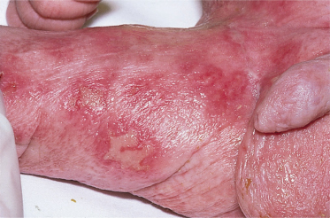
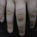
Except for HSV, VZV and rubella, few congenital viral infections are associated with specific cutaneous signs; in fact, most congenital viral infections are probably completely asymptomatic. Grouped vesicles on an erythemtatous base are typical of HSV (Fig. 8.2). Two recently reported cases indicate that congenital HSV infections can likewise present with widespread erosions without any vesicles or vesiculopustules [1]. Varicella-zoster virus typically presents with disseminated vesicles, vesiculopustules and papules (Fig. 8.3). Disseminated vesicles have likewise been reported with coxsackievirus infections [2–4], which may ultimately ulcerate or become nodular [5]. Molluscum contagiosum can be transmitted vertically and presents at birth with either congenital or early onset (6 weeks of age) typical molluscum lesions [6]. Table 8.2 lists congenital viral infections associated with cutaneous signs.
Table 8.2 Cutaneous signs of congenital viral infection
CF, complement fixation text; ELISA, enzyme-linked immunosorbent assay; HI, haemagglutination inhibition; HSV, herpes simplex virus; IgE and IgM, immunoglobulin E and M; LA, latex agglutination; PCR, polymerase chain reaction.
References
1 Koch LH, Fisher RG, Chen C et al. Congenital herpes simplex virus infection: two unique cutaneous presentations associated with probable intrauterine transmission. J Am Acad Dermatol 2009;60:312–15.
Related posts:


Stay updated, free articles. Join our Telegram channel

Full access? Get Clinical Tree


