Erythema gyratum repens is the only annular erythema covered in this chapter that has not yet been reported in children. Erythema migrans is the most frequently encountered annular erythema in the pediatric population covered in this chapter.
Erythema annulare centrifugum
History.
Darier first used the term erythema annulare centrifugum in 1916 [1,2]. This eruption is considered a reactive migratory annular erythema which occurs more commonly in adults. However, cases have been reported in newborn infants, children and families [3–7]. Some authors divide erythema annulare centrifugum into two clinically and histologically distinct variants: a superficial form and a deep form [8,9].
Aetiology.
Most authors believe erythema annulare centrifugum is a cutaneous hypersensitivity reaction to an underlying agent [2]. Unfortunately, an aetiological agent is often not identified [10]. Erythema annulare centrifugum has been associated with several infections including Epstein–Barr virus, herpes zoster, molluscum contagiosum, candidiasis, dermatophytosis, ascariasis and tuberculosis [1,5,7,11–16]. Drugs that have been associated with erythema annulare centrifugum include etizolam, piroxicam, amitriptyline, hydroxycholoroquine sulfate, hydrochlorothiazide and cimetidine [1,17–22]. Other associated conditions include sarcoidosis, liver disease, thyroid disorders and hypereosinophilic syndrome [23–26]. Rarely, malignancies such as Hodgkin disease, multiple myeloma, leukemia, prostate carcinoma, nasopharyngeal carcinoma and squamous cell carcinoma have been associated with erythema annulare centrifugum [1,27–31]. Interestingly, there has been one report of an associated food hypersensitivity to Penicillium mould found in blue cheese [32].
Pathology.
Histologically, erythema annulare centrifugum has been separated into superficial and deep variants on the basis of the distribution of the perivascular infiltrate [1,9,33]. The superficial variant displays focal spongiosis, parakeratosis and a lymphohistiocytic perivascular infiltrate surrounding the superficial vascular plexus [9,33]. The deep variant lacks epidermal change and has a tightly cuffed, ‘coat-sleeve’, superficial and deep perivascular lymphohistiocytic infiltrate [9,33].
Laboratory Studies.
There are no specific laboratory features. If laboratory abnormalities are present, they may be reflective of an associated underlying disorder [2].
Clinical Features.
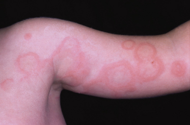
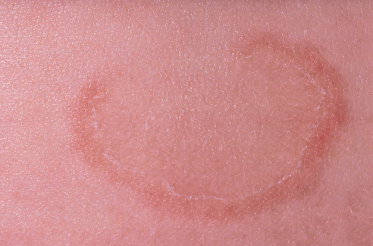
Erythema annulare centrifugum begins as an erythematous papule that slowly migrates (2–3 mm/day) and peripherally enlarges into an arcuate or polycyclic lesion with central clearing (Fig. 76.2) [2,34]. Lesions can reach up to 10 cm in diameter and are located predominantly on the trunk and proximal extremities [2]. The superficial variant of erythema annulare centrifugum may have a pruritic border with trailing scale and rare vesicles [2,8,27]. The deep variant lacks scale or pruritus and has a firm indurated border [2,8,27]. Individual lesions may resolve within days to weeks while new lesions continue to emerge [10,27].
Fig. 76.2 Erythema annulare centrifugum with fine scaling in the inner aspect of the advancing palpable border.
Courtesy of Professor J. Harper.
Prognosis.
Erythema annulare centrifugum may be persistent, lasting from several weeks to several years, until eventual spontaneous regression occurs [10,27]. Annually recurring erythema annulare centrifugum has been reported in adults [35]. The clinical course may also parallel that of an associated underlying disorder [2].
Treatment.
Symptomatic treatment with antihistamines or topical steroids may provide some relief; however, treatment will not affect the chronic and recurrent nature of the condition [1,27]. Case reports of erythema annulare centrifugum responding to topical calcipotriol or topical tacrolimus have been reported [36,37]. Treatment of an associated underlying condition may lead to resolution of the eruption [2].
Differential Diagnosis.
The differential diagnosis of the superficial variant of erythema annulare centrifugum includes: tinea corporis, subacute cutaneous lupus erythematosus, neonatal lupus erythematosus, pityriasis rosea, erythema gyratum repens, drug eruption and psoriasis [1,34,38]. The differential diagnosis of the deep variant of erythema annulare centrifugum includes erythema migrans, erythema marginatum, urticaria, granuloma annulare, erythema multiforme, sarcoidosis, borderline and lepromatous leprosy, and annular erythema of infancy [1,34,38].
References
1 Hurley HJ, Hurley JP. The gyrate erythemas. Semin Dermatol 1984;3:327–36.
2 Tyring S. Reactive erythemas: erythema annulare centrifugum and erythema gyratum repens. Clin Dermatol 1993;11:135–9.
3 Metzker A, Frumkin A. Erythema annulare of infancy. Cutis 1988;42:353–5.
4 Fried R, Schonberg IL, Litt JZ. Erythema annulare centrifugum (Darier) in a newborn infant. J Pediatr 1957;50:66–7.
5 Hammar H. Erythema annulare centrifugum coincident with Epstein–Barr virus infection in an infant. Acta Paediatr Scand 1974;63:788–92.
6 Watsky KL, Hansen T. Annular erythema in identical twins. Cutis 1989;44:139–40.
7 Vasily DB. Erythema annulare centrifugum and molluscum contagiosum. Arch Dermatol 1976;114:1853.
8 Bressler GS, Jones RE. Erythema annulare centrifugum. J Am Acad Dermatol 1981;4:597–602.
9 Ackerman AB. Histologic Diagnosis of Inflammatory Skin Diseases. Lea and Febiger 1978:174–5,231–3.
10 Mahood JM. Erythema annulare centrifugum: a review of 24 cases with special reference to its association with underlying disease. Clin Exp Dermatol 1983;8:383–7.
11 Jillson OF. Allergic confirmation that some cases of erythema annulare centrifugum are dermatophytids. AMA Arch Derm Syphilol 1954;70:355–9.
12 Schmid MH, Wollenberg A, Sander CA, Bieber T. Erythema annulare centrifugum and intestinal Candida albicans infection-coincidence or connection? Acta Derm Venereol 1997;77:93–4.
13 Shelley WB. Erythema annulare centrifugum due to Candida albicans. Br J Dermatol 1965;77:383–4.
14 Furue M, Akasu R, Ohtake N, Tamaki K. Erythema annulare centrifugum induced by molluscum contagiosum. Br J Dermatol 1993;129:646–7.
15 Hendricks AA, Lu C, Elfenbein GJ, Hussain R. Erythema annulare centrifugum associated with ascariasis. Arch Dermatol 1981;117:582–5.
16 Sugita K, Kabashima K, Tokura Y. Erythema annulare centrifugum associated with herpes zoster. Eur J Dermatol 2008;18:205–6.
17 Hogan DJ, Blocka KL. Erythema annulare centrifugum associated with piroxicam. J Am Acad Dermatol 1985;13:840–1.
18 Hudson LD. Erythema annulare centrifugum: an unusual case due to hydroxychloroquine sulfate. Cutis 1985;36:129–30.
19 Merrett AC, Marks R, Dudley FJ. Cimetidine induced erythema annulare centrifugum: no cross sensitivity with ranitidine. Br Med J 1981;283:698.
20 Goette DK, Beatrice E. Erythema annulare centrifugum caused by hydrochlorothiazide-induced interstitial nephritis. Int J Dermatol 1988;27:129–30.
21 Kuroda K, Yabunami H, Hisanaga Y. Etizolam-induced superficial erythema annulare centrifugum. Clin Exp Dermatol 2002;27:34–6.
22 Garcia-Doval I, Peteiro C, Toribio J. Amitriptyline-induced erythema annulare centrifugum. Cutis 1999;63:35–6.
23 Altomare GF, Capella GL, Frigerio E. Sarcoidosis presenting as erythema annulare centrifugum. Clin Exp Dermatol 1995;20:502–3.
24 Tsuji T, Kadoya A. Erythema annulare centrifugum associated with liver disease. Arch Dermatol 1986;122:1239–40.
25 Shelley WB, Shelley ED. Erythema annulare centrifugum as the presenting sign of the hypereosinophilic syndrome: observations on therapy. Cutis 1985;35:53–5.
26 Braunstein B. Erythema annulare centrifugum and Graves’ disease. Arch Dermatol 1982;118:623.
27 Kim KJ, Chang SE, Choi JH et al. Clinicopathologic analysis of 66 cases of erythema annulare centrifugum. J Dermatol 2002;29:61–7.
28 Leimert JT, Corder MP, Skibba CA, Gingrich RD. Erythema annulare centrifugum and Hodgkin’s disease. Arch Intern Med 1979;139:486–7.
29 Yaniv R, Shpielberg O, Shpiro D, Feinstein A, Ben-Bassat I. Erythema annulare centrifugum as the presenting sign of Hodgkin’s disease. Int J Dermatol 1993;32:59–61.
30 Helbling I, Walewska R, Dyer MJ, Bamford M, Harman KE. Erythema annulare centrifugum associated with chronic lymphocytic leukaemia. Br J Dermatol 2007;157:1044–5.
31 Stokkermans-Dubois J, Beylot-Barry M, Vergier B, Bouabdallah K, Doutre MS. Erythema annulare centrifugum revealing chronic lymphocytic leukaemia. Br J Dermatol 2007;157:1045–7.
32 Shelley WB. Erythema annulare centrifugum. Arch Dermatol 1964;90:54–8.
33 Weedon D. Skin Pathology, 2nd edn. Churchill Livingstone, 2002:246.
34 Harrison P. The annular erythemas. Int J Dermatol 1979;18:282–90.
35 Muret MP, Pujol RM, Gimenez-Arnau AM et al. Annually recurring erythema annulare centrifugum: a distinct entity? J Am Acad Dermatol 2006;54:1091–5.
36 Gniadecki R. Calcipotriol for erythema annulare centrifugum. Br J Dermatol 2002;146:317–9.
Related posts:


Stay updated, free articles. Join our Telegram channel

Full access? Get Clinical Tree


