Key Management Features
- Rest, ice, elevation
- Topical corticosteroids
- Oral antihistamines for mild systemic reactions
- Epinephrine for severe reactions
Mild reactions may be treated with ice and topical corticosteroids. Mild systemic effects may respond to oral antihistamines. The treatment of choice for anaphylaxis is intramuscular or intravenous epinephrine. Subcutaneous injection produces less reliable absorption. Systemic corticosteroids may also be helpful in patients with severe reactions but only as adjunctive therapy.
Unexpected stings are frequent. Therefore, any patient with life-threatening hymenopterid allergy should carry an epinephrine autoinjector at all times. The EpiPen® contains 0.3 mL of epinephrine 1:1000, while the EpiPen® Jr contains 0.3 mL of epinephrine 1:2000. The devices are identical and include a grey safety cap at one end which must be removed in order to activate the device. Patients should receive instruction regarding use of the kit while still in the office. The kit should be maintained at room temperature and out of the sun. The contents should be inspected periodically for evidence of brown discoloration. Patients at risk for anaphylaxis should wear a medical alert bracelet in case they are discovered unconscious. Desensitization can improve quality of life and should be discussed with an allergist. Rush desensitization regimens are gaining popularity and appear to be safe [7,8], even in patients with underlying mastocytosis [9]. Oral desensitization regimens are being studied [10].
Differential Diagnoses
If the stinging insect is not observed, hymenopterid stings may be confused with stings of other insects. Skin testing can verify the diagnosis of allergy but not all allergen preparations are equivalent [11]. Anaphylactoid responses may be a sign of underlying mastocytosis [12]. Assays for tryptase are a more reliable means of establishing a diagnosis of systemic mastocytosis than those for histamine.
References
1 Sicherer SH, Leung DY. Advances in allergic skin disease, anaphylaxis and hypersensitivity reactions to foods, drugs and insects in 2008. J Allergy Clin Immunol 2009;123:319–27.
2 Cox R. Evaluation of the patterns of potentially toxic exposures in Mississippi following Hurricane Katrina. Clin Toxicol 2008;46:722–7.
3 Bilò BM. Epidemiology of insect-venom anaphylaxis.Curr Opin Allergy Clin Immunol 2008;8:330–7.
4 Yanagawa Y. Cutaneous hemorrhage or necrosis findings after Vespa mandarinia (wasp) stings may predict the occurrence of multiple organ injury: a case report and review of the literature. Clin Toxicol 2007;45:803–7.
5 Hoffman DR. Fire ant venom allergy. Allergy 1995;50:535–44.
6 Stafford CT. Hypersensitivity to fire ant venom. Ann Allergy Asthma Immunol 1996;77:87–95.
7 Judd CA. Successful administration of a 1-day imported fire ant rush immunotherapy protocol. Ann Allergy Asthma Immunol 2008;101:311–15.
8 Gorska L. Analysis of safety, risk factors and pretreatment methods during rush hymenoptera venom immunotherapy. Int Arch Allergy Immunol 2008;147:241–5.
9 Bonadonna P. Allergen specific immunotherapy is safe and effective in patients with systemic mastocytosis and Hymenoptera allergy. J Allergy Clin Immunol 2008;121:256–7.
10 Patriarca G. Sublingual desensitization in patients with wasp venom allergy: preliminary results. Int J Immunopathol Pharmacol 2008;21:669–77.
11 Golden DB. Dialyzed venom skin tests for identifying yellow jacket-allergic patients not detected using standard venom. Ann Allergy Asthma Immunol 2009;102:47–50.
12 Müller UR. The problem of anaphylaxis and mastocytosis. Curr Allergy Asthma Rep 2009;9:64–70.
Bed Bugs
Key Diagnostic Criteria
- Breakfast, lunch and dinner pattern of bites, often along lines of clothing
- Examine mattress seams, cracks and crevices for evidence of bugs, ova and faeces
The recent epidemic of bed bug bites in the United States and Europe has reawakened interest in this humble hemipterid. The order Hemiptera consists of insects with wings that are half membranous and half sclerotic. Two families within the order, Cimicidae (bed bugs) and Reduviidae (reduviid bugs), are of medical importance. Cimicidae are blood-sucking ectoparasites of mammals or birds. Any of them can find their way into homes and bite humans when their preferred host is no longer available.
Bed bugs have flat oval bodies and retroverted mouthparts best visible from the side. Unlike other hemipterids, the membranous portion of the wing is absent. Cimex lectularius (the temperate bed bug) and C. hemipterus (the tropical bed bug) are those most commonly implicated in bites. C. lectularius also parasitizes bats, chickens and other domestic animals. It ranges in size from 5 mm to 7 mm. Females are slightly longer than males and tropical bed bugs are longer than their temperate counterparts. All are red-brown in colour with widely separated compound eyes. The female deposits eggs along the seams of mattresses and on rough surfaces of cracks and crevices. The eggs are white to off-white in colour and 1 mm in length. They are accompanied by blood-stained faeces.
Bed bugs are nocturnal and the best way to demonstrate them is to throw back the covers and search just before dawn when they emerge to feed. Bites present as erythematous papules, often in groups of three (breakfast, lunch and dinner) along clothing lines.
Key Management Features
- Individual bites respond to potent topical corticosteroids
- Definite management requires identification and elimination of bed bugs
- A professional exterminator should be contacted
Various insecticides demonstrate activity against bed bugs, dichlorvos being among the best. The residual activity of the insecticide varies by the surface, with organic surfaces showing the poorest persistence. Microencapsulation of insecticides has been shown to enhance persistence.
Differential Diagnoses
A widespread urticarial and bullous eruption may be seen in children and may mimic an immunobullous disorder. Fleas produce a similar pattern of bites in a row but tend to affect the lower extremities.
Caterpillars and Moths
Key Diagnostic Criteria
- A variety of caterpillars and moths can produce clinical dermatitis
- Urticating hairs are commonly present
- Hairs embedded in the cornea and conjunctivae produce ophthalmia nodosa
- Megalopyge caterpillars produce immediate pain and a haemorrhagic railroad-track pattern
- South American Lonomia caterpillars can produce a fatal haemorrhagic diathesis
Lepidopterids (moths and caterpillars) usually produce mild and self-limited reactions. However, some can be severe and even life-threatening.
Hylesia moths produce epidemics of dermatitis in Latin American cities and ports. Lymantria dispar, the gypsy moth caterpillar, produces similar epidemics in the eastern United States. Wind dispersion of stinging hairs from Hylesia and processionary caterpillars can result in large numbers of affected individuals and increase the likelihood of ocular involvement.
Caterpillar exposures occur as occupational injuries and in children who try to catch or pet soft furry caterpillars. Signs and symptoms are often non-specific and the diagnosis is made by temporal association. Caterpillars of medical importance are highly regional. In parts of South America, cutaneous haemorrhage and laboratory evidence of coagulation anomalies suggest lonomism. In Spain and Israel, less commonly in other European countries. epidemics of dermatitis are caused by processionary caterpillars that form long rows along the limbs of pine or oak trees. In the southern and south-western United States, pain and a haemorrhagic train-track pattern suggest Megalopyge envenomation. In the eastern United States, the Io moth caterpillar, Automeris io, causes a self-limited pruritic eruption. Closely related Hemileuca caterpillars are distributed in the United States and Canada and cause more persistent symptoms. The buck moth caterpillar (H. maia) causes pain, vesiculation, oedema and ecchymosis. The saddleback caterpillar, Acharia stimulea, causes urticarial lesions with stinging and vesiculation.
Key Management Features
- Symptomatic management with ice and topical corticosteroids is often sufficient
- Tape stripping may remove embedded hairs
- Antivenin is available for Lonomia envenomation
- Patients with ophthalmia nodosa should be evaluated by an ophthalmologist
Most reactions to moths and caterpillars are self-limited. Tape stripping can be used to remove embedded hairs and washing with soap and water can also be helpful. Clothing should be laundered separately. Ice is helpful for painful reactions. For pruritic reactions, ice, topical corticosteroids, topical camphor and menthol and topical anaesthetics such as pramoxine may be helpful.
In the case of ocular exposure, referral to an ophthalmologist is advised. Patients with suspected Lonomia envenomation are at risk for death and should be managed by those familiar with the envenomations. Antivenin is available for some species [1].
Differential Diagnoses
The manifestations of many caterpillars and moths are non-specific. Contact with tarantulas may also produce ophthalmia nodosa. The haemorrhagic effects of Lonomia envenomation could be confused with other haemorrhagic syndromes. The diagnosis is best established by history or by identification of the offending caterpillar or moth.
Reference
1 Caovilla JJ, Barros EJ. Efficacy of two different doses of antilonomic serum in the resolution of hemorrhagic syndrome resulting from envenoming by Lonomia obliqua caterpillars: a randomized controlled trial. Toxicon 2004;43:811–18.
Spiders
Key Diagnostic Criteria
- Brown recluse spiders cause dermonecrotic reactions
- Widow spiders produce tetany and abdominal pain
Few spider venoms are well characterized, as only a handful of species are known to be dangerous to humans. In addition to bite reactions, many species of tarantula have urticating hairs and defend themselves by flicking hairs into the eyes of a perceived attacker. The most severe forms of arachnidism are caused by Australian funnel web spiders (Atrax sp), brown spiders of the family Loxoscelidae (e.g. the brown recluse spider, Loxosceles reclusa) and widow spiders (Latrodectus sp).
Loxosceles Spiders
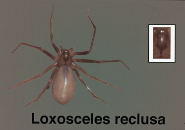
These spiders are dull yellow to light brown. The brown recluse spider has a dark-brown, violin-shaped marking on the dorsum of the cephalothorax (Fig. 73.2). Loxosceles spiders are shy and active mainly at night. They favour attics, cupboards, garages, basements and wood piles. These are not aggressive spiders and the bite is defensive in nature.
Fig. 73.2 Brown recluse spiders demonstrate a characteristic violin-case pattern on the cephalothorax.
There are a number of species of Loxosceles in North and South America. Of the 13 known species in the USA, five are known to induce human skin necrosis: L. reclusea, L. laeta, L. deserta, L. arizonica and L. rufescens. L. laeta is the most important species in South America and L. rufescens is widespread in southern Australia.
Related posts:


Stay updated, free articles. Join our Telegram channel

Full access? Get Clinical Tree


