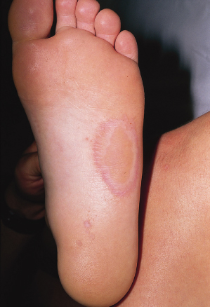
Although most cases are symptom free, painful lesions have been described in children [9,15] (Fig. 56.2). In the painful variant, erythematous to violaceous tender plaques are often present in addition to the typical pitted lesions. There is no evidence for deeper invasion into the stratum corneum by the bacterium in these symptomatic cases. Therefore, the exact reason for the pain in some patients is not known. Predisposing factors for painful lesions include occlusive footwear such as tennis shoes made of synthetic materials and prolonged exposure to moisture.
Fig. 56.2 Large area of pitted keratolysis on the lateral sole in an adolescent boy. Smaller, more typical lesions, are seen near the heel of the foot.
Courtesy of Dr Neil Prose.
The organism may be rarely seen on a potassium hydroxide preparation but is more easily demonstrated on Gram-stained scrapings or thin tissue sections [14]. The organism can be cultured on appropriate medium if specific identification is desired, but this is usually not necessary.
Prognosis.
The condition is rapidly responsive to an adjustment in the contributing environmental factors (prolonged exposure to moisture, occlusive footwear and hyperhidrosis), various topical preparations and systemic antibiotics.
Differential Diagnosis.
The characteristic clinical presentation is easily recognized and diagnosed but isolated hyperhidrosis and tinea pedis (dermatophyte) infection must be considered as a cause of maceration and bromhidrosis. Pitting associated with the basal cell naevus syndrome can be distinguished by the presence of other specific clinical features seen in this syndrome.
Treatment.
Therapy for pitted keratolysis is simple and efficacious. Although, in the past, various treatments (20% aluminium chloride, formalin ointment, various topical antibiotics and imidazoles) were used with some success, topical antibiotics such as erythromycin and clindamycin are safe and rapidly effective and should be the first line of treatment. Mupirocin ointment has been found to be effective when conventional therapy has failed [16], but its use should be restricted to elimination of MRSA carriership. Systemic treatment with erythromycin has been successful. The painful variant also responds well to these therapies [9,11,15,17]. Therapy should also include an effort to reduce predisposing factors such as hyperhidrosis, frequent and prolonged contact with water and wearing of occlusive footwear.
References
1 Acton HW, McGuire C. Keratolysis plantare sulcatum; lesion due to actinomycetic fungus. Ind Med Gaz 1930;65:61–5.
2 Nordstrom KM, McGinley KS, Cappiello L et al. Pitted keratolysis: the role of Micrococcus sedentarius. Arch Dermatol 1987;123:1320–5.
3 Gill KA, Buckels LJ. Pitted keratolysis. Arch Dermatol 1968;98:7–11.
4 Gillum RL, Qadri SM, Al-Ahdal MN et al. Pitted keratolysis: a manifestation of human dermatophilosis. Dermatologica 1988;177:305–8.
5 Woodgyer AJ, Baxter M, Rush-Munro FM et al. Isolation of Dermatophilus congolensis from two New Zealand cases of pitted keratolysis. Australas J Dermatol 1985;26:29–35.
6 Zaias N, Taplin D, Rebell G. Pitted keratolysis. Arch Dermatol 1965;92:151–4.
7 Castellani A. Keratoma plantare sulcatum. J Ceylon Br Med Assoc 1920;1:12–14.
8 De Almeida HL Jr, de Castro LA, Rocha NE et al. Ultrastructure of pitted keratolysis. Int J Dermatol 2000;39:698–701.
9 Shah AS, Kamino H, Prose NS. Painful, plaque-like pitted keratolysis occurring in childhood. Pediatr Dermatol 1992;9:251–4.
10 Takama H, Tamada Y, Yokochi K et al. Pitted keratolysis: a discussion of two cases in non-weight-bearing areas. Acta Dermatol Venereol 1998;78:225–6.
11 Zaias N. Pitted and ringed keratolysis: a review and update. J Am Acad Dermatol 1982;7:787–91.
12 Lee HJ, Roh KY, Ha SJ et al. Pitted keratolysis of the palm arising after herpes zoster. Br J Dermatol 1999;140:974–5.
13 Takama H, Tamada Y, Yano K et al. Pitted keratolysis: clinical manifestations in 53 cases. Br J Dermatol 1997;137:282–5.
14 Sehgal VN, Ramesh V. Crateriform depression – an unusual clinical expression of pitted keratolysis. Dermatologica 1983;166:209–11.
15 Lamberg SI. Symptomatic pitted keratolysis. Arch Dermatol 1969;100:10–11.
16 Vazquez-Lopez F, Perez-Oliva N. Mupirocine ointment for symptomatic pitted keratolysis. Infection 1996;24:55.
17 Burkhart CG. Pitted keratolysis: a new form of treatment [letter]. Arch Dermatol 1980;116:1104.
Erythrasma
Definition.
Erythrasma is a mild chronic superficial infection of the skin resembling dermatophyte infection. It is caused by Corynebacterium minutissimum. The skin lesions show a characteristic ‘coral-red’ fluorescence when viewed under a Wood’s light.
History.
Before 1960, the aetiological agent of erythrasma was presumed to be a fungal organism. Lagana first indicated that a diphtheroid bacterium isolated from an erythrasma lesion was the cause of this condition. In 1961, Sarkany et al. [1] confirmed that C. minutissimum was the cause of erythrasma.
Aetiology and Pathogenesis.
A warm humid climate is believed to be a predisposing factor for erythrasma and therefore more extensive involvement is seen in the tropics and during the hot summer months. The incidence of the disease is also higher among diabetic subjects [2,3]. Hyperhidrosis, obesity, advanced age and compromised host status are additional risk factors. Poor hygiene has historically been thought to be a predisposing factor, but Somerville et al. [4] found the incidence to be unaffected by the state of personal cleanliness, the use of deodorants and antibacterial soaps.
Because C. minutissimum, the bacterium most commonly implicated in erythrasma, has been grown from normal human skin flora, it seems probable that some local or systemic alteration must occur before this normal bacterium can act as a pathogen. The exact nature of these alterations has not been elucidated. An unusual Corynebacterium species, C. afermentans, was the only organism isolated from the skin of an individual with disseminated erythrasma involving the abdomen, arms, thighs, legs, neck and back, as well as the axillae and groin [5].
Pathology.
On histological examination, corynebacteria are seen in the horny layer in small amounts as Gram-positive rods and filaments. Electron microscopic examination of thin-sectioned skin from patients with erythrasma has also demonstrated ‘diphtheroids’ in the horny layer [6]. Skin biopsy is rarely performed to diagnose erythrasma as the diagnosis is easily made by Wood’s light examination.
Clinical Features.
Related posts:


Stay updated, free articles. Join our Telegram channel

Full access? Get Clinical Tree


