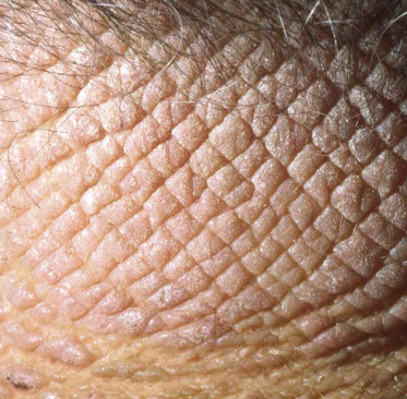
Fig. 42.2 Lichen simplex chronicus on the back of the neck, a very typical location. This close up view of lichenified skin in LSC shows the characteristic skin thickening and exaggeration of the normal skin markings.
The areas most commonly affected are those within easy reach of the patient to scratch, such as the lower legs, forearms, ankles, wrists, nape of the neck and genitalia. However, any area can be affected.
Although LSC may appear at any age, it is uncommon in childhood. LSC most often begins in young adulthood, and its incidence increases with age [12]. LSC is more common in women than in men [11].
Prognosis.
The course of LSC is chronic, with multiple exacerbations and partial remissions. These clinical fluctuations are the result of changes in the amount of scratching and rubbing. It is a benign condition that can be extremely uncomfortable and difficult to treat, leading to significant emotional distress in affected patients and their families. Morbidity is related to the occurrence of superimposed infection.
Differential Diagnosis.
The clinical appearance of LSC is usually characteristic. However, other common pruritic dermatoses, such as atopic dermatitis (AD) and lichen planus, must be considered in the differential diagnosis. Lichen planus can present with lichenified plaques or hypertrophic lesions. However, the presence of typical papules at other locations supports the diagnosis. It may be difficult to distinguish lichenified plaques of AD from LSC, and in this instance a history of AD and the distribution of the lesions can help distinguish the two entities. Plaques of psoriasis may resemble lesions of LSC, but the presence of silvery scale and an underlying deep-red hue aid in the diagnosis of psoriasis. Tinea corporis, especially in the form of a Majocchi granuloma, may resemble a plaque of LSC but can be diagnosed by potassium hydroxide examination or fungal culture. Skin biopsy may be necessary to definitively distinguish LSC from each of these skin conditions.
Treatment.
Therapy should focus on treating the cause of pruritus and eliminating the itch–scratch cycle. Predictably, lesions will not resolve if rubbing and scratching persists. It is essential that the patient understand their role in the perpetuation of LSC and make an attempt to stop the scratching.
There are no evidence-based guidelines for the treatment of LSC in children. Xerosis should be controlled with proper bathing habits and the use of emollients. Oral antihistamines may impart a modest improvement in pruritus and help with bedtime sedation. Topical corticosteroid preparations, generally in the mid-to-upper potency ranges, help alleviate pruritus and decrease inflammation. Occlusion, with or without a topical steroid, is very effective. DuoDERM®, Unna wraps, ace bandages and saran wrap have been used with good results, and flurandrenolide tape (Cordran® tape) is a commercially available topical steroid within an occlusive barrier. The addition of 0.25% menthol and 0.25% camphor to the topical corticosteroid may also help alleviate the itch [1]. Intralesional injection of triamcinolone (5–10 mg/mL injected into the dermis and not subcutaneous fat) is sometimes helpful in adults but certainly not well accepted by children. An open-label trial of pimecrolimus 1% cream improved signs and symptoms of vulvar LSC in all 12 patients studied [13]. Tacrolimus 0.1% ointment has been successful in treating LSC in children and, like pimecrolimus, provides a therapeutic alternative to topical corticosteroids [14]. Doxepin 5% cream has been used successfully for LSC [15], with one report of its use in a 3-year-old child [16]. Due to transcutaneous resorption of the drug, sedation can be a limiting side-effect of topical doxepin.
Therapies for refractory LSC include topical aspirin solution [17] and intradermal botulinum toxin type A [18]. In the presence of underlying psychopathology, which may include attention deficit or hyperactivity disorder, consultation with a child psychologist or psychiatrist may be necessary. Biofeedback, cognitive behavioural therapy and hypnosis have all been used with variable success to help control the itch–scratch cycle [19]. Topical or systemic antibiotics are indicated in the case of superimposed bacterial infection. The systemic therapies discussed in the section on prurigo nodularis (below) are seldom needed.
References
1 Krafchik BR, Halbert A, Yamamoto K et al. Eczematous dermatitis. In: Schachner LA, Hansen RC (eds) Pediatric Dermatology, 3rd edn. London: Elsevier, 2003: 642.
2 Koo J, Lee CS. Psychocutaneous diseases. In: Bolognia JL, Jorizzo JL, Rapini RP (eds) Dermatology, 2nd edn. London: Elsevier, 2008: 111.
3 Gerritsen MJ, Gruintjes FW et al. Lichen simplex chronicus as a complication of herpes zoster. Br J Dermatol 1998;138:921–2.
4 Gupta MA, Gupta AK. Psychodermatology: an update. J Am Acad Dermatol 1996;34:1030–46.
5 Konuk N, Koca R, Atik L et al. Psychopathology, depression and dissociative experiences in patients with lichen simplex chronicus. Gen Hosp Psych 2007;29:232–5.
6 Lynch PJ. Lichen simplex chronicus (atopic/neurodermatitis) of the anogenital region. Dermatol Ther 2004;17:18–19.
7 Greaves MW. Recent advances in pathophysiology and current management of itch. Ann Acad Med Singapore 2007;36:788–92.
8 Schmelz M, Hilliges M, Schmidt R et al. Active ‘itch fibers’ in chronic pruritus. Neurology 2003;61:564–6.
9 Weedon D. The psoriasiform reaction pattern. In: Weedon D, Strutton G (eds) Skin Pathology, 2nd edn. Philadelphia: Elsevier, 2002: 86.
10 Burton JL, Holden CA. Eczema, lichenification and prurigo. In: Champion RH, Burton JL, Burns DA et al (eds) Textbook of Dermatology, 6th edn. Oxford: Blackwell Science, 1998: 629–80.
11 Lotti T, Buggiani G, Prignano F. Prurigo nodularis and lichen simplex chronicus. Dermatol Ther 2008;21:42–6.
12 Goh C, Akarapanth R. Epidemiology of skin disease among children in a referral skin clinic in Singapore. Pediatr Dermatol 1994;11:125–8.
13 Goldstein AT, Parneix-Spake A, McCormick CL et al. Pimecrolimus cream 1% for treatment of vulvar lichen simplex chronicus: an open-label, preliminary trial. Gynecol Obstet Invest 2007;64:180–6.
14 Aschoff R, Wozel G. Topical tacrolimus for the treatment of lichen simplex chronicus. J Dermatol Treat 2007;18:115–7.
15 Drake LA, Millikan LE. The antipruritic effect of 5% doxepin cream in patients with eczematous dermatitis. Doxepin Study Group. Arch Dermatol 1995;131:1403–8.
16 Thomson KF, Highet AS. 5% Doxepin cream to treat persistent lichenification in a child. Clin Exper Dermatol 2001;26:100.
17 Yosipovitch G, Sugeng MW, Chan YH et al. The effect of topically applied aspirin on localized circumscribed neurodermatitis. J Am Acad Dermatol 2001;45:910–13.
18 Heckmann M, Heyer G, Brunner B et al. Botulinum toxin type A injection in the treatment of lichen simplex. An open pilot study. J Am Acad Dermatol 2002;46:617–19.
19 Shenefelt PD. Biofeedback, cognitive-behavioral methods, and hypnosis in dermatology: is it all in your mind? Dermatol Ther 2003;16:114–22.
Prurigo
Prurigo is a widely used, imprecise dermatological term representing a papular (papular prurigo) or nodular (nodular prurigo) reaction pattern to intense pruritus and focal scratching seen predominantly in atopics. In tropical countries such as Mexico, the most common cause is insect bites. Other causes include photosensitivity (actinic prurigo), psychological factors and, rarely, malignancy, usually Hodgkin lymphoma.
This section deals with prurigo nodularis, Sutton’s summer prurigo and prurigo pigmentosa. Other types of prurigo are covered elsewhere as follows: atopic prurigo or Besnier’s prurigo (Chapter 28), papular urticaria (Chapter 71) and actinic prurigo (Chapter 106).
Prurigo Nodularis
Definition.
Hyde originally described prurigo nodularis (PN) in 1909 [1]. It is a chronic dermatitis, characterized by intensely pruritic nodules that are difficult to treat, occurring mainly on the extensor aspect of the lower extremities. It is more common in adults but may affect both children and adolescents. Prurigo nodularis has also been called picker’s nodule or chronic circumscribed nodular lichenification [2].
Aetiology.
The aetiology of PN remains unknown. The nodule is induced by picking and scratching which is usually triggered by itch. PN may be a primary cutaneous disease or a secondary reaction to pruritus and scratching provoked by a separate cause [3]. Lesions of PN have selectively activated, itch-dedicated C-neurones which may create a lower threshold for itch [4].
Secondary causes of pruritus in some patients include local trauma, arthropod bites or other localized inflammation such as folliculitis. PN is frequently reported in children with atopic dermatitis. In this cohort, PN is occasionally accompanied by cutaneous hypersensitivity to various environmental allergens [5].
Systemic illnesses associated with pruritus and prurigo nodularis include [6]:
- obstructive biliary disease (intrinsic, extrinsic or drug induced)
- anaemia
- chronic renal failure
- Hodgkin disease
- leukaemia
- polycythaemia rubra vera
- systemic mastocytosis
- solid tumours
- carcinoid syndrome
- hypo- and hyperthyroidism
- diabetes mellitus
- infections: parasites, hepatitis B or C, human immunodeficiency virus, mycobacterial infections
- drug reactions
- illicit drug use
- gluten enteropathy [7]
- other forms of malabsorption [8].
In children the most common systemic association is Hodgkin lymphoma [9,10].
Related posts:


Stay updated, free articles. Join our Telegram channel

Full access? Get Clinical Tree


