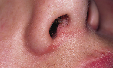
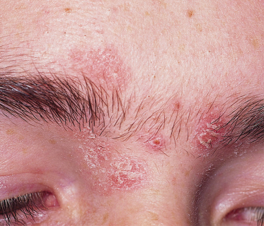
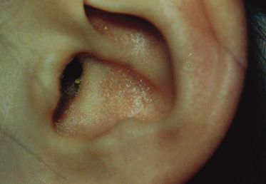
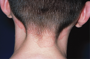
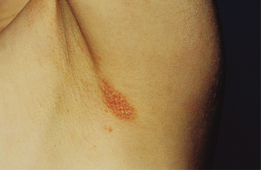
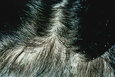
With a prevalence of between 2% and 5%, seborrhoeic dermatitis is more common in males than in females. As discussed above, it usually starts during puberty, together with the increased sebaceous gland activity, with a peak incidence around 40 years of age. The rash is typically symmetrical, involving the nasolabial creases, nasofacial sulci and adjacent cheek, glabella, hair-bearing skin of the eyebrows and scalp, scalp hair margin, periauricular skin and the central chest and upper back. Seborrhoeic intertrigo is the involvement of the main body flexures and usually occurs in individuals with more severe disease. Sequential involvement generally occurs with the scalp, eyebrows, nasolabial folds, cheeks, anterior chest, ears and body folds. In the scalp, it is characterized by a diffuse, yellowish greasy scale and in milder cases by fine, dry loose flakes, often referred to as dandruff. Dandruff and seborrhoeic dermatitis are regarded as part of a spectrum and not as separate conditions. On the central chest, seborrhoeic dermatitis often has a petaloid pattern. Itching may occur, especially in the scalp, but is not a conspicuous symptom.
Seborrhoeic dermatitis is more common in patients with acquired immune deficiency syndrome (AIDS), in whom it is also often more severe.
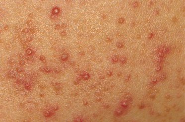
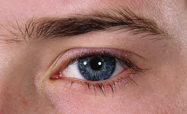
Pityrosporum ovale is also associated with pityriasis versicolor, Pityrosporum folliculitis (Fig. 41.8), seborrhoeic blepharitis (Fig. 41.9) and some forms of atopic dermatitis. The forms of seborrhoeic dermatitis recognized include pityriasiform seborrhoeide, seborrhoeic eczematide, intertriginous seborrhoeic dermatitis, disseminated seborrhoeic dermatitis and seborrhoeic erythroderma.
Differential Diagnosis.
Related posts:


Stay updated, free articles. Join our Telegram channel

Full access? Get Clinical Tree


