23 Partial Breast Reconstruction Using Distant Flaps
Surgical options for the treatment of breast cancer have evolved to incur less morbidity while optimizing breast aesthetics. 1 Recent advances in imaging and adjuvant therapy have led to detection at earlier stages and have increased the number of patients who are candidates for breast-conserving therapy (BCT). 2 , 3 BCT leaves the patient with more overall native breast tissue than mastectomy; however, patients may be left with a significant tissue deficit or deformity, and reconstructive surgeons face a larger subset of patients who have complex partial breast deformities. 4 The reconstructive options for these deformities fall into two general groups. Since the breast is a paired organ, one way to achieve balance when tissue is removed from one breast is to rearrange the remaining tissue and to reduce the contralateral side. If this is not indicated, additional tissue needs to be transferred to the affected breast with either local or distant flaps. For some of these patients, the best option for breast reconstruction will be a distant flap. In those cases, we prefer to use muscle-preserving abdominal flaps, either the superficial inferior epigastric artery (SIEA) flap, or the deep inferior epigastric perforator (DIEP) flap.
Patient Assessment and Indications
With BCT, the NAC and its sensation are usually preserved, but a significant number of women will have resulting deformity and asymmetry that ultimately affect breast cosmesis. 4 The percentage of patients who require reconstruction after BCT varies widely in the literature. This is probably because of limitations in the length of follow-up, differences in the proportion of breast size versus tissue excised, and variations in postoperative radiotherapy. 5 In one prospective study by Clough et al, 6 the median time between the initial breast conservation surgery and correction of cosmetic sequelae was 7 years. In this cohort, they reported that 20% to 30% of patients who underwent BCT developed significant deformity that could warrant reconstruction; others have reported rates as high as 60%. 7 For many women, preservation of the NAC is an important aspect of breast reconstruction. In the past, this was achieved with BCT. However, in the past 10 years surgeons have been performing an increasing number of of nipple-sparing mastectomies, preserving the NAC. Consequently, in the senior author’s practice (A.S.), the rate of BCT has decreased, whereas that of nipple-sparing mastectomy has increased. Over time, there may be an evolving trend toward nipple-sparing mastectomy and away from BCT. 8
Timing of Reconstruction
To date, there is no reliable prognostic classification of defects from BCT. The reconstructive surgeon should discuss the anticipated defect with the breast surgeon and radiation oncologist.
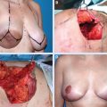
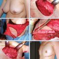
We prefer to allow the breast surgeon to perform the lumpectomy and primary closure. After final pathologic clear margins are verified 1 to 2 weeks later, we reconstruct the defect in a second-stage procedure. This prevents positive margins requiring reresection with local tissue rearrangement. The other consideration in immediate partial reconstruction is the need for adjuvant radiotherapy. Although it is well accepted that breast reconstruction after total mastectomy should be delayed after radiotherapy, we have found that adjuvant radiation dosage in BCT is administered at a lower dose and therefore causes significantly less deleterious effects on the affected tissue. 9 Radiotherapy protocols differ among institutions, and discussion regarding local protocols may help with preoperative planning of the reconstruction.
Immediate Reconstruction
The availability of a large amount of tissue allowed by the transfer of a free flap, especially in locations not amenable to local tissue flaps, such as the medial breast, allows the breast surgeon’s patient to undergo a wide local excision. This improves the patient’s oncologic care, because positive surgical margins and ipsilateral breast tumor recurrence are associated with an increased risk of systemic recurrence and decreased disease-specific survival. 10 , 11 Furthermore, various reports in the literature have recognized that there is a trend toward lower rates of local recurrence, with an increase in the peritumor free margin width. 12 – 14
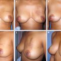
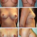
Immediate reconstruction is most often favored by the patient, because this means she has no residual defect rather than “living with a deformity” for several years. If a patient initially presents to a multidisciplinary BCT team, then once the final pathology has demonstrated clear margins of resection, immediate reconstruction should be considered to improve the eventual aesthetic outcome. A well-perfused flap prevents cavitation and skin shrinkage in the lumpectomy site by providing well-vascularized tissue when radiotherapy is planned. We have seen evidence of excellent results in these patients, and usually there is little to no delay in initiating radiotherapy.
Delayed Reconstruction
Often the reconstructive surgeon does not have the luxury of evaluating the patient before the initial excision. Some patients present to the surgeon months or even years after BCT. These patients often have a scarred and tethered defect from past irradiation of the breast. This is caused when the breast parenchyma has not been approximated and a seroma formed that was subsequently replaced by fibrosis. This in turn is further affected by irradiation of the tissue, which increases the volume loss, distortion, and fibrosis. 15 , 16 Reconstruction in these patients involves release of this contracture, which often leads to re-creation of a rather large defect. The reconstructive surgeon should be aware that these patients often have an unexpectedly large tissue deficit that is more likely to require a free tissue transfer for adequate volume replacement.
The need cannot be overstated for a thorough physical examination and imaging to confirm the absence of residual disease or recurrence before reconstruction.
Patient Selection
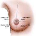
Certain tumor locations, such as medial quadrants of the breast, are deficient in surrounding breast tissue to fill the resultant dead space, leading to excessive undermining of the adjacent tissues. 17 Rearrangement of local tissue from the back or from the area inferior to the breast is the next option. Local island perforator flaps, the thoracodorsal artery perforator (TDAP) flap and the intercostal artery perforator (ICAP) flap, are excellent choices. However, the location and volume of the defect, as well as limitations in the arc of rotation of these local perforator flaps, may reduce these options. The latissimus dorsi myocutaneous flap is another choice for a local flap because of its ability to fill in large deficits and reach the lateral breast, but it is not easily transposed to the medial quadrants. 18
Distant flaps represent a solution for patients with partial breast deformities that are not amenable to local flaps because of location, defect size, or patient preference.
When discussing the option of a local versus a distant flap, the patient’s expectations and willingness to undergo treatment should be explored. Patients with a significant deformity and a strong desire for BCT are candidates for free tissue transfer of a muscle-preserving flap. Patients view the opportunity to improve their overall appearance as an overwhelming benefit of the abdominal perforator free flap surgery. This is accomplished through the concurrent flattening of the abdominal contour at the donor site. Furthermore, in lieu of a local flap that creates an extended incision adjacent to the breast, this surgery allows a less conspicuous scar location, which may even overlie an old cesarean scar. Published reports have demonstrated high patient satisfaction with their abdominal contour after lower abdominal perforator harvest for breast reconstruction. 19 , 20 Even on relatively slim patients who have had a full-term pregnancy, there is usually a small amount of excess abdominal tissue that is adequate for a mini-SIEA or a mini-DIEP flap.
Treatment Decisions
The patient with a large breast defect who is considering BCT and reconstruction rather than a complete mastectomy must be informed about the possibility of recurrence and demonstrate the means and motivation to continue with follow-up breast cancer surveillance. These patients should be informed that screening may be obscured by scarring as a result of autologous reconstruction. Free tissue transfer may be complicated by fat necrosis, which must be differentiated from recurrent breast cancer, often by imaging and occasionally by biopsy. However, the incidence of fat necrosis in these small flaps is rare, and improving radiologic imaging has improved monitoring in these patients.
The reconstructive surgeon should inform the patient of prognostic factors affecting her outcome. Healthy patients with adequate abdominal tissue who do not have severe comorbidities such as diabetes mellitus or coronary arterial disease are generally suitable candidates. Some patients may have other relative contraindications, such as age greater than 60 years, obesity defined as a BMI of 30 or more, prior abdominal surgery with extensive scarring, extensive prior abdominal liposuction, an active smoking history, or a coagulation disorder. Therefore each patient should be evaluated on a case-by-case basis.
Preoperative Planning
The choice of donor site for reconstruction warrants discussion. Although critics state that sacrificing the lower abdominal tissue for only a partial breast defect is contraindicated, there are many women who choose to undergo a cosmetic abdominoplasty, knowing that the average woman’s chances of developing breast cancer is at least 13%. The chances of local recurrence vary from 9% to 15%, so if the same logic applies to spare the abdominal tissue in the case of breast cancer, then should the average woman be counseled against having an elective abdominoplasty? We contend that there are now many described reconstructive options for autologous breast reconstruction that also offer minimal patient morbidity if a patient later requires total breast reconstruction because of a recurrence. These include the superior and inferior gluteal perforator (SGAP and IGAP) flaps, in addition to the transverse upper gracilis (TUG) flap and the anterior lateral thigh flap (ALT), which may be suitable even for large-volume breast reconstruction. 21
We prefer to preserve the thoracodorsal vessels to preserve the latissimus dorsi myocutaneous flap as a salvage option for patients when microsurgical tissue transfer is unsuccessful.
Related posts:
Stay updated, free articles. Join our Telegram channel

Full access? Get Clinical Tree


