Current laser technologies for depilation are primarily based upon mechanisms of selective photothermolysis [1]. By targeting and heating melanin-rich structures in the hair follicle, it is possible to affect the stem cells of the bulge and/or the matrix cells of the bulb. The goal is to maximize the ratio of bulb/bulge damage to epidermal heating. Individuals with dark hair and pale skin are optimal responders; conversely, patients with grey or blond hair tend to respond less well because there may be insufficient melanin to absorb laser energy. A variety of techniques may improve treatment response, including varying pulse durations, wavelengths, and application of epidermal cooling.
The safety and tolerability of laser hair removal in children remain less well characterized in the literature and are extrapolated from experience in adults. In adults, laser hair removal is an established treatment for hypertrichosis that is safe, well tolerated and may be associated with improvement in quality of life. One meta-analysis showed that laser hair removal may be more effective short term than shaving, waxing, electrolysis and epilation [2].
A broad set of lasers and light sources have been utilized for hair removal in adults, while the specific paediatric literature remains limited. In general, the long-pulsed ruby (694 nm), long-pulsed alexandrite (755 nm) and diode (800–810 nm) lasers have been used with Fitzpatrick skin types II–IV with appropriate surface cooling. The long-pulsed alexandrite with cryogen cooling at fluences of 16–32 J/cm2 and long-pulsed alexandrite with continuous chilled-air cooling at fluences of 16–27 J/cm2 have both been used in paediatric patients with lighter skin. YAG lasers are ideal for treating dark skin or hair that is very thick and black. The long-pulsed Nd:YAG (1064 nm) with a chilled contact sapphire tip at fluences of 20–35 J/cm2 or a long-pulsed Nd:YAG with cryogen cooling air fluences of 16–26 J/cm2 have been used to successfully treat children with Fitzpatrick skin types IV–VI [3]. Intense pulsed light (IPL) has also been used.
Patients should be warned that high-power, high-energy treatments may cause transient crusting of the skin. Long-term complications include paradoxical laser-induced hair growth, pain, scarring and postinflammatory pigment changes (especially in patients with darker skin types), though the incidence of these side-effects remains low. Normal hair regrowth occurs quite commonly and should be anticipated; 4–6 treatments per year is usually sufficient to effectively control hair growth in the majority of patients. Even without retreatments, hair regrowth is often less dense (i.e. returning individual hairs are thinner), and it may take several years before hair regrowth fully returns to its previous state. Unlike most adults, younger children may not be able to tolerate hair laser treatment without anaesthesia, and special care with pain relief is encouraged. General anaesthesia is appropriate for younger children with extensive treatment areas. The administration of long-acting local anaesthetic nerve blocks after treatment may also be helpful in the recovery phase.
The speed and effectiveness of treatment vary with the machine, the patient’s adherence to surgical pre- and postoperative protocols, and the experience of the laser surgeon. Overall, depilation by laser is well tolerated in children if administered appropriately.
Pseudo-folliculitis Barbae
Numerous laser technologies have been used to treat pseudo-folliculitis barbae (PFB), a chronic inflammatory disease of hair-bearing areas induced by plucking or shaving of curved hairs. In this condition, the sharpened hair remnants may penetrate the skin and incite a foreign body reaction that can lead to formation of papules, papulopustules and, rarely, hypertrophic scars and keloids. Postinflammatory hyperpigmentation is a common sequela.
Much of the reported use of lasers as therapy for PFB comes from dermatological surgeons treating the condition in the US military. The 810 nm diode, long-pulsed Nd:YAG and long-pulsed alexandrite lasers have been utilized for this purpose. The long-pulsed Nd:YAG (40–100 J/cm2 fluences) remains the modality of choice for Fitzpatrick skin types IV, V and VI [4].
References
1 Anderson RR, Parrish LA. Selective photothermolysis: precise microsurgery by selective absorption of pulsed radiation. Science 1983;220:224–7.
2 Haedersdal M, Wulf HC. Evidence based review of hair removal using lasers and light sources. J Eur Acad Dermatol Venerol 2006;20:9–20.
3 Rajpar SF, Hague JS, Abdullah A, Lanigan SW. Hair removal with the long pulse alexandrite and long pulse ND:YAG lasers is safe and well tolerated in children. Clin Exp Dermatol 2009;34(6):684–7.
4 Ross E, Cooke L, Timko A, Overstreet KA, Graham BS, Barnette DJ. Treatment of pseudofolliculitis barbae in skin types IV, V, and VI with a long-pulsed neodymium:yttrium aluminum garnet laser. J Am Acad Dermatol 2002;47:263–70.
Pigmented Lesions [1]
Melanin, the main chromophore of the epidermis, possesses a wide absorption spectrum that slowly decreases from the ultraviolet to the near-infrared. This makes it well suited for treatment by a number of highly selective Q-switched lasers, such as the Q-switched frequency-doubled Nd:YAG (532 nm), Q-switched ruby (694 nm), Q-switched alexandrite (755 nm) and Q-switched Nd:YAG (1064 nm). Less pigment-selective lasers, such as the long-pulsed ruby (694 nm), variable-pulsed diode (800–810 nm) and long-pulsed Nd:YAG lasers, are particularly useful for targeting larger pigmented structures. Ablative lasers, such as the carbon dioxide (10,600 nm) and erbium:YAG (2940 nm) lasers, are not pigment specific and have been used to remove superficial pigmented lesions via non-specific destruction.
Naevus of Ota and Naevus of Ito
Naevus of Ota, involving areas of the face supplied by the first and second divisions of the trigeminal nerve, and naevus of Ito (naevus fuscocoeruleus acromiodeltoideus), usually presenting on the neck, shoulder or upper arm, are anatomic-specific presentations of the same underlying condition. They consist, histologically, of elongated dendritic melanocytes scattered among collagen bundles in the upper portion of the dermis. Unlike dermal melanocytosis, these lesions do not disappear spontaneously. In fact, darkening of lesions has been noted during and after puberty. These disorders are almost always benign, though rare transformation to malignant melanoma has been reported in naevus of Ota, most frequently among white patients. Because several studies suggest that treatment of children for these lesions produces complete clearance faster than treatment of similar lesions in adults, many paediatric laser surgeons advocate early intervention [2].
The concentration of pigment in naevus of Ota and Ito is reduced as compared to lentigines or café-au-lait macules. Consequently, it is difficult for these pigmented dermal lesions to become heated enough using long-pulsed technologies such that sufficient pigment reduction can be achieved. Short pulses of Q-switched technologies, however, are capable of delivering the energy required for selective heating of these lesions. Q-switched lasers utilize high energy delivered in short pulses, allowing significant fragmentation of pigment; they are standardly utilized for naevus of Ota and Ito.
Several specific lasers have been utilized to treat these lesions, including the Q-switched ruby, alexandrite and Nd:YAG lasers [3]. The Q-switched Nd:YAG (1064 nm) laser is particularly useful for lesions on darker skin types (Figs 189.2, 189.3). In patients with lighter skin types, the Q-switched ruby laser (1–7 treatments at fluences of 9–10 J/cm2 with 5 mm spot size) has been demonstrated to be a very safe and effective treatment modality [4]. The risk of postinflammatory pigment change should be addressed, and the need for multiple treatment sessions should be anticipated. Lesions that involve the periorbital areas and those of a predominantly blue-green colour (reflecting a greater depth than the predominantly brown lesions) tend to require more treatment sessions. Acquired dermal melanocytosis (aka Hori naevus) is less amenable to laser treatment than true naevi of Ota and Ito. In adults, initial topical bleaching with tretinoin 0.1% and hydroquinone 5% ointment containing 7% lactic acid has been used to help reduce epidermal melanin prior to laser surgery. This has not been fully investigated in the paediatric age group.
Fig. 189.2 Naevus of Ota. Typical blue-grey appearance prior to a series of treatments with a Q-switched Nd:YAG laser.
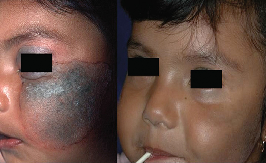
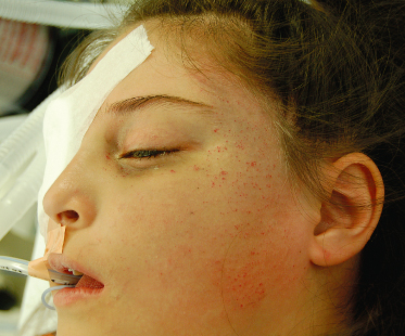
Fig. 189.3 Naevus of Ota. Intraoperative image showing punctate petechiae immediately after laser treatment.
Café-au-lait Macules
Café-au-lait macules are superficial, discrete, homogenously coloured, light tan to dark brown macules or patches, which develop in childhood and can increase with age. While single lesions are common in healthy people, multiple café-au-lait macules may be associated with systemic conditions (e.g. neurofibromatosis, McCune–Albright, Bloom, Silver–Russell, Watson, and Westerhof syndromes). These lesions have no reported tendency toward malignant transformation, so treatment usually focuses on minimizing deformity.
Several lasers have been reported to lighten café-au-lait macules with variable responses. ‘Long-pulsed’ lasers (i.e. on the order of milliseconds) mostly in the visible light spectrum have been used with some reported success. Intense pulsed light devices and long-pulsed alexandrite, diode and KTP lasers have also been used to gently heat static epidermal pigmented lesions. These lasers possess an excellent safety profile, but multiple treatment sessions are usually required and complete clearance does not always occur. Treatment with a Q-switched ruby or Q-switched alexandrite works well in patients with lighter skin types (Fitzpatrick I–IV); whitening at the lowest fluences is a clinical sign of effective treatment. Other options include the 510 pigmented dye laser, frequency-doubled Nd:YAG (532 nm) laser, especially in darker skin types, Er:YAG resurfacing, and the carbon dioxide (CO2) laser used at low power.
Café-au-lait macules may recur after laser therapy and in some cases, hyperpigmentation – occasionally permanent – may occur in treated areas. Other studies have reported clearing without recurrence with various laser technologies. Most follow-up studies to date have demonstrated that recurrence of pigmentation may occur and multiple treatments are to be expected.
Dermal Melanocytosis (Mongolian Spots)
These deep brown to bluish-grey macules or patches typically occur in the lumbosacral areas, buttocks and, less commonly, the lower limbs, flanks and shoulders of otherwise normal infants. Their distinctive slate blue to blue-black colour is a result of the Tyndall effect, a phenomenon of light scattering in the skin. Light rays with long wavelengths (e.g. red, orange and yellow) are less scattered after striking melanin and pass through the skin relatively free from disruption. Colours with shorter wavelengths (e.g. blue, purple and violet), however, strike melanin and scatter back to the skin’s surface, creating the slate grey colour characteristic of these lesions.
Related posts:


Stay updated, free articles. Join our Telegram channel

Full access? Get Clinical Tree


