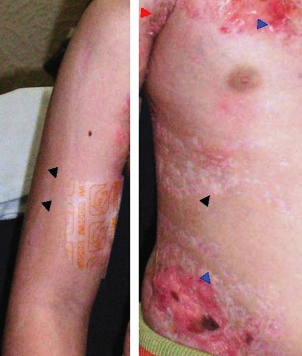
Fig. 133.2 Skin lesions in focal dermal hypoplasia. The left panel demonstrates longitudinal streaks of hypopigmented skin (black arrowheads) on trunk and arm, where a Blaschkolinear pattern can be observed. The right panel shows details of the skin features: focal dermal hypoplasia (blue arrowheads) and yellow-pink areas of fat tissue (red arrowheads).
Papillomas
These can appear at any time on the skin and mucous membranes, but are rarely present at birth. They are most common around the lips and eyes and on vulvar, perianal and perineal areas where they can be confused with condylomata acuminata. They have also been reported at other sites including the oesophagus and larynx where they may result in breathing and swallowing difficulties [14,15]. Recurrence after excision has been observed and they can occasionally take on giant proportions [16].
Skeletal System
Focal dermal hypoplasia patients have typical skeletal findings that are asymmetrical and variable, with predominant involvement of hands and feet in 70% of cases [1]. These include syndactyly, polydactyly, ectrodactyly and absence or hypoplasia of digits. Lobster claw deformity (split hand or split foot with syndactyly and absence of rays) is a major distinct feature of this condition [5] (see Fig. 133.1). In rare affected cases, the hands or feet and part of long bones may be missing in an asymmetrical pattern. Scoliosis occurs in 15–20% of cases and asymmetry in the size and shape of the face, trunk or limbs in 30% of cases [5]. In 20% of cases, there is osteopathia striata, which are longitudinal linear striations in the metaphyses of long bones seen on radiography [1,17]. It is highly characteristic of FDH, but has also been seen in other disorders. A separate X-linked dominant condition, osteopathia striata with cranial sclerosis (OSCS), was hypothesized to be allelic with FDH but recently, mutations in the WTX gene in Xp have been shown to be responsible for this disorder, establishing it as distinct from FDH [18].
Other findings include spina bifida occulta, diastasis pubis [12,19], joint hyperlaxity, deformities of ribs and clavicles, split sternum, reduction in bone density, osteoporosis, fibrous dysplasia of the bone, cystic lesions and expanding lesions including giant cell tumours of bone [20] and osteochondroma in one case [21].
Face
Focal dermal hypoplasia patients have a typical facial appearance with asymmetry, a pointed chin, maxillary hypoplasia, broad nasal tip with a narrow bridge and sometimes notching of the alae nasi (see Fig. 133.1). Papillomas of the lips, perioral fissures, leucokeratosis, gum hyperplasia, notching of the alveolar ridge, bifid or notched uvula and tongue clefts have been reported in several patients. Cleft lip and cleft and high-arched palate are present in a few percent and some severe cases have complex extensive facial clefts [11]. The ears are often thin and protruding and can be low-set, asymmetrical, small or deformed with poor development of cartilage, and presence of auricular appendages, cholesteatoma and hearing deficit.
Teeth
Approximately 40% of people with FDH have dental abnormalities [5,12]. Findings include typical longitudinal grooving of teeth, taurodontia, enamel defects with caries, anodontia, hypoplastic or dysplastic teeth, delayed eruption, irregular spacing, malocclusion and notched incisors.
Hair
The hair in FDH is often sparse and brittle. Localized areas of absent scalp and pubic hair have been reported [5,12].
Eyes
In 20–40% of reported cases there is significant eye involvement [1,5], mostly chorioretinal colobomas [22] but microphthalmia, microcornea and ectopia lentis are also frequently present. In addition, anophthalmia, papillomas of the eyelids, aniridia, heterochromia, microcornea, corneal clouding, keratoconus, cataracts, optic atrophy [2], retinal neovascularization and vitreous haemorrhage [23], irregularity of pupils and lacrimal duct abnormalities and cysts [2,13] have also been reported. These can be associated with strabismus or nystagmus, or result in blindness or significant visual handicap in some patients [5].
Nails
Dystrophy, atrophy, anonychia, grooving (see Fig. 133.1) and spooning of both fingernails and toenails is very frequently present [5,12].
Other Rarer Features
There can be respiratory and digestive tract abnormalities, including, duodenal atresia, intestinal malrotation [24], anterior placement of anus [19,24], anal stenosis [25], omphalocoele [5], inguinal, diaphragmatic [26], epigastric and hiatus hernias. In the genitourinary tract, renal agenesis [11], hypoplastic kidney [25], horseshoe kidney [12,25,27], cystic renal dysplasia [5] and hydronephrosis [24,25] have all been reported. In addition, a bicornuate uterus, asymmetrical labia and vaginal canal were reported in one patient [25]. Cardiovascular system abnormalities associated with FDH include mediastinal dextroposition, patent ductus arteriosus [24], total anomalous pulmonary venous return, truncus arteriosus with truncal origin of hypoplastic pulmonary arteries, cardiac ventricular septal defect and hypoplasia of the lungs and pulmonary veins [11,25]. Recurrent tonsillitis, respiratory infections, conjunctivitis, urinary tract infections and otitis media have been observed [5]. However, no immunological deficiency has been reported and these infections are probably related to predisposing underlying structural defects of these organ systems. Rare cases with absent nipples are also reported.
Central Nervous System
About 15% of patients have mental deficiency with varying severity, which may be overestimated due to sensory deprivation and difficulty assessing mental function in more severely affected individuals who may have combined hearing and vision loss [5]. Agenesis of the corpus callosum [28], meningomyelocoele, hydrocephalus, Arnold–Chiari malformation [29], gyral abnormalities and absent cerebellar vermis [25] have been reported but overall, CNS anomalies are uncommon in FDH and there is no increased incidence of seizures.
Growth and Lifespan
Growth retardation in utero and mild short stature have been described, but poorly characterized. The lifespan is normal in the majority of cases [1].
Histopathology
Atrophic, Striate and Lipomatous Lesions
The hallmark features of the typical atrophic, striate and lipomatous lesions of FDH syndrome are the diminished thickness of the dermis with evidence for defective collagen formation [12], widened blood vessels in the papillary dermis and islands of mature adipose tissue deposits scattered within the reticular and papillary dermis [12] that are thought to initiate around these capillaries [9], but can be restricted initially to the reticular dermis. It has been suggested that the lipomatous lesions represent different degrees of the same process [9].
In early-stage skin lesions, the epidermis is usually normal. The papillary dermis shows increased numbers of small blood vessels and, sometimes, a perivascular lymphohistiocytic infiltrate [13,30]. The dermis is thinned, with attenuated collagen fibres. The elastic tissue can be increased or decreased [31]. Adipocytes or adipocyte-like cells can be found in small deposits 3–4 cells thick around blood vessels in the dermis, but not in continuity with the subcutaneous fat layer. These aggregates tend to enlarge and result in fat nodule formation. When large lobulated fat masses have developed in later stages, the relationship with blood vessels can be lost. The fat masses can be separated from the epidermis by only a few collagen fibres [12,31]; there is usually a layer of dermal connective tissues beneath the fat nodules, separating them from the subcutaneous layer. Atrophic lesions have a paucity of appendages [30]. Hyperpigmented areas can be associated with epidermal acanthosis and papillomatosis and increased melanin pigment in the epidermal basal layer [30].
Electron microscopy reveals loosely arranged collagen bundles composed of scattered abnormally formed and oriented fibres [30,31]. Elastic fibres are scarce but of normal morphology, but the fibroblasts in affected areas appear more oval, larger and have reduced organelle content with vacuoles, and irregular thickening below the nuclear lamina [30,31]. In these cells, the Golgi is enlarged and the rough endoplasmatic reticulum (RER) cisternae are dilated and contain amorphous material [31]. Both mature, uniloculated and immature, multiloculated adipocytes can be found in the dermal fat nodules [30,31].
Congenital Aplasia
Absence of epidermis and sometimes dermis is observed in the lesions of congenital aplasia. The subcutaneous fat may be deficient. On the scalp, sometimes only the appendages fail to develop.
Inflammatory Lesions
The inflammatory lesions [10] with marked oedema in the papillary dermis, perivascular lymphocytic infiltration, increased numbers of fibroblasts and clusters of lipocytes at the mid-dermis occur in some patients at birth and diminish with time. It has been suggested that these histological findings represent a transitional phase and may contribute to the process of fat deposition.
Papillomas
The papillomas consist of a fibrovascular stalk covered by a layer of acanthotic stratified squamous epithelium. Hyperkeratosis and parakeratosis are often present [12]. A case of a papilloma with lymphocytic infiltration has been reported [32].
Genetics.
Focal dermal hypoplasia is an X-linked dominant disorder that affects predominatly females; all males with FDH have been demonstrated to have somatic mosaicism [5]. Such males have never been shown to transmit FDH to their sons, but can have severely affected daughters if they have gonadal tissue involvement [1,4].
In 2007, mutations in PORCN were demonstrated to cause FDH [4,8]. PORCN is currently the only known gene to cause FDH. To date, nearly 70 different point mutations are known [4,7,8,33,34]. PORCN, the human homologue of the fruit fly (Drosophila melanogaster
Related posts:


Stay updated, free articles. Join our Telegram channel

Full access? Get Clinical Tree


