Immunoelectron microscopy indicates that some desmosomal glycoproteins are quantitatively less concentrated in perilesional epidermis [18]. Lesional keratinocytes express keratins and extracellular matrix components characteristic of hyperproliferation [19,20]. Involucrin is also expressed prematurely [21]. In acantholytic cells, the extracellular domains of desmogleins are lost, whereas intracellular domains of desmogleins and desmosomal plaque proteins are distributed diffusely in the cytoplasm, where they may be trapped in tonofilament aggregates [18,22–25].
Clinical Features.
Expression of the disease varies, but males and females are affected with equal frequency. More than 60% of patients develop signs between the ages of 6 and 20, with onset peaking between the ages of 11 and 15. Congenital Darier disease has been reported but is rare [26].
More than 95% of patients have acral involvement and children may have hand involvement before any other signs of the disease. In one study, seven of 13 children had nail involvement, eight had palmar pits and ten had acrokeratosis verruciformis. Only three of these children had the typical rash of Darier disease [2].
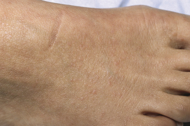
The most specific acral sign of Darier disease is a combination of longitudinal red and white lines extending from the base to the free edge of the nail plate. These may be associated with V-shaped notching of the free edge of the nail, subungual hyperkeratosis or splinter haemorrhages (Fig. 125.2). The nails are fragile so they break and split, but at first these changes may be attributed to nail biting. Most patients have pits or punctate keratotic papules on palms and soles. The fine palmar pits are easier to detect in children by using palm prints [2]. Children may develop haemorrhagic macules or blisters on the hands and feet, but these are uncommon [1]. Discrete flat-topped warty papules (acrokeratosis verruciformis) are an early sign on the backs of the hands and feet (Fig. 125.3). The skin-coloured papules are always bilateral.
Fig. 125.2 Longitudinal red and white lines in the nail are an early sign of Darier disease. These may be associated with nail fragility, notching and splitting.
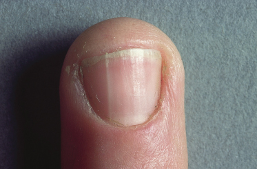
Fig. 125.3 Small flesh-coloured papules of acrokeratosis verruciformis are scattered over the dorsum of both feet in this patient with Darier disease.
The greasy, crusted papules in Darier disease are flesh-coloured, yellow or brown (Fig. 125.4) but in pigmented skin, Darier disease may present as hypopigmented macules and papules (Fig. 125.5) [27–29]. Papules appear on seborrhoeic areas of the trunk, the supraclavicular fossae, the sides of the neck, the forehead, the ears and the scalp. At first the lesions are discrete but later may coalesce into crusted, malodorous plaques. Itching is common but not invariable. Pain is unusual in uncomplicated disease. Most patients have some flexural involvement; occasionally this is severe.
Fig. 125.4 The crusted papules on the chest are yellowish-brown and rather greasy. Some are coalescing into plaques.
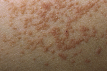
Fig. 125.5 Darier disease may present with hypopigmented lesions in children with pigmented skin. This child had typical hyperpigmented papules in addition to hypopigmented macules and papules.
The condition is usually mild in children, so the rash is often overlooked until summer, when symptoms and signs are exacerbated by heat or sweating. Lesions may develop 1–2 weeks after an episode of sunburn [30,31].
Mucosal involvement has been described in some patients, including salivary gland obstruction [32]. Papules or verrucous plaques develop on the palate, alveolar ridges, buccal mucosa or tongue.
Children mosaic for the Darier mutation (see above) may present with localized disease [7,33–35]. Keratotic papules appear on one side of the body in streaks or whorls following Blaschko’s lines. Palmar pits and nail changes may be present on the same side of the body. Heat, sweating and sunburn also aggravate segmental disease.
Complications.
Patients are prone to widespread cutaneous infections, but no specific or consistent abnormality in immune function has been demonstrated [36]. Herpes simplex infection causes erythema, vesiculation, erosions, crusting and pain. The possibility of herpetic infection should be considered in any patient with a painful exacerbation, even if vesicles are not obvious. Infection with Staphylococcus aureus may also cause blisters. Secondary bacterial overgrowth is common in the keratotic debris and may contribute to malodour.
Associated Conditions.
Bipolar affective disorder and mental impairment have been described in some patients, but the prevalence of these conditions in Darier disease is low [1]. A gene that confers susceptibility to bipolar affective disorder may be present in the Darier region [37–39]. Lithium exacerbates Darier disease and if possible should be avoided for the treatment of bipolar affective disorder in these patients [40,41].
Prognosis.
Darier disease is chronic and life-long, but severity is unpredictable and fluctuates. Some patients always have relatively mild disease, whereas in others the condition worsens inexorably. Occasionally patients do improve in old age [1].
Differential Diagnosis
Clinical
Crusted plaques on the trunk, flexural papules and scaling in the scalp may suggest seborrhoeic dermatitis. Comedones with keratotic papules on the face or chest may be misdiagnosed as acne. Acrokeratosis verruciformis may simulate plane warts. Solitary longitudinal red or white lines in the nails may indicate a subungual tumour.
Related posts:


Stay updated, free articles. Join our Telegram channel

Full access? Get Clinical Tree


