Clinical Features.
The primary lesions are papulovesicular. In time they evolve from vesicular or haemorrhagic pink papules to encrusted, scaly and dyschromic lesions. The papule of PL is red–brown in colour and, because of its haemorrhagic origin, does not blanch. The evolution of the papule is characteristic [17]. Within 3–4 weeks it enlarges to 3–4 mm and becomes flatter, with less well-defined margins. There is often a single overlying ‘mica’ scale that can be easily removed by a scratch. The hyperpigmented residual lesion may become hypopigmented after sun exposure. This latter phenomenon is a hallmark of disease.
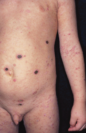
When the inflammatory changes are more acute, frank blisters or varioliform vesicopustules are seen. When haemorrhage and necrosis predominate, black necrotic lesions may appear (Fig. 100.2). The well-circumscribed necrotic lesions may be as large as 1 cm in diameter. Atrophic scarring may occur. The simultaneous presence of papules, vesicles, pustules and necrotic lesions gives a polymorphic appearance to the eruption. A febrile ulceronecrotic variant [18] has been reported to occur in children and adults [19]. In adult patients, it is reported to cause death in up to 25% of cases [20].
Fig. 100.2 Pityriasis lichenoides: large necrotic lesions associated with micropapules at different stages.

The distribution of the lesions is variable. However, involvement of the scalp, hands and feet is rare. The clinical course is variable. Usually, especially in the chronic variant, recurring eruptions lead to clinical polymorphism. Lesions in different phases of evolution, from small purpuric papules to larger papules, crusted scales and dyschromic healed lesions, are present. In these cases, the presence of small purpuric papules is indicative of active disease.
There may be just a few episodes and complete resolution within 6–8 weeks. More commonly, new crops occur for many months or, sometimes, years. During the summer months, sun exposure leads to a more rapid regression of the lesions and seems to prevent new lesions appearing. However, in autumn the eruption may recur.
There is no definite relationship between the acute character of the lesions and the duration of the disease. Although the necrotic variant is often acute and self-limited, recurrent eruptions of large necrotic lesions may occur for years. The median duration – 20 months – of PLEVA and PLC is not significantly different [21].
Laboratory investigations are usually within normal limits in PL, although the erythrocyte sedimentation rate and C-reactive protein may be elevated in the acute episode [3]. Exceptionally, a consumption of complement (C3–C4) can be shown.
Differential Diagnosis.
Stay updated, free articles. Join our Telegram channel

Full access? Get Clinical Tree


