Chapter 38 State of the Art Flexor Tendon Rehabilitation
Outline
In 1917, Harmer1 published his results of uncontrolled early active flexion and extension following flexor tendon repair. Over the course of the past 100 years, we have come full circle in our approach: For a long period, injured flexor tendons in the hand were immobilized or resected, with later grafting. In the 1960s, passive mobilization programs were in vogue, and today we have returned with greater sophistication to active mobilization as the preferred approach. Nonetheless, there is limited high-quality evidence supporting one form of early mobilization over another.2,3
Immobilization
The more conservative approaches of immobilization and/or resection and grafting were a response to the complexity of the flexor tendons in zone 2, known as “no man’s land.” Surgeons were understandably hesitant to perform a repair at that level because of the unacceptably high risk of rupture or development of restrictive adhesions. In 1941, Mason and Allen4 found that in immobilized repairs there was an initial drop in repair strength, followed by a gradual increase. At 3 weeks, repair strength was equivalent to that on the first postoperative day. Because of the observed dip in strength, it was considered necessary to protect the repair with complete immobilization for 3 weeks. Since then research has supported the value of controlled early mobilization of repairs. Even though immobilization is no longer the preferred approach, there are many cases in which this is the treatment of choice (e.g., in young children).
When a repair must be immobilized, the cast or splint is designed to keep the repaired flexor tendon on slack by positioning the wrist at neutral to 20° to 30° of flexion, the metacarpophalangeal (MCP) joints in 40° to 70° of flexion, and the interphalangeal (IP) joints at 0°. Following 3 weeks of immobilization, there are two predictable problems. Since immobilized repairs grow weaker before they return to their immediate postoperative strength, we can expect a higher risk of rupture with active motion in these patients. At the same time, frustratingly, restrictive tendon adhesions will have developed. Therapy in these cases focuses on increasing tendon excursion while protecting the repair from excessive stress. A 1991 article by Cifaldi Collins and Schwarze5 describes a systematic approach to regaining tendon function following initial immobilization of repairs in zones 2 and 3. They provide timing guidelines for initiating tendon gliding exercises, blocking and other specifically targeted interventions to restore gliding without overstressing the immobilized repair.
Early Passive Mobilization
Before any significant research had been performed in early mobilization of tendon repairs, forward-thinking surgeons such as Kleinert6 and Duran7 had designed postoperative management programs incorporating controlled passive motion of the repair in the first 3 to 4 weeks. These programs involved protecting the tendon repair by placing it on slack in a dorsal protective splint in some degree of wrist and MCP joint flexion as described above. For both programs, elastic traction held the fingers in flexion. Therapy consisted of passive flexion and either active or passive extension, to provide passive tendon gliding of the flexor tendon with minimal stress to the repair.
Kleinert’s technique, designed for zone 2 and which he also advocated for zone 1, involved hourly active extension against dynamic traction, followed by relaxation of the finger and passive flexion by means of the elastic traction, premised on the theory that the flexors would relax with resisted extension. However, later research8,9 has shown that the desired relaxation of the flexors does not occur consistently.
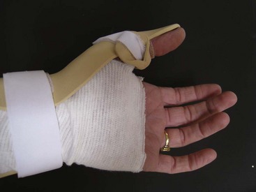
Over the years various authors have proposed modifications of the Kleinert approach. To prevent or minimize the risk of proximal interphalangeal (PIP) joint flexion contractures, elastic resistance to extension can be decreased by manually releasing rubber band traction during active extension or by using a lighter rubber band for exercises as in the Washington regimen (Figure 38-1).10 Some immobilize the interphalangeal joints in extension at night, and others use the splint design proposed by May and colleagues,11 with the dorsal finger component ending at the PIP joint for optimal PIP joint extension. At night a separate volar piece is strapped on to the splint to keep the IP joints in extension.11 In the same study, May and colleagues examined the issue of using dynamic flexion for all four fingers versus only the involved finger.11 They concluded that incorporation of all four fingers contributed to improved results by making passive flexion easier to attain. Other authors10 have recommended incorporating only the involved finger in traction. Some practitioners apply traction to only the involved finger(s) for flexor digitorum profundus (FDP) repairs, in order to facilitate inadvertent “cheating” with active motion of the uninvolved providing limited active motion of the involved finger(s).

To provide greater passive excursion of the FDP through flexion of the distal interphalangeal (DIP) joint, a palmar pulley can be added to redirect the line of traction to the distal palmar crease (Figure 38-2).10,12,13 To mobilize the flexor pollicis longus (FPL), the thumb MCP joint is immobilized and the IP joint must be passively flexed, with active or passive extension (Figure 38-3).14

Duran’s approach7 involved less frequent mobilization entirely through passive flexion and extension. His exercises were designed to provide passive excursion of the repair relative to surrounding tissues and differential glide between the FDP and flexor digitorum superficialis (FDS) in zones 2 and 3. In 1980 Strickland and Glogovac15 published a study showing improved results in zone 2 repairs mobilized passively compared with immobilized repairs. For that study they used a modified version of the Duran approach, now known as modified Duran. They eliminated the rubber band traction, splinting fingers in IP joint extension between exercises, modified the wrist flexion angle in the splint, and added composite passive flexion and active extension to the original exercises. Over the years the Strickland and Glogovac approach has been further modified to include more frequent mobilization and other variations.
A study by Cooney and colleagues16 found that incorporating synergistic wrist motion (SWM) would increase passive flexor tendon excursion in zones 2, 3, and 5: extending the wrist assists passive digit flexion via tenodesis, while wrist flexion during digit extension uses tenodesis to decrease stress on the repaired flexor(s) and allows greater digit extension. Since then a number of authors have looked at the stresses placed on the tendon and the amount of excursion attained with SWM.9,17,18 Amadio and Tanaka19,20 have proposed a further modification on a passive SWM exercise. The wrist is in flexion initially, with the fingers in extension, followed by passive finger flexion, wrist extension and MCP joint extension (holding the IP joints in flexion). The combination of wrist extension and MCP joint hyperextension with the DIP and PIP joints fully flexed produces an increase in flexor tendon tension. They hypothesize that in vivo this small increase in tendon tension may increase FDP excursion. An added benefit would be control of any intrinsic muscle tightness developing in response to blocking the MCP joints in flexion in the splint.
Early Active Mobilization
Although early passive mobilization produced improved results,15 surgeons and therapists questioned whether they could attain even better functional results with techniques producing greater excursion of the repair site. The model of passive excursion is logical and clearly safe in theory, but in practice the repaired tendon and adjacent tissues may be edematous, with added bulk from sutures and related injuries. In those cases passive flexion may simply fold the tendon or cause it to bunch up due to the gliding resistance it encounters.
Hitchcock and colleagues21 showed that controlled active mobilization actually increased the strength of the repair in the first few postoperative days, in contrast to the accepted dip in strength demonstrated in immobilized repairs.
Savage22 studied the effect of wrist position on active tension on the flexor tendon, or the force required to flex the interphalangeal joints against the passive resistance of the extensors. He found that the smallest amount of force was required with the wrist in 45° of extension, and the greatest amount of force was required when the wrist was in 45° of flexion. Since then, other authors23–28 have explored this problem in greater depth, taking into account the total force exerted by the repaired tendon, due to edema, joint stiffness, and the gliding resistance at the tendon–pulley interface. In practice, therapists must deal with the total work of flexion with all of its components.
Savage’s work22 supported the concept of either positioning the wrist in extension during flexor mobilization or incorporating synergistic wrist motion with finger flexion and extension. As a result, early mobilization programs were adapted to bring the wrist into less flexion, and in some cases, into extension. More recently a study by Kursa and his colleagues9 provided additional evidence, at least for FDS, for which greater forces were recorded with finger flexion and extension when the wrist was at 30° of flexion.
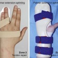
Active mobilization programs can be divided into three basic approaches: active,29–32 assisted active,33 and place-hold (active-hold).34–36 The first two programs are self-explanatory. In place-hold programs the involved fingers are placed in flexion and the patient holds the position with a gentle muscle contraction. Assisted active and place-hold flexion exercises are designed to decrease the work of flexion by overcoming passive resistance manually. All of these approaches assume that the tendon will be protected through splint position, and that work of flexion will be reduced through edema control and passive exercises performed prior to active flexion. Most published programs include a dorsal protective splint with the MCP joints in some degree of flexion, IP joints allowed to extend to 0° and the wrist in slight flexion, neutral or slight extension to decrease the work of flexion.29,32,33,35,36 One published program, the Indianapolis program,34 adds an exercise splint with a hinged wrist, allowing wrist extension to 30°, to incorporate SWM and thus both increase excursion and decrease work of flexion (Figures 38-4 to 38-6)

Stay updated, free articles. Join our Telegram channel

Full access? Get Clinical Tree


