IN THIS CHAPTER
- •
Introduction 55
- •
Patient Selection 55
- •
Preoperative history and considerations 56
- •
Operative approach 57
- •
Postoperative care 69
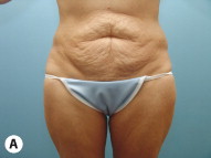
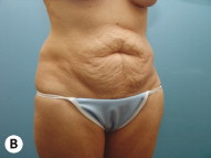
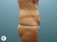
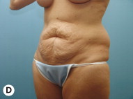
The full abdominoplasty procedure is suitable for the majority of patients seeking abdominal contouring. This procedure addresses and corrects excess abdominal adiposity and soft-tissue laxity, rectus diastasis and abdominal wall laxity, and skin striae. A significant number of full abdominoplasty candidates present after having had children as well as after failed attempts at weight loss. Patients often have the desired goal of returning to their pre-pregnancy or pre-weight gain abdominal contour.
Introduction
The full or complete abdominoplasty is the most commonly performed method of abdominoplasty. This is because the majority of abdominal contouring patients present with a combination of excess adiposity, significant soft-tissue laxity, diastasis recti, and abdominal striae. This procedure offers the most significant correction for these problems in the majority of cases. A full or complete abdominoplasty incision usually extends across the abdomen laterally to a point corresponding to each anterior superior iliac spine. This length is necessary to achieve the best results by facilitating complete removal of the infraumbilical skin and soft-tissue laxity that bothers these patients. Undermining the abdominal soft-tissue apron to the xiphoid process allows for correction of rectus diastasis and strengthening of the myofascial components of the abdominal wall. This procedure can offer a lifetime improvement, particularly for patients who maintains their weight following the procedure.
This chapter will emphasize a number of technical points that can enhance the final result, including the concurrent use of tumescent liposuction, subscarpal fat resection, and wide rectus abdominis muscle plication techniques.
Patient Selection ( Box 7.1 )
The majority of patients presenting for abdominal body contouring are candidates for full abdominoplasty. They are usually good candidates if they are not significantly overweight and do not present with prohibitory health issues. Healthy patients with infraumbilical striae, moderate excess adiposity, skin and soft-tissue laxity, and rectus diastasis or myofascial laxity are ideal candidates for full abdominoplasty. Patients who are significantly overweight, with a BMI >30–35, may also be candidates, but would benefit from better preoperative weight management. Patients more than 100 lb over ideal body weight, referral for bariatric surgery should be considered.
- •
Smoking
- •
Diabetes mellitus
- •
Malnutrition
- •
Various wound-healing disorders
- •
Bowel/bladder dysfunction
- •
Immunodeficiency
- •
Medications that inhibit blood coagulation
- •
A significant history of pulmonary or deep vein thrombosis
- •
Lower extremity lymphedema/venous insufficiency
- •
Significant medical problems, including COPD/pulmonary issues, renal insufficiency, anemia, and other systemic issues that may make and abdominal tightening surgery dangerous
Note: Age alone should not be a contraindication to abdominoplasty procedures if the patient is in good health. Patients who have a history of chronic pain should also be approached cautiously, as postoperative care may be significantly affected.
Diabetes is a significant relative contraindication to this procedure, but with careful planning, limited liposuction, and attention to detail, a safe and successful result can be achieved. Patients with respiratory difficulties are potentially also of increased concern, because myofascial plication results in a transient increase in intra-abdominal pressure which may be a source of postoperative respiratory difficulties. For these patients, muscle plication could be deferred and a more conservative approach to soft-tissue resection may also be considered.
Smoking is of great concern for all abdominoplasty procedures, particularly when thorough concurrent tumescent liposuction, subscarpal fat resection, and strong muscle plication is being considered. Active smokers are at increased risk of ischemia and necrosis, particularly midline in the infraumbilical area above the transverse incision. Also at risk is the remaining subcutaneous fat, which can undergo necrosis due to vasoconstriction-induced ischemia secondary to smoking. In our practice, we suggest complete smoking cessation for 6 weeks preoperatively for patients wishing to undergo the complete full abdominoplasty. This will allow the patient’s physiology to return to a more normal level and hopefully eliminate many of the deleterious effects on healing that smoking creates. The changes associated with long-term chronic smoking can be improved but may not be completely corrected, and we stress that there is always some continued increased risk for patients with a history of smoking.
Possible future pregnancy should be discussed with the patient and, if this is anticipated, it is suggested that abdominoplasty be performed following the last pregnancy. Bowel or bladder dysfunction should be considered a relative contraindication to myofascial plication, as this could worsen the patient’s preoperative condition.
Preoperative History and Considerations: ( Box 7.2 )
Abdominal scars from previous operations should be carefully examined and discussed with the patient (see Chapter 14 , Management of Pre-existing Abdominal Scars). A right subcostal cholecystectomy scar should be treated with great care, because undermining to the level of the scar can result in ischemia and/or necrosis of the residual tissue medially, from the cholecystectomy scar to the midline. It is important to explain to the patient that limited dissection is necessary to preserve perforating vessels to the tissue medial to the subcostal scar. Because of this, the amount of undermining will be reduced and the final tension on the incision will also be less, so as to minimize the risk of ischemia and/or necrosis. This may result in some residual laxity at the end of the procedure. It is easy for the patient to accept this when a proper explanation has been given preoperatively, but very difficult if the explanation and discussion take place postoperatively.
- •
Smoking cessation/avoidance of nicotine exposure for 4–6 weeks prior to surgery
- •
Multivitamin daily
- •
Stop aspirin/other blood thinning products with primary care doctor’s permission
- •
Abstain from all dietary/herbal supplements not approved by the surgeon
- •
Basic laboratory tests should be tailored to each patient and may include CBC, Chem 7, Factor V Leiden, and PT/PTT/INR
- •
Medical clearance if needed
- •
Wearing abdominal binder for 2 weeks before surgery
- •
Shower with a gentle antimicrobial soap the night before and day of surgery
A vertical midline incision is not a contraindication to surgery and can often be improved with scar revision. Importantly, if transverse laxity exists, additional tissue transversely can be removed, further enhancing the procedure with no increased risk for ischemia. This allows for transverse tightening during abdominoplasty, in addition to the vertical tightening associated with lower abdominal soft-tissue excision. The vertical tissue excision also allows for removal of some of the most poorly perfused tissue in the midline following undermining. If significant transverse laxity exists it is possible to remove a large amount of central abdominal tissue, which can minimize or virtually eliminate areas for potential fat necrosis or ischemia.Appendectomy or laparoscopy incisions are usually of minimal concern. The appendectomy scar is often included in the tissue resection. Laparoscopic surgery scars are usually short and do not interfere with undermining, but they can be associated with an unrecognized hernia discovered intraoperatively (see Chapter 15 , Complications). This should be taken into account during liposuction and with soft-tissue elevation. All scars, particularly a vertical midline scar, can be associated with an incisional hernia. The most common location for an incisional hernia is in the midline, particularly in patients following massive weight loss. Hernias not associated with an incision are most commonly seen in the periumbilical region and should be evaluated for during the preoperative examination. These hernias are easy to deal with intraoperatively, but should be kept in mind during liposuction and soft-tissue elevation.
Preoperative laboratory analysis is dependent on the facility where the procedure is to be performed, surgeon and anesthesiologist preference, and existing health issues. For an otherwise healthy patient, basic preoperative laboratory analysis should be considered and may include a pregnancy test, complete blood count (CBC), basic metabolic panel (BMP/Chem7), and standard coagulation profiles (PT/PTT/INR).
Prophylaxis for pulmonary embolism (PE) and deep vein thrombosis (DVT) is an important consideration. Patients taking birth control medications or hormone replacement therapy are at an increased risk for DVT and PE, and this should be discussed preoperatively. Discontinuing such medications for 2 weeks preoperatively will reduce the associated risks to a normal level. In some cases, however, it may not feasible to stop these medications and the patient needs to be informed of the increased risk of DVT/PE. A personal or family history of DVT or PE is of even greater concern. If there is such a history, preoperative laboratory analysis to evaluate the patient’s coagulation profile including a factor V Leiden and to search for possible familial tendencies for hypercoagulability is recommended. A hematology consultation should be considered to evaluate for this.
The abdominal wall plication performed with full abdominoplasty can result in increased intra-abdominal pressure and potential interference with venous return and pulmonary ventilation volumes. Because of this, we recommend placing all abdominoplasty patients in a tight abdominal binder for 2 weeks preoperatively. We ask them to wear the binder both day and night, which may help them accommodate to the increased intra-abdominal pressure changes associated with myofascial plication, and have documented a consistent reduction in abdominal circumference at the level of the anterior superior iliac crest, umbilicus, and xiphoid in patients who consistently wore the binder.
Operative Approach
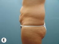
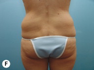
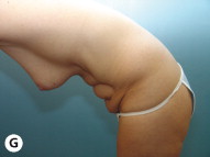
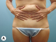
Preoperative photographs are taken in nine cardinal views ( Fig. 7.1 A–G) anterior, posterior, left and right oblique, left and right lateral, and left and right posterior oblique, and diver’s view. These last two views are more appropriate if posterior or posterolateral work is done, such as when concurrent liposuction is performed. These views are important because the reduction of the fullness at the waistline is often most evident from a posterior or posterolateral view.
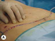
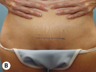
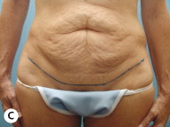
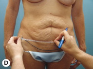
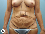
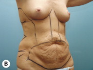
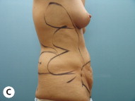
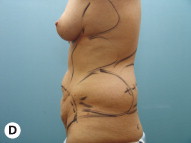
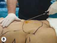
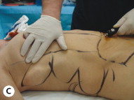
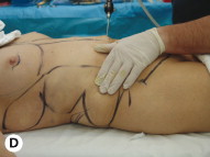
Markings are made with the patient standing. We ask the patient to place their hands together and strongly lift the lower abdominal skin vertically ( Fig. 7.2 A ). The central portion of the initial transverse incision is then marked at the superior level of the pubic symphysis. This normally removes the upper third of the hair-bearing mons ( Fig. 7.2 B ). This mark is continued laterally, following a natural skin fold ( Fig. 7.2 C ). The length and location of this proposed incision is then measured to ensure symmetry ( Fig. 7.2 D ).
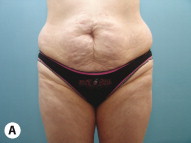
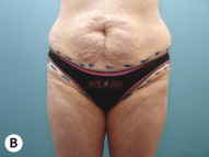
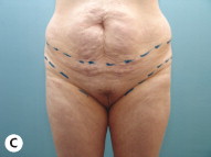
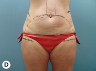
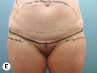
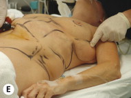
It is often helpful to ask the patient to bring in their most revealing swimwear or lingerie. This will allow the incision to be placed and concealed within the borders of the garment ( Fig. 7.3 A–E ). The lateral extent of the incision is marked and agreed upon by the patient. This avoids any postoperative concerns about the location and length of the incision.
If the patient has a ‘long’ waist, if they have only modest abdominal soft-tissue laxity, or if they have previously undergone an abdominoplasty and present for revision, the umbilical defect may not be incorporated within the resection segment and an inverted T closure may be necessary. The lower the initial transverse incision, the more likely an inverted T closure will be needed because of the longer vertical distance to the umbilicus. For patients with greater skin and soft-tissue laxity, the full abdominoplasty incision can be extended to go posterior to the anterior axillary line to address this without creating lateral dog-ears. Doing so results in a technique more appropriately referred to as an extended abdominoplasty (see Chapter 8 , Extended Abdominoplasty).
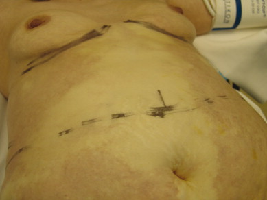
After the lower incision is marked, the proposed upper incision is drawn which estimates the amount of tissue to be resected ( Fig. 7.4 A ). The true extent of tissue resection will be determined intraoperatively. All areas for liposuction are marked at this time. This includes the entire central abdomen, lateral axilla and flanks, hip rolls, and mons ( Fig. 7.4 A–D ).
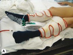
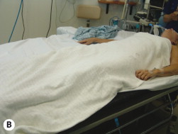
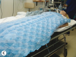
The patient is brought to the operating room after preoperative markings and photographs are completed. They are placed in the supine position and compression stockings are placed ( Fig. 7.5 A ). The patient is covered with warm blankets to prevent heat loss, and an intravenous line is established ( Fig. 7.5 B ). A warming blanket is used intraoperatively to maintain optimal body temperature ( Fig. 7.5 C ). General anesthesia is administered without the use of nitrous oxide to reduce the risk of intra-abdominal distension. Intravenous antibiotics (Ancef 1 g) and steroids (Decadron 4 mg) are routinely administered unless contraindicated by the patient’s medical history.
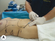
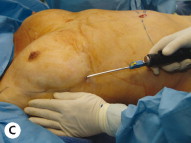
To minimize bleeding, all incision lines are injected with a solution of lidocaine and epinephrine ( Fig. 7.6 A ). The regions to be liposuctioned are then infiltrated with tumescent solution ( Fig. 7.6 B–E ). Normally the central abdomen, upper abdomen, lateral abdomen, flanks, hip rolls, and mons are infiltrated. The loose areolar plane between the abdominal wall and the subcutaneous tissue is also infiltrated to facilitate rapid dissection during tissue elevation.
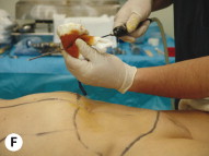
For a full or complete abdominoplasty, it is standard practice to infiltrate between 4 and 6 L of fluid, as this facilitates complete exsanguination of the tissue to be suctioned. Vessel compression and tissue exsanguination prevent stagnation of blood in the abdominoplasty flap. Blood stagnation may result in the formation of vascular microthrombi and the release of inflammatory mediators detrimental to flap viability.This infiltration thoroughly hydrates the tissue, allows cannulas to be passed with minimal trauma, and compresses the vessels, rendering them a smaller target for trauma and thus protecting them. It is important to calculate the total dose of lidocaine and modify the volume of tumescent fluid as needed, prior to infiltration. Large-volume infiltration requires more dilute lidocaine concentration (see Chapter 3 , Liposuction in Abdominal Contouring).
Infiltration is performed before full prepping and draping both for efficient use of time as well as to allow sufficient time for vasoconstruction to tale effect. Entry sites are prepared with Betadine and the cannula is frequently wiped with Betadine during the infiltration process ( Fig. 7.6 F ). Fluid is infiltrated until tissue turgor is achieved. Following infiltration, the patient is prepared and draped. This allows sufficient time for the epinephrine in the tumescent fluid to achieve the maximum vasoconstrictive effect ( Fig. 7.7 ). The interval between infiltration and liposuction is approximately 20 minutes, which is ideal for achieving excellent vasoconstriction.
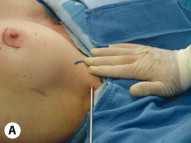
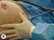
Liposuction is performed on all infiltrated areas. Suctioning of the lateral axilla and fullness above the breast is best performed through an axillary entry site ( Fig. 7.8 A, B ). Occasionally an inframammary entry site is used, which provides efficient access to the lateral breast, flank, and upper abdomen ( Fig. 7.8 C ). The umbilical entry site is ideal for access to the central abdomen ( Fig. 7.8 D, E ). In the area of proposed undermining, liposuction is performed on the fat superficial to Scarpa’s fascia, because subscarpal fat resection will be performed which will remove the fat deep to Scarpa’s fascia throughout the entire area.
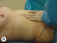
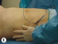
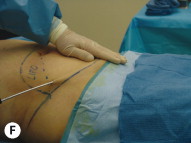
Additional entry sites within the marked tissue to be excised can used to treat the flanks, hip rolls, and mons ( Fig. 7.8 E, F ). All liposuction is performed on the areas outside the central abdominal region where subscarpal fat resection will be performed.
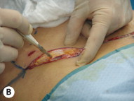
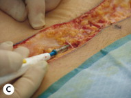
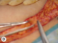
The low transverse incision, previously marked, is made with a number 10 blade into, but not entirely through the dermis ( Fig. 7.9 A ). Electrocautery is then used to complete the incision, sealing the blood vessels within the subdermal plexus ( Fig. 7.9 B ). Electrocautery is also used to deepen the incision through the superficial fascia to reach the deep subcutaneous tissue. The superficial inferior epigastric vessels are identified and controlled ( Fig. 7.9 C, D ). This is important, because these vessels can bleed postoperatively and lead to a hematoma (see Chapter 15 , Complications).