, Shimin Chang2, Jian Lin3 and Dajiang Song1
(1)
Department of Orthopedic Surgery, Changzheng Hospital Second Military Medical University, Shanghai, China
(2)
Department of Orthopedic Surgery, Yangpu Hospital Tongji University School of Medicine, Shanghai, China
(3)
Department of Microsurgery, Xinhu Hospital Shanghai Jiao Tong University, Shanghai, China
As the name suggests, digital artery perforator (DAP) flaps are based off the digital arteries. The concept of DAP flaps arose from the observation that many arterial branches from the digital artery originate in the lateral aspect of the finger. These branches perforate the thin fascia and adiposal tissue, terminating in multiple arterioles in the subdermal layer [1, 2].
Koshima et al. originally reported the utility of DAP flaps for fingertip reconstruction [3].
16.1 Vascular Anatomy
The reverse adipofascial dorsal digital (RADD) flaps are nourished by dorsal communicating branches of the digital arteries, which are numerous and constant (Fig. 16.1); venous drainage depends on the parallel venous system surrounding the arterial pedicle [2, 4–14].
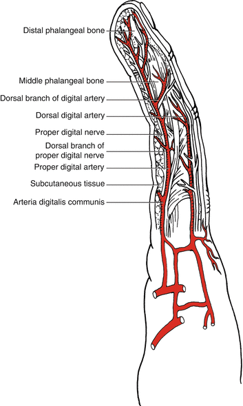
Fig. 16.1
Vascular anatomy of the digital artery
16.2 Illustrative Case
Case 1
A 39-year-old woman suffered a crush amputation of her right thumb fingertip following an accident with a metal press machine. The defect size was 3.0 × 2.5 cm (Fig. 16.2).

Fig. 16.2
Preoperative view
Flap Design
The flap is designed on the radial dorsal metacarpal artery perforator that located close to the defect (Fig. 16.3). The selected perforator is marked at the level of the metacarpal neck (Fig. 16.4).


Fig. 16.3
Flap design
Fig. 16.4
Schematic drawing of the flap design
Flap Elevation
The flap is elevated from proximal to distal under tourniquet control in the loose areolar tissue plane superficial to the extensor tendon paratenon (Fig. 16.5). Large veins at the edge of the flap are avoided because these veins only add to stasis within the flap (Fig. 16.6).


Fig. 16.5
Flap elevation
Fig. 16.6
Schematic drawing of the flap elevation
Flap Transfer and Insetting
The flap is then rotated into the defect and anchored loosely to the edges of the defect with the interphalangeal and metacarpophalangeal joints in extension (Fig. 16.7).

Fig. 16.7
Flap rotation
Follow-Up

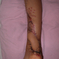
Case 2
A 27-year-old man suffered crush injury of the dorsum of his left thumb (Fig. 16.9).

Fig. 16.9
Preoperative view
The defect size was 3.0 × 2.0 cm.
Flap Design
A distally based flap was designed (Fig. 16.10). The pedicle was based just medial and proximal to the defect (Fig. 16.11).


Fig. 16.10
Flap design
Fig. 16.11
Schematic drawing of the flap design
Flap Elevation
The designed flap is elevated superficial to the digital neurovascular bundle (Fig. 16.12). At the side of the flap most proximal to the defect, the DAP closest to the defect is preserved as the pedicle vessel. Because the pedicle was very narrow in diameter, it was necessary to include peri-areolar soft tissue at the base of the pedicle (Fig. 16.13).


Fig. 16.12
Flap elevation
Fig. 16.13
Schematic drawing of the flap elevation
Flap Transfer
The perforator-based island flap is then rotated 90° to cover the defect (Fig. 16.14).

Fig. 16.14
Flap rotation
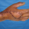
Flap Insetting
The donor site is closed with split-thickness skin graft (Fig. 16.15).

Fig. 16.15
Postoperative view
Follow-Up

Case 3
A 30-year-old man suffered crush amputations of his right middle and index fingers following an accident with a book press machine.
Related posts:

Stay updated, free articles. Join our Telegram channel

Full access? Get Clinical Tree


