, Shimin Chang2, Jian Lin3 and Dajiang Song1
(1)
Department of Orthopedic Surgery, Changzheng Hospital Second Military Medical University, Shanghai, China
(2)
Department of Orthopedic Surgery, Yangpu Hospital Tongji University School of Medicine, Shanghai, China
(3)
Department of Microsurgery, Xinhu Hospital Shanghai Jiao Tong University, Shanghai, China
A skin flap from the medial leg based on the posterior tibial artery and its cutaneous branches was first described by Zhang and colleagues in 1983 [1].
24.1 Vascular Anatomy
The posterior tibial artery is almost always present and is normally the largest terminal branch of the popliteal artery or tibioperoneal trunk [2].
As it reaches the ankle, the posterior tibial artery lies between the flexor digitorum longus and soleus muscles and Achilles tendon to almost assume a somewhat subcutaneous path [3].
Cutaneous perforators arise from the posterior tibial artery at unpredictable intervals (Fig. 24.1).
Fig. 24.1
Vascular anatomy of the posterior tibial artery
There are three distinct clusters of perforators for the posterior tibial artery at the following distances proximal to the medial malleolus: (1) 4–9 cm, (2) 13–18 cm, and (3) 21–26 cm.
Perforators with the largest caliber were typically present in the proximal two-thirds of the leg (Fig. 24.2).
Fig. 24.2
The perforators of the posterior tibial artery
24.2 Case 1: Distally Based Perforator Propeller Flap for Plate Exposure
A 60-year-old female suffered from an open Pilon fracture (Fig. 24.3). The skin over the medial malleolar area was necrotic after plate fixation, although MIPPO technique was applied (Fig. 24.4).
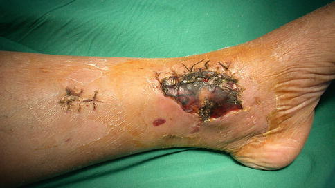
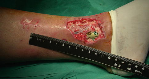
Fig. 24.3
Preoperative view
Fig. 24.4
The wound after thorough debridement
After debridement, the defect was measured 4 × 5 cm with plate exposure. We designed a posterior tibial artery perforator flap to cover the wound (Fig. 24.5).
Fig. 24.5
Schematic drawing of the flap design
Related posts:

Stay updated, free articles. Join our Telegram channel

Full access? Get Clinical Tree


