, Shimin Chang2, Jian Lin3 and Dajiang Song1
(1)
Department of Orthopedic Surgery, Changzheng Hospital Second Military Medical University, Shanghai, China
(2)
Department of Orthopedic Surgery, Yangpu Hospital Tongji University School of Medicine, Shanghai, China
(3)
Department of Microsurgery, Xinhu Hospital Shanghai Jiao Tong University, Shanghai, China
Lovie et al. [1] originally described the ulnar artery forearm flap as a free flap in 1984.
The true ulnar artery perforator (UAP) flap includes skin from the ulnar forearm or hand based on an ulnar artery perforator, with preservation of the ulnar artery. In the distal third of the forearm, an island flap based on a single dorsal perforator of the ulnar artery has been used. Becker and Gilbert [2] described a flap from the ulnar border of the distal dorsal forearm based on the dorsal branch of the ulnar artery.
11.1 Vascular Anatomy
The ulnar artery territory passes around the ulnar border of the forearm, extending onto the dorsal surface to the subcutaneous border of the ulna. Most of the perforators longitudinally anastomose with each other along the course of the main artery. These perforators form a deep fascial plexus and are arranged longitudinally along the main arterial axis.
In the distal third of the forearm, the ulnar artery gives off its dorsal branch 2–7 cm proximal to the pisiform bone. The distally based dorsal UAP flap is outlined on the dorsal ulnar aspect of the distal forearm. The potential area of the flap is 6 by 16 cm, with the length extending from the distal third of the forearm to the distal third of the dorsal surface of the hand [3]. The flap can be used for reconstructing the palm, dorsum, or ulnar border of the hand [4–8].
The dorsal branch of the ulnar artery runs obliquely from the palmar to the dorsal surface, passing deep to the flexor carpi ulnaris muscle and superficial to the ulnar nerve. It then gives off a fasciocutaneous branch that divides into an ascending branch and a descending branch (Fig. 11.1).
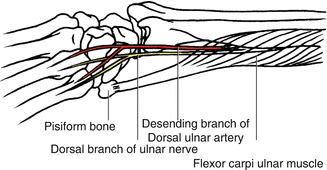
Fig. 11.1
Vascular anatomy of ulnar artery and nerve
11.2 Illustrative Case
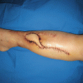
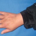
This 35-year-old man’s right hand was crushed in an industrial ice machine and was managed elsewhere with dressings for several weeks (Fig. 11.2).
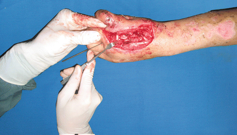
Fig. 11.2
Preoperative view
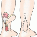
Flap Design
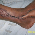
The flap measured 10 × 4.5 cm. The incision site was outlined on the ulnar side of the wrist and forearm overlying the tendon of flexor carpi ulnaris muscle (Fig. 11.3). The pisiform was identified and the pedicle was located emerging between 2 and 5 cm from it (Fig. 11.4).
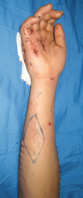
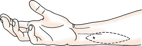


Fig. 11.3
Flap design
Fig. 11.4
Schematic drawing of flap design
Related posts:

Stay updated, free articles. Join our Telegram channel
Full access? Get Clinical Tree
