Key Words
warts, subungual, periungual, molluscum contagiosum, herpeticum, debridement, varicella, chickenpox, postherpetic neuralgia, herpes simplex
Warts
Warts are benign epidermal neoplasms that are caused by human papillomaviruses (HPVs), which are small DNA viruses. There are more than 100 different types of HPVs, and new types are discovered each year. HPVs infect epithelial cells of the skin, mouth, esophagus, larynx, trachea, and conjunctiva and cause both benign and malignant lesions. They induce a variety of infections ( Table 12.1 ).
| Clinical Manifestation | HPV Types |
|---|---|
| Plantar warts | 1, 2, 3, 4, 27, 29, 57 |
| Common warts | 1, 2, 3, 4, 27, 57 |
| Flat warts | 3, 10, 28 |
| Epidermodysplasia verruciformis | 5, 8, 9, 12, 14, 15, 17, 19–25, 36, 47, 50 |
| Genital warts, laryngeal papillomas | 6, 11 |
| Butcher’s warts | 7 |
| Focal epithelial hyperplasia (Heck disease) | 13, 32 |
| Anogenital dysplasias and neoplasms (rarely laryngeal carcinomas) | 16, 18, 31, 33, 35, 42, 43, 44, 45, 51, 52, 56, 58, 59, 68 |
| Keratoacanthoma | 37 |
| Cutaneous squamous cell carcinoma | 38, 41, 48 |
| Oral papillomas, inverted nasal and papillomas | 57 |
| Buschke–Löwenstein tumors | 6, 11 |
| Bowenoid papulosis | 16, 18, 33, 39 |
| Cystic warts | 60 |
| Pigmented wart | 65 |
| Vulvar papilloma | 70 |
| Oral papillomas (in HIV-infected patients) | 72, 73 |
| Common wart in renal allograft recipient | 75–77 |
| Cutaneous wart | 78 |
Clinical Infection
Warts commonly occur in children and young adults, but they may appear at any age. Warts are transmitted simply by touch; it is not unusual to see warts on adjacent toes (“kissing lesions”). Warts commonly appear at sites of trauma, on the hands, in periungual regions as a result of nail biting, and on plantar surfaces. Plantar warts may be acquired from moist surfaces in communal swimming areas. Their course is highly variable; most resolve spontaneously in weeks or months, and others may last years or a lifetime. Infection with HPV can be latent, subclinical, or clinical. Latent infections are detected with molecular biologic techniques. Subclinical infections are found with a colposcope or microscope. HPVs induce hyperplasia and hyperkeratosis.
Immunologic Response
The regression of virus-infected cells involves a multifactorial response that includes cell-mediated immunity and induction of interferons. Individual variations in cell-mediated immunity may account for differences in severity and duration. Warts develop on many immunosuppressed patients. Warts occur more frequently, last longer, and appear in greater numbers in immunosuppressed individuals such as those with HIV, transplants, lymphoma, and those taking immunosuppressive medications. Individuals with atopic dermatitis, especially children are at increased risk for warts.
Treatment
Some types of warts respond quickly to routine therapy, whereas others are resistant. It should be explained to patients that warts often require several treatment sessions before a cure is realized. Because warts are confined to the epidermis, they can be removed with little, if any, scarring. To avoid scarring, treatment should be conservative. Treatment that results in a hand with many scars is not worthwhile for lesions that undergo spontaneous resolution.
Warts obscure normal skin lines; this is an important diagnostic feature. When skin lines are re-established, the warts are gone. Warts vary in shape and location and are managed in several different ways.
Warts: the Primary Lesion
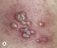
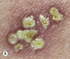
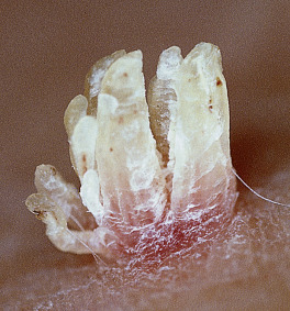
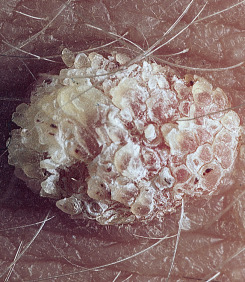
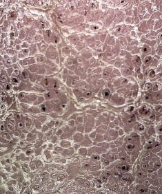
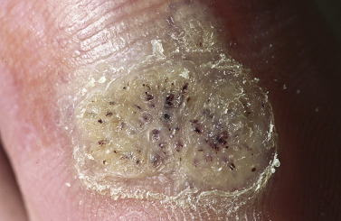
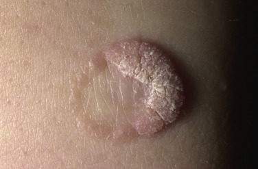
Warts are tumors initiated by a viral infection of keratinocytes. The cells proliferate to form a mass but the mass remains confined to the epidermis. There are no “roots” that penetrate the dermis. Several types of warts form cylindrical projections. These projections are clearly seen in digitate warts that occur on the face ( Fig. 12.1 ). The projections fuse in common warts on thicker skin ( Fig. 12.2 ); this produces a highly organized mosaic pattern on the surface. This pattern is unique to warts and is a useful diagnostic sign ( Fig. 12.3 ). Thrombosed black vessels become trapped in these projections and are seen as black dots on the surface of some warts ( Fig. 12.4 ). Although warts remain confined to the epidermis, the growing mass can protrude deeper into the skin and displace the dermis. Blunt dissection of a wart shows that the under-surface is smooth ( Fig. 12.5 ).

Common Warts
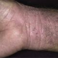
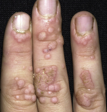
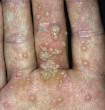
Common warts (verruca vulgaris) begin as smooth, skin-colored papules and evolve into dome-shaped, gray-brown, hyperkeratotic growths with black dots on the surface ( Figs. 12.6 and 12.7 ). The black dots, which are thrombosed capillaries, are a useful diagnostic sign and may be exposed by paring the hyperkeratotic surface with a #15 surgical blade. The hands are the most commonly involved areas, but warts may be found on any skin surface. In general, warts are few in number, but it is not unusual for common warts to become so numerous that they become confluent and obscure large areas of normal skin.
Treatment.
Topical salicylic acid preparations, liquid nitrogen ( Figs. 12.8 and 12.9 ), and very light electrocautery are the best methods of initial therapy. Blunt dissection is used for resistant or very large lesions. The technique for the application of salicylic acid is described in the treatment section for plantar warts. Duct tape occlusion is not effective.
Cryotherapy.
Cryotherapy is effective for common warts and is reasonable first-line therapy. The hyperkeratotic surface should be pared if possible and liquid nitrogen applied with either a spray or a cotton-tipped applicator so that a 1- to 2-mm zone of frozen tissue is created and maintained around lesional skin for about 5 seconds. A small blister, sometimes hemorrhagic, is expected. Excessive freezing causes massive swelling, hemorrhagic blisters, hypopigmentation or hyperpigmentation, and scarring. Sharp pain lasts for minutes and sometimes hours; some children tolerate the pain. Freezing may be repeated every 2 to 4 weeks until resolution of the wart.
Treatment of Recalcitrant Warts
Imiquimod.
Nightly application of the immunomodulatory drug imiquimod 5% cream may be effective. The patient is instructed to soak the wart to soften the keratin surface. Some patients respond well to soaking warts in hot water at 113° F for 30 minutes at least 3 times per week. Removal of the keratin with an abrasive material, such as a pumice stone, and application of tape to cover the area facilitate penetration of the imiquimod cream.
Apple Cider Vinegar.
Many people claim success with this technique. Dip the wart in vinegar for 20 minutes. Apply Vaseline around the wart to protect the skin. Soak a small piece of cotton in vinegar and tape it to the wart overnight. Repeat each night until the wart is gone.
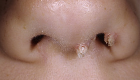
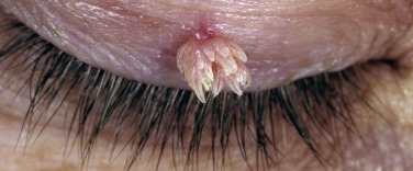
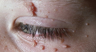
Filiform and Digitate Warts
These growths consist of a few or several finger-like, skin-colored projections emanating from a narrow or broad base. They are most commonly observed about the mouth, beard, eyes, and ala nasi ( Figs. 12.10 to 12.13 ).

Treatment.
These are the easiest warts to treat. Those with a very narrow base do not require anesthesia. A firm base is created by retracting the skin on either side of the wart with the index finger and thumb. A curette is then firmly drawn across the base, removing the wart with one stroke. Bleeding is controlled with gauze pressure rather than by using Monsel’s solution, which is painful. This technique is particularly useful for young children who refuse local anesthesia with a needle. Light electrocautery is an alternative.
Flat Warts
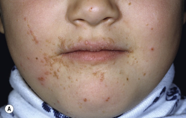
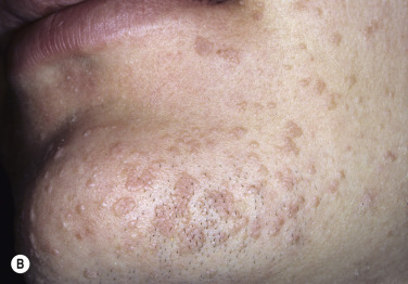
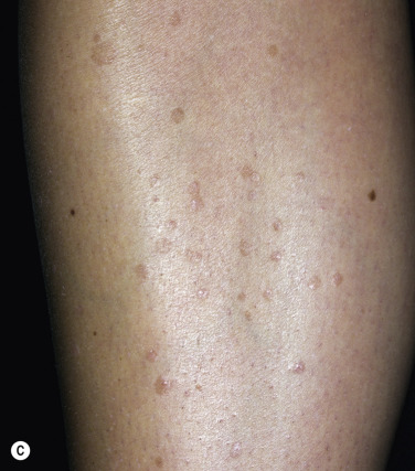
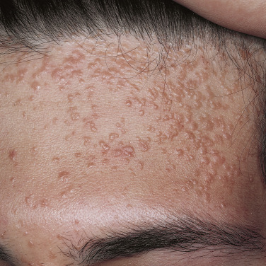
Flat warts (verruca plana) are pink, light brown, or light yellow and are slightly elevated, flat-topped papules that vary in size from 0.1 to 0.5 cm. There may be only a few, but in general they are numerous. Plana warts are mainly caused by HPV-3 and HPV-10. Typical sites of involvement are around the mouth ( Fig. 12.14A ), on the forehead ( Fig. 12.15 ), on the backs of the hands, and on shaved areas such as the beard area in men and the lower legs in women. A line of flat warts may appear as a result of scratching these sites.
Treatment.
Flat warts present a special therapeutic problem. Their duration may be lengthy, and they may be very resistant to treatment. In addition, they are usually located in cosmetically important areas where aggressive, scarring procedures are to be avoided. Imiquimod 5% cream applied every day or every other day may be effective. Freezing of individual lesions with liquid nitrogen or applying a very light touch with the electrocautery needle may be performed for patients who are concerned with cosmetic appearance and desire quick results. Treatment with 5-fluorouracil cream applied once or twice a day for 3 to 5 weeks may produce dramatic clearing of flat warts; it is worth the attempt if other measures fail. Persistent hyperpigmentation may occur following 5-fluorouracil use. This result may be minimized by applying the ointment to individual lesions with a cotton-tipped applicator. Warts may reappear in skin inflamed by 5-fluorouracil.
A study of patients with recalcitrant facial plana (age range from 5 to 35 years) reported that oral isotretinoin 0.5 mg/kg/day prescribed for 2 months resulted in a complete response in 73% of patients .
Plantar Warts
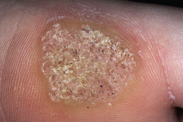
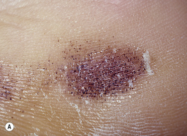
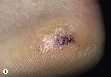
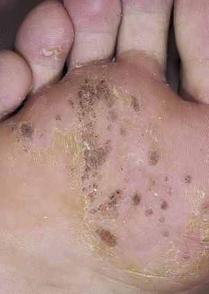
Warts of the soles are called plantar warts. Patients may, incorrectly, refer to warts on any surface as plantar warts. Plantar warts frequently occur at points of maximum pressure, such as over the heads of the metatarsal bones or on the heels ( Fig. 12.16 ). A thick, painful callus forms in response to pressure and the foot is repositioned while walking. This may result in distortion of posture and pain in other parts of the foot, leg, or back. A little wart can cause a lot of trouble.
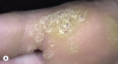
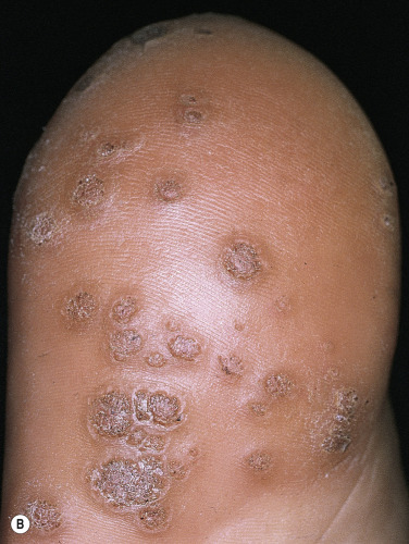
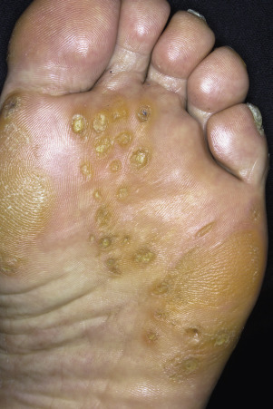
Warts may appear anywhere on the plantar surface. A cluster of many warts that appears to fuse is referred to as a mosaic wart ( Figs. 12.17 to 12.19 ).
Differential Diagnosis
Corns.
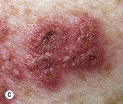
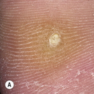
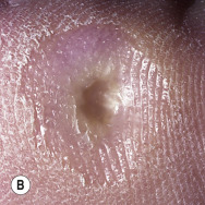
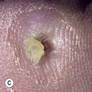
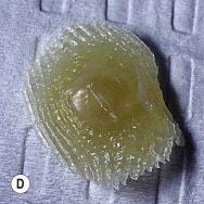
Corns are a mechanically induced lesion that forms over or under a weight-bearing surface or structure ( Fig. 12.20 ). Corns (clavi) over the metatarsal heads are frequently mistaken for warts. The two entities can be easily distinguished by paring the callus with a #15 surgical blade. Warts lack skin lines that cross their surface and have centrally located black dots that bleed with additional paring. Examination with a hand lens shows a highly organized mosaic pattern on the surface. Corns also lack skin lines crossing the surface, but they have a hard, painful, well-demarcated, translucent central core ( Fig. 12.21A ). The core or kernel can be removed easily by inserting the point of a #15 surgical blade into the cleavage plane between normal skin and the core, holding the scalpel vertically, and smoothly drawing the blade circumferentially ( Figs. 12.21B–D ). The hard kernel is freed by drawing the blade horizontally through the base to reveal a deep depression. Pain is greatly relieved by this simple procedure. Lateral pressure on a wart causes pain, but pinching a plantar corn is painless.
The treatment of corns is targeted at reducing the friction or pressure at a specific location. This can be accomplished with orthotic therapy and/or surgical correction of the osseous deformity creating the mechanical pressure point. Podiatric or orthopedic surgeons familiar with biomechanics and reconstructive surgery perform these corrective procedures.
Black Heel.
Horizontally arranged clusters of blue-black dots (ruptured capillaries or petechiae) may appear on the upper edge of the heel or anywhere on the plantar surface following the shearing trauma of sports that involve sudden stops or position changes ( Fig. 12.22A ). It is caused by the shearing force of the epidermis sliding over the rete pegs of the papillary dermis. At first glance, this may be confused with a wart or acral–lentiginous melanoma, but closer examination reveals normal skin lines, and paring does not cause additional bleeding ( Fig. 12.22B ). The condition resolves spontaneously in a few weeks.
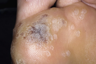
Black Warts.
Warts in the process of undergoing spontaneous resolution, particularly on the plantar surface, may turn black ( Fig. 12.23 ) and feel soft when pared with a blade. Cell-mediated immunity against virus-infected keratinocytes may take place in the process of regression of some warts.
Treatment.
Plantar warts do not require therapy as long as they are painless. Although their number may increase, it is sometimes best to explain the natural history of the viral infection and wait for resolution rather than subject the patient to a long treatment program. Minimal discomfort can be relieved by periodically removing the callus with a blade or pumice stone.
Patients many times seek treatment because the pain limits daily weight-bearing activities such as walking, running and sports.
Debridement.
It is very important to debride the hyperkeratotic tissue over and around plantar warts to ensure penetration of the medication. This may require seeing the patient every 2 to 3 weeks.
Combination Therapy.
Multiple simultaneous techniques are often required to successfully treat plantar warts and may include the following regimens.
Keratolytic Therapy (Salicylic Acid Liquid).
Keratolytic therapy with salicylic acid (over-the-counter) is conservative initial therapy for plantar warts. The treatment is nonscarring and relatively effective but requires persistent application of medication once each day for many weeks.
The wart is pared with a blade, pumice stone, or sandpaper (emery board). The affected area is soaked in warm water to hydrate the keratin surface; this facilitates penetration of the medicine. A drop of solution is applied with the applicator and allowed to dry. Solution may be added as needed to cover the entire surface of the wart. Penetration of the acid mixture is enhanced if the treated wart is covered with a piece of adhesive tape. Inflammation and soreness may follow tape occlusion, necessitating periodic interruption of treatment; consequently, the patient may be satisfied with the longer, more comfortable process of simply applying the solution at bedtime. White, pliable keratin forms in a few days and should be pared with a blade or worn away with abrasives such as sandpaper or a pumice stone. Ideally, the white keratin should be removed to expose pink skin; to accomplish this, an occasional visit to the office may be necessary.
Keratolytic Therapy (40% Salicylic Acid Plasters).
This is a safe, nonscarring treatment similar to keratolytic therapy with salicylic acid liquid except the salicylic acid has been incorporated into a pad. Salicylic acid plasters are particularly useful in treating mosaic warts that cover a large area.
The plaster is cut to the size of the wart. The backing of the plaster is removed and the sticky surface is applied to the wart and secured with tape. The plaster is removed in 24 to 48 hours, the pliable white keratin is reduced in the manner previously described, and another plaster is applied. The treatment requires many weeks, but it is effective and less irritating than salicylic acid and lactic acid liquid. Pain is relieved because a large amount of keratin is removed during the first few days of treatment.
Apple Cider Vinegar.
Many people claim success with this technique. Dip the wart in vinegar for 20 minutes. Apply Vaseline around the wart to protect the skin. Soak a small piece of cotton in vinegar and tape it to the wart overnight. Repeat each night until the wart is gone.
5-Fluorouracil (5-FU).
Application of 5-FU cream 5% under tape over 12 weeks resulted in an 85% clearance rate. The average time to cure occurred at 9 weeks of treatment.
Blunt Dissection.
Blunt dissection is a surgical alternative that is fast, effective (90% cure rate), and usually nonscarring. It is superior to both electrodesiccation–curettage and excision because normal tissue is not disturbed. (See Chapter 27 for surgical techniques.)
Imiquimod.
The immunomodulating drug imiquimod is more effective on thicker keratinized (nongenital) skin when occluded and used in combination with cryotherapy or a keratolytic agent. It is essential to debride the thick scale before applying imiquimod. The patient applies the cream daily and covers with tape (for ≥12 hours) to enhance penetration. Response to the use of imiquimod on the plantar surface is usually not preceded by an inflammatory reaction.
Suggestive Therapy.
Suggestive therapy generally works through the age of 10 years. A banana peel, potato eye, or a penny applied to the skin and covered with tape for a 1 to 2 week period has been effective in young children. Another technique is to draw the body part on a piece of paper and then draw a picture of the wart on the diagram. Crumble the pictures and then discard them.
Cantharidin.
Cantharidin mixtures are very effective for plantar warts. At the clinician’s office, apply Canthacur-PS (cantharone plus podophyllin 5% plus salicylic acid 30%) and allow the solution to dry. Avoid touching unaffected skin. Cover with occlusive tape (e.g., Blenderm) or moleskin and remove in 24 hours, or earlier if there is significant discomfort. A blister usually appears. Patients can relieve pain by breaking the blister. Patients may apply moleskin or felt padding around but not over the lesion to reduce pressure. In 2 to 3 days, remove the blister by excising with a scissors or using a curette after administering a local anesthetic. Repeat weekly if necessary.
Laser.
Various lasers (e.g., long-pulsed Nd-YAG, CO 2 , and pulsed dye lasers) are available for treating resistant warts. The procedure is expensive and painful, often requiring local anesthesia.
Chemotherapy.
For years a variety of acids have been successfully used to treat plantar warts. This technique is occasionally used to treat warts that have recurred after treatment with other techniques and is occasionally used as initial therapy. Like keratolytic therapy, repeated application is required. Home application of acids is too dangerous; therefore weekly or biweekly visits to the clinician’s office are required. A number of acids may be used (bichloroacetic acid, trichloroacetic acid).
Treatment is as follows: The excess callus is pared. The surrounding area is protected with petrolatum. The entire lesion is coated with acid, and the acid is worked into the wart with a sharp toothpick. This procedure is repeated every 7 to 10 days.
Formalin.
This may be considered for resistant cases. Mosaic warts or other large involved areas may be treated with daily soaking for 30 minutes in 4% formalin solution. The firm, fixed tissue is pared before subsequent soaking. Lazerformaldehyde solution (10% formaldehyde) is commercially available for direct application to warts. There is a risk of inducing sensitization to formalin.
Cryosurgery.
Cryosurgery on the sole may produce a deep, painful blister and interfere with mobility. Repeated light applications of liquid nitrogen are preferred to aggressive treatment. Cryotherapy is equally effective when applied with a cotton wool bud or by means of a spray. A surgical blade is used to debulk the wart before freezing. Liquid nitrogen is applied until ice-ball formation has spread from the center to include a margin of 2 mm around each wart. A double or triple freeze–thaw cycle may be more effective than a single freeze. Treatment is given every 2 to 4 weeks for up to 3 months.
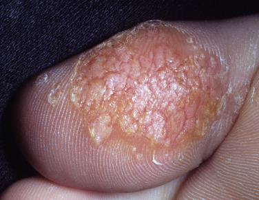
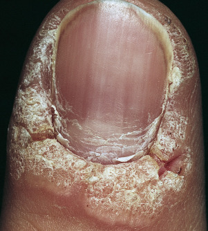
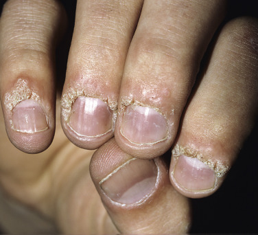
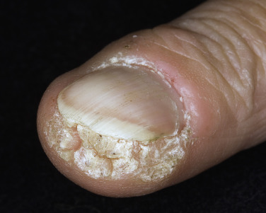
Subungual and Periungual Warts
Subungual and periungual warts ( Figs. 12.24 to 12.26 ) are more resistant to both chemical and surgical methods of treatment than are warts located in other areas. A wart next to the nail may simply be the tip of the iceberg; much more of the wart may be submerged under the nail.
Treatment.
The tips of the fingers and toes are a confined area. Therapeutic measures that cause inflammation and swelling, such as cryosurgery, may produce considerable pain.
Cryosurgery.
Small periungual warts respond to conservative cryosurgery; warts that extend under the nail do not respond. The use of aggressive cryosurgery over superficial nerves on the volar or lateral aspects of the proximal phalanges of the fingers has caused neuropathy. Permanent nail changes may occur if the nail matrix is frozen.
Cantharidin.
Cantharidin (Cantharone) causes blister formation at the dermoepidermal junction but does not cause scarring. Adverse effects are postinflammatory hyperpigmentation, painful blistering, and dissemination of warts to the area of blistering.
In treatment, the solution is applied to the surface and allowed to dry and then occluded with tape. The patient is seen 1 week later for evaluation. Blisters are opened and the remaining wart is retreated. If blistering does not occur, then cantharidin is applied in one to three layers and covered with tape for 48 hours. Each layer should be dry before the next application of cantharidin. The treatment is very effective for some patients, but there are some warts that do not respond to repeated applications.
Keratolytic Preparations.
The same procedures described for treating plantar warts with salicylic acid and lactic acid paint and salicylic acid plasters are useful for periungual warts.
Blunt Dissection.
When conventional measures fail, blunt dissection offers an excellent surgical alternative (see Chapter 27 ). Local anesthesia is induced with 2% lidocaine without epinephrine around and under small warts. A digital block is required for larger warts. Hemostasis during the procedure is maintained by firm pressure over the digital arteries or with a rubber-band tourniquet. The nail should be removed only if the wart is very large and imbedded. The procedure is exactly the same as that described for blunt dissection of plantar warts.
Duct Tape Occlusion.
Duct tape occlusion therapy may be more effective than cryotherapy for common warts. To completely cover the wart, the tip of the finger is wrapped with duct tape. The tape remains in place for 6 days, is removed at home, is then reapplied in a similar manner 12 hours later, and remains in place for an additional 6 days. This procedure is repeated for up to 2 months.
Molluscum Contagiosum
Clinical Manifestations.
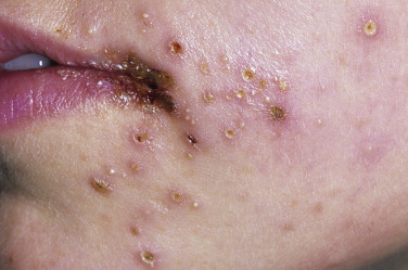
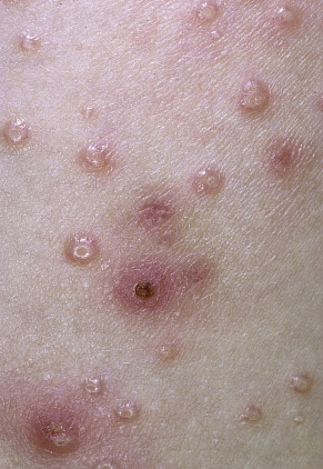
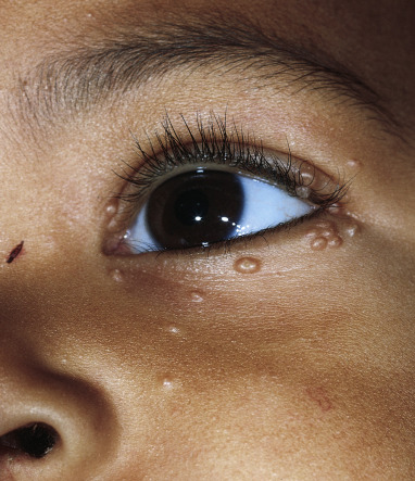
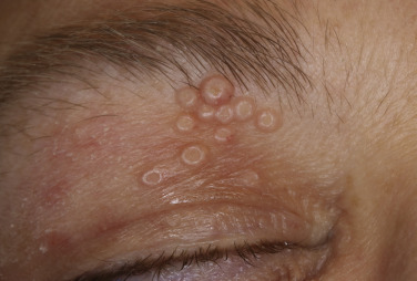
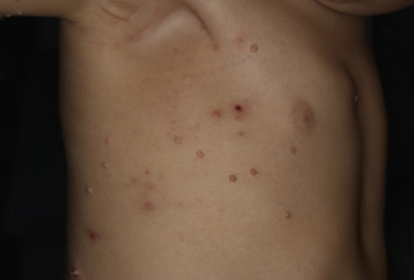
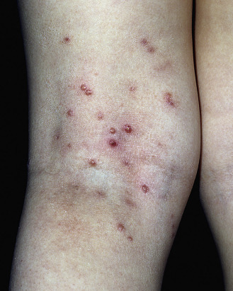
Molluscum contagiosum is a very common viral infection of the skin characterized by discrete, 2- to 5-mm, slightly umbilicated, skin-colored, dome-shaped papules ( Fig. 12.27 ). It spreads via autoinoculation, scratching, or touching a lesion and fomites. The areas most commonly involved are the face ( Fig. 12.28 and Fig. 12.29 ), trunk ( Fig. 12.30 ), axillae, extremities in children, and the pubic and genital areas in adults. Lesions are frequently grouped; there may be few or many covering a wide area. Unlike warts, the palms and soles are not involved. It is not uncommon to see erythema and scaling at the periphery of a single lesion or several lesions. This may be the result of inflammation from scratching or may be a hypersensitivity reaction. Lesions spread to inflamed skin, such as areas of atopic dermatitis ( Fig. 12.31 ). The individual lesion begins as a smooth, dome-shaped, white to skin-colored papule. With time, the center becomes soft and umbilicated. Most lesions are self-limiting and clear spontaneously in 6 to 9 months; however, they may last 2 to 4 years or longer. Genital molluscum contagiosum may be a manifestation of sexual abuse in children.
Inflammatory Reactions.
Approximately 20% of patients develop inflamed lesions. They typically present as erythematous, edematous papules and papulonodules that may become pustular or fluctuant. Molluscum dermatitis presents as a pruritic eczematous eruption in the skin surrounding molluscum lesions. The dermatitis can be diffuse or nummular. Patients with a history of atopic dermatitis are more likely to have dermatitis associated with their lesions. Inflammation usually leads to regression of affected lesions and sometimes heralds clearance of the entire eruption, including lesions that do not develop inflammation. Patients with inflamed lesions are less likely to have an increased number of lesions during the next few months than those without inflamed lesions.
Molluscum Contagiosum in HIV-Infected Patients.
Molluscum contagiosum is a common and at times severely disfiguring cutaneous viral infection in patients with HIV. Atypical facial lesions occur with either multiple small papules or giant nodular tumors. Cutaneous cryptococcosis may resemble molluscum contagiosum in patients with HIV. Cytologic examination of skin brushing reveals encapsulated budding yeasts. An inverse relation between CD4+ count and the number of molluscum contagiosum lesions is observed.
Diagnosis.
If necessary the diagnosis can be established easily with laboratory methods. The virus infects epithelial cells, creating very large intracytoplasmic inclusion bodies and disrupting cell bonds by which epithelial cells are generally held together. This lack of adhesion causes the central core of the lesion to be soft. Rapid confirmation can be made by removing a small lesion with a curette and placing it with a drop of potassium hydroxide between two microscope slides. The preparation is gently heated and then crushed with firm, twisting pressure. Larger umbilicated papules have a soft center, the contents of which can be obtained by scooping with a needle. This material contains only infected cells and can be examined directly in a heated potassium hydroxide preparation. The infected cells are dark and round and disperse easily with slight pressure, whereas normal epithelial cells are flat and rectangular and tend to adhere to each other in sheets. Virions streaming out of the amorphous mass can be seen if Sedi-Stain, a supravital stain used to stain urine sediments, is used. Toluidine blue gives the same results. Viral inclusions (large, eosinophilic, round, intracytoplasmic bodies) are easily seen in a fixed and stained biopsy specimen.
Treatment.
Treatment must be individualized. Conservative nonscarring methods should be used for children who have many lesions. Genital lesions in adults should be definitively treated to prevent spread by sexual contact (see Chapter 11 ). New lesions that are too small to be detected may appear after treatment and may require additional attention. Topical corticosteroids are used to treat both nearby dermatitis and dermatitis involving the lesions.
Curettage.
Small papules can be quickly removed with a curette and without local anesthesia in adults. Children might tolerate curettage after a lidocaine/prilocaine cream (EMLA) is applied for analgesia. The cream is applied 30 to 60 minutes before treatment. Bleeding is controlled with gauze pressure. Monsel’s solution is painful to use in an unanesthetized area in children. Curettage is useful when there are a few lesions because it provides the quickest, most reliable treatment. A small scar may form; therefore this technique should be avoided in cosmetically important areas.
Forceps.
Forceps may be utilized to gently pinch the molluscum lesions to extract the central core. This method can be utilized with eyelid lesions and children.
Cryosurgery.
Cryosurgery is the treatment of choice for patients who do not object to the pain. Most children will not tolerate cryosurgery. The papule is touched lightly with a nitrogen-bathed cotton swab or spray until the advancing, white, frozen border has progressed to form a 1-mm halo on the normal skin surrounding the lesion. This should take approximately 5 seconds. This conservative method destroys most lesions in one to three treatment sessions at 1- or 2-week intervals and rarely produces a scar.
Cantharidin.
Cantharidin, a chemovesicant extract from the blister beetle, is very effective, well tolerated, and safe in children. It penetrates the epidermis and induces vesiculation through acantholysis. Cantharidin is sparingly applied to each nonfacial lesion with the blunt wooden end of a cotton-tipped applicator. Contact with surrounding skin is avoided, and a maximum of 20 lesions are treated per visit. The treated areas are washed with soap and water after 4 to 6 hours, or sooner if burning, discomfort, or vesiculation occurs; therapy is repeated at 2- to 4-week intervals. Lesions blister and may clear without scarring. Occasionally, new lesions appear at the site of the blister created with cantharidin. Blistering and pain are mild to moderate. Pitted shallow depressions sometimes occur.
Potassium Hydroxide 5%.
Parents are instructed to apply the pharmacist-prepared solution twice daily with a cotton swab. A brief stinging may occur shortly after the application. Most lesions clear in 4 weeks.
Podophyllotoxin 0.5%.
The daily application of podophyllotoxin 0.5% may be tried for cases resistant to other therapies.
Hypoallergenic Surgical Adhesive Tape.
Tape is applied once each day after showering and is used each day until the lesion ruptures and the core is discharged. The average time to clearance is 16 weeks.
Salicylic Acid.
Salicylic acid solution applied each day without tape occlusion may cause irritation and encourage resolution.
MolluscumRx.
This topical solution and others like it are a natural treatment comprised of plant extracts. They are nonprescription treatments available through the Internet and sold in some physicians’ offices. Their efficacy has not been proved.
Laser Therapy.
Lesions on the genital area may be treated with the pulsed dye or the carbon dioxide laser.
Trichloroacetic Acid Peel in Immunocompromised Patients.
Patients with HIV infection who have extensive facial molluscum contagiosum infection were treated with trichloroacetic acid peels. Peels were performed with 25% to 50% trichloroacetic acid (average, 35%) and were repeated every 2 weeks as needed. A total of 15 peels were performed with an average reduction in lesion counts of 40.5% (range, 0% to 90%).
Herpes Simplex
Genital herpes simplex virus (HSV) infections are discussed in Chapter 11 .
HSV infections are caused by two different virus types (HSV-1 and HSV-2), which can be distinguished by laboratory and office tests. HSV-1 is generally associated with oral infections, and HSV-2 is associated with genital infections. HSV-1 genital infections and HSV-2 oral infections are becoming more common, possibly as a result of oral–genital sexual contact. Both types seem to produce identical patterns of infection. Many infections are asymptomatic, and evidence of previous infection can be detected only by an elevated immunoglobulin G (IgG) antibody titer. HSV infections have two phases: the primary infection, after which the virus becomes established in a nerve ganglion; and the secondary phase, characterized by recurrent disease at the same site. The rate of recurrence varies with virus type and anatomic site. Genital recurrences are nearly six times more frequent than oral–labial recurrences; genital HSV-2 infections recur more often than genital HSV-1 infections; and oral–labial HSV-1 infections recur more often than oral HSV-2 infections. Infections can occur anywhere on the skin. Infection in one area does not protect the patient from subsequent infection at a different site. Lesions are intraepidermal and usually heal without scarring.
Primary Infection
Many primary infections are asymptomatic and can be detected only by an elevated IgG antibody titer. Like most viral infections, the severity of disease increases with patient age. The virus may be spread via respiratory droplets, direct contact with an active lesion, or contact with virus-containing fluid such as saliva or cervical secretions in patients with no evidence of active disease. Symptoms occur from 3 to 7 or more days after contact. Tenderness, pain, mild paresthesias, or burning occurs before the onset of lesions at the site of inoculation. Localized pain, tender lymphadenopathy, headache, generalized aching, and fever are characteristic prodromal symptoms. Some patients have no prodromal symptoms.
Lesions
Grouped vesicles on an erythematous base appear and subsequently umbilicate ( Fig. 12.32 ). The vesicles in primary herpes simplex ( Figs. 12.33 to 12.36 ) are more numerous and scattered than those in the recurrent infection ( Figs. 12.37 to 12.40 ). The vesicles of herpes simplex are uniform in size in contrast to the vesicles seen in herpes zoster, which vary in size. Mucous membrane lesions accumulate exudate, whereas skin lesions form a crust. Lesions last for 2 to 4 weeks unless secondarily infected and heal without scarring.