Key Words
acneiform, drug eruptions, exfoliative erythroderma, lichenoid, erythematosus-like drug eruption, vasculitis, exanthems, measles, Kawasaki, enterovirus, roseola, rubella, scarlet fever, toxic shock syndrome, echovirus, coxsackievirus
The word exanthem means a skin eruption that bursts forth or blooms. Exanthematous diseases are characterized by widespread, symmetric, erythematous, discrete, or confluent macules and papules that initially do not form scale. Exanthematous disease is one of the few diseases for which the term maculopapular is an appropriate descriptive term. Other lesions, such as pustules, vesicles, and petechiae, may form, but most of the exanthematous diseases begin with red macules or papules. Widespread red eruptions such as guttate psoriasis or pityriasis rosea may have a similar beginning and are often symmetric, but these conditions have typical patterns of scale and are therefore referred to as papulosquamous eruptions. Diseases that begin with exanthems may be caused by bacteria, viruses, or drugs. Most have a number of characteristic features such as a common primary lesion, distribution, duration, and systemic symptoms. Some are accompanied by oral lesions that are referred to as enanthems.
Table 14.1 describes the characteristic features of some viral exanthems that help distinguish them from drug eruptions.
| Diagnosis | Description and Distinguishing Features |
|---|---|
| Measles (rubeola) | The rash is morbilliform (meaning “measles-like”), a term often used to describe exanthematous drug eruptions, and is usually itchy. Unlike most drug eruptions, the rash seen in measles often begins on the head and neck and spreads rapidly. It usually begins a few days after the onset of fever, cough, coryza, and conjunctivitis. White spots on the buccal mucosa (Koplik spots) help establish the diagnosis. Typical or atypical rash may occur in previously vaccinated adults, principally those who received only older, killed vaccine or who were incompletely vaccinated. |
| Rubella | Symptoms are usually milder than those seen in measles, with a similar rash that usually resolves within 3 or 4 days. The rash is often accompanied by fever, adenopathy, and arthralgias. |
| Roseola infantum (exanthema subitum) | Young children have a high temperature for 3 to 5 days; it usually resolves around the time of onset of subitum, with the rash a pink, short-lived eruption. Human herpesvirus 6 is the most frequent cause. Adults have cervical adenopathy, with variable rash and fever that may last for months. The rash usually starts on the trunk and spreads to the face and extremities. |
| Erythema infectiosum (fifth disease) | In young children, fever (with characteristic “slapped cheeks”) develops 2 to 4 days before generalized rash or disease, which begins on proximal extremities and spreads both centrally and peripherally. In adults, arthralgias, which may persist for many weeks, and fever are prominent. The rash often has a livedo pattern. Facial involvement is less prominent in adults than in children. The disease is caused by parvovirus B19. |
| Infectious mononucleosis | In adolescents and adults, the rash is usually associated with aminopenicillin administration, with an onset within 3 days after administration (a more rapid onset than is usual for drug eruptions). Patients are unlikely to have rash with readministration of aminopenicillin after recovery. |
| Acute graft-versus-host disease | The rash typically occurs 2 to 4 weeks after transplantation. It may be pruritic. If generalized, the rash is often difficult to distinguish clinically from an exanthematous drug eruption. |
| Acute human immunodeficiency | The rash has an acute onset 1 to 6 weeks after infection and is usually accompanied by fever, malaise, virus seroconversion myalgias, arthralgias, and lymphadenopathy. It is a symmetric exanthematous rash that involves the face, palms, and soles. Oral and genital aphthous-type ulcers may occur. |
| Other viral exanthems | Causative agents include echoviruses, coxsackie virus, togavirus, and others. |
* Other diagnostic aids may include viral culture, skin biopsy, detection of virus by means of polymerase chain reaction assay, and serologic tests for antibodies (especially IgM antibody in acute infections).
Exanthems were previously consecutively numbered according to their historic appearance and description: first disease, measles; second disease, scarlet fever; third disease, rubella; fourth disease, “Dukes disease” (probably coxsackievirus or echovirus); fifth disease, erythema infectiosum; and sixth disease, roseola infantum.
Exanthems
Measles
Transmission and Risk.
Measles (rubeola or morbilli) is a highly contagious viral disease transmitted by contact with droplets from infected individuals ( Box 14.1 ). The virus is spread by coughing and sneezing, close personal contact, or direct contact with infected nasal or throat secretions. The virus remains active and contagious in the air or on infected surfaces for up to 2 hours. It can be transmitted by an infected individual from 4 days before the onset of the rash to 4 days after the onset. If one person has the disease, a high proportion of their susceptible close contacts will also become infected. Unimmunized young children are at highest risk for measles and its complications, including death. Measles can be particularly deadly in countries experiencing or recovering from war, civil strife, or a natural disaster. Infection rates soar because damage to health services interrupts routine immunization. Overcrowding in camps for refugees and internally displaced people greatly increases the risk of infection.
Clinical description – an acute illness characterized by:
- •
Generalized, maculopapular rash lasting ≥3 days; and
- •
Temperature ≥101° F or 38.3° C; and
- •
Cough, coryza, or conjunctivitis
Confirmed – an acute febrile rash illness *
* Temperature does not need to reach ≥101° F/38.3° C and rash does not need to last ≥3 days.
with:- •
Isolation of measles virus †
† Not explained by MMR vaccination during the previous 6 to 45 days.
from a clinical specimen; or
- •
Detection of measles virus–specific nucleic acid * from a clinical specimen using polymerase chain reaction; or
- •
Immunoglobulin G (IgG) seroconversion † or a significant rise in measles IgG antibody † level using any evaluated and validated method; or
- •
A positive serologic test for measles immunoglobulin M antibody ‡
‡ Not otherwise ruled out by other confirmatory testing or more specific measles testing in a public health laboratory.
; or
- •
Direct epidemiologic linkage to a case confirmed by one of the methods above
Complications.
Most cases have a benign course.
Severe measles is likely in poorly nourished young children, especially if they do not receive sufficient vitamin A, or are immunocompromised by human immunodeficiency virus/acquired immunodeficiency syndrome (HIV/AIDS) or other diseases.
Children die from complications. Complications are more common in children younger than age 5 years or adults older than age 20 years. The most serious complications include blindness, encephalitis, severe diarrhea, and ear infections. Pneumonia is the most common cause of death associated with measles. Encephalitis occurs in 1 out of 1000 cases; otitis media is reported in 5% to 15% of cases and pneumonia in 5% to 10% of cases. Survivors of encephalitis often have permanent brain damage and subsequent mental disability. The case death rate in developing countries is 1% to 5%, but may be as high as 25% in populations where high levels of malnutrition and poor access to health care exist. People who recover from measles are immune for life. Measles occurring during pregnancy may affect the fetus. Most commonly, this involves premature labor and moderately increased rates of spontaneous abortion and low-birth-weight infants. Measles infection in the first trimester of pregnancy may be associated with an increased rate of congenital malformation.
Incidence.
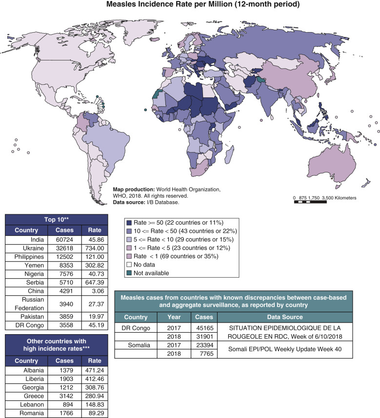
Measles continues to be a major health concern in many parts of the world, including select countries in Africa, Asia, the Pacific and Europe ( Fig. 14.1 ![]() ). The World Health Organization (WHO) estimates that between 2000 and 2017, measles vaccination prevented 21.1 million deaths, which represents an 80% reduction from previous years. In 2017, 110,000 people died from measles; most were under the age of 5. In the United States, large measles outbreaks occur when travelers return to the United States infected with measles. Most people who become infected with measles are not immunized. In 2014, the United States experienced the largest number of reported cases (667 cases from 27 states), since measles was eliminated from the United States in 2000. Communities at greatest risk are those with high numbers of unimmunized individuals (e.g., Amish communities), highlighting the importance of immunizing children.
). The World Health Organization (WHO) estimates that between 2000 and 2017, measles vaccination prevented 21.1 million deaths, which represents an 80% reduction from previous years. In 2017, 110,000 people died from measles; most were under the age of 5. In the United States, large measles outbreaks occur when travelers return to the United States infected with measles. Most people who become infected with measles are not immunized. In 2014, the United States experienced the largest number of reported cases (667 cases from 27 states), since measles was eliminated from the United States in 2000. Communities at greatest risk are those with high numbers of unimmunized individuals (e.g., Amish communities), highlighting the importance of immunizing children.
Vaccine.
The recommended age of first vaccination varies from 6 to 15 months and is a balance between the optimum age for seroconversion and the probability of acquiring measles before that age. The proportions of children who develop protective concentrations of antibody after measles vaccination are about 85% at age 9 months and 95% at 12 months. Two doses of measles vaccine are needed to achieve sufficiently high levels of population immunity to interrupt transmission. WHO recommends that the first dose of measles vaccine be administered at age 9 months, although countries in which the risk of measles is low often provide the first dose at age 12 to 15 months. The duration of vaccine-induced immunity is at least several decades. Secondary vaccine failure rates are estimated to be about 5% at 10 to 15 years after immunization, but are probably lower when vaccination is given after 12 months of age. Decreasing antibody concentrations do not necessarily imply a complete loss of protective immunity, because a secondary immune response usually develops after re-exposure to measles virus, with a rapid rise in antibody titers without overt clinical disease. Fever to 39.4° C (103° F) occurs in about 5% of seronegative vaccine recipients.
Laboratory Confirmation.
The detection of measles virus–specific immunoglobulin M (IgM) in a specimen of serum or oral fluid is diagnostic of acute infection and is the most commonly used serologic test. Acute infection can be confirmed with a four times or greater increase in measles virus–specific IgG antibody concentrations between acute and convalescent sera. The presence of IgG antibodies to measles virus in a single serum specimen is evidence of previous infection or immunization, which cannot be distinguished serologically. Measles virus–specific IgM antibodies might not be detectable until 4 days or more after rash onset and usually fall to undetectable concentrations within 4 to 8 weeks of rash onset. Measles RNA by real-time polymerase chain reaction (RT-PCR) also confirms measles infection.
Typical Measles
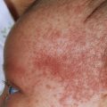
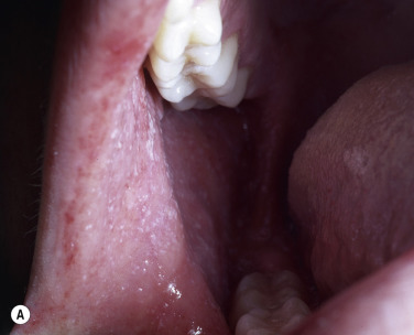
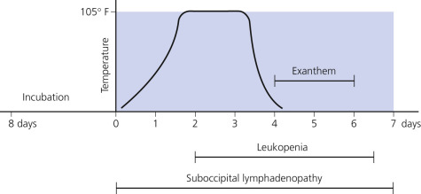
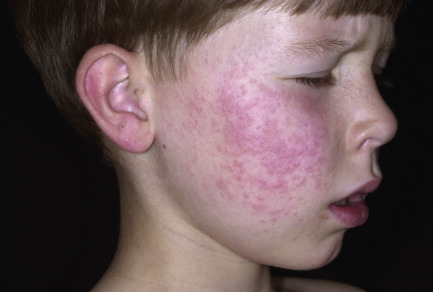
The incubation period of measles (rubeola) ( Fig. 14.2 ) averages 10 to 12 days from exposure to prodrome and 14 days from exposure to rash (range, 7 to 18 days). The disease is spread by respiratory droplets and can be communicated from slightly before the beginning of the prodromal period to 4 days after appearance of the rash; communicability is minimal after the second day of the rash. Prodromal symptoms of severe, brassy cough; nasal congestion; conjunctivitis; photophobia; and fever appear 3 to 4 days before the exanthem and increase daily in severity. The nose and eyes run continuously: the classic sign of measles. Koplik spots (blue-white spots with a red halo) appear on the buccal mucous membrane opposite the premolar teeth 24 to 48 hours before the exanthem and remain for 2 to 4 days.
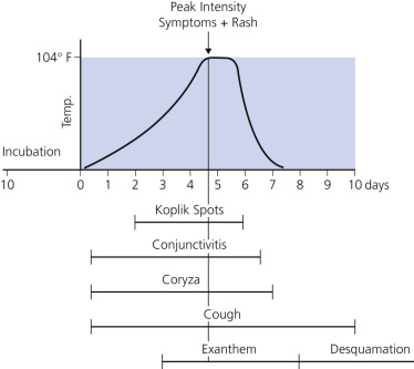
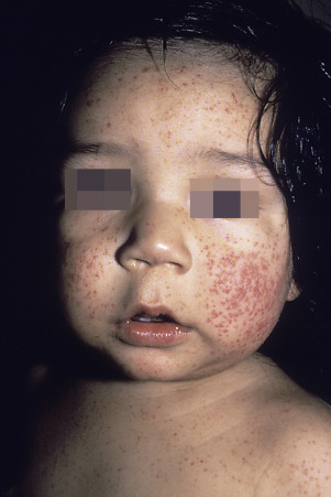
Eruptive Phase.
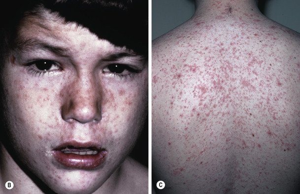
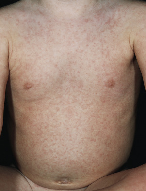
The rash begins on the fourth or fifth day on the face and behind the ears, but in 24 to 36 hours it spreads to the trunk and extremities ( Fig. 14.3 ). It reaches maximum intensity simultaneously in all areas in approximately 3 days and fades after 5 to 10 days. The rash consists of slightly elevated maculopapules that vary in size from 0.1 to 1.0 cm and vary in color from dark red to a purplish hue. They are frequently confluent on both the face and the body, a feature that is such a distinct characteristic of measles that eruptions of similar appearance in other diseases are termed morbilliform (looks like measles). The early rash blanches on pressure; the fading rash is yellowish brown with a fine scale, and it does not blanch. Supportive treatment is the only necessity unless complications, such as bacterial infection or encephalitis, appear.
Management of Measles
Nutritional support and prevention of dehydration with oral rehydration are necessary. Antibiotics treat eye and ear infections and pneumonia. Vitamin A deficiency impairs epithelial integrity and systemic immunity and increases the incidence and severity of infections during childhood. Vitamin A supplementation is effective in reducing total mortality and complications from measles infections; it is likely to be more effective in populations with nutritional deficiencies.
Vitamin A Treatment.
Vitamin A, retinol-binding protein (RBP), and albumin levels are significantly reduced early in the exanthem. Treatment with vitamin A reduces morbidity and mortality in measles, and all children with severe measles should be given vitamin A supplements regardless of whether they are thought to have a nutritional deficiency.
This can help prevent eye damage and blindness. Vitamin A–treated children recover more rapidly from pneumonia and diarrhea, have less croup, and spend fewer days in the hospital. Treated patients have an increase in the total number of lymphocytes and measles IgG antibody. Also, for treated patients the risk of death or a major complication during a hospital stay is half that of untreated patients.
WHO recommends administration of once-daily doses of 200,000 international units of vitamin A for 2 consecutive days to all children aged 12 months or older who have measles.
Lower doses are recommended for younger children: 100,000 international units per day for children aged 6 to 12 months and 50,000 international units per day for children younger than 6 months. In children with clinical evidence of vitamin A deficiency, a third dose is recommended 2 to 4 weeks later.
Immunity.
Persons are considered immune if they have written documentation of adequate immunization with live measles vaccine on or after the first birthday, physician-diagnosed measles, or laboratory evidence of measles immunity. Most persons born before 1957 are likely to have been naturally infected and generally need not be considered susceptible. Routine serologic screening to determine measles immunity is not recommended.
Individuals Exposed to Disease.
Live vaccine, if given within 72 hours of measles exposure, may provide protection and is preferable to the use of human immunoglobulin in persons at least 12 months of age if there is no contraindication.
Use of Human Immunoglobulin.
Human immunoglobulin can be given to prevent or modify measles in a susceptible person within 6 days after exposure. The following children and adults who come into contact with measles should be considered for treatment with human normal immunoglobulin (HNIG):
- 1.
Those with compromised immunity.
- 2.
Infants aged 5 to 12 months (those younger than age 5 months will usually have maternal antibodies). Measles–mumps–rubella (MMR) vaccine may be preferable for some children 6 months of age because it will provide longer-term protection from measles than HNIG.
- 3.
Infants of mothers who develop measles, because such infants will not have maternally derived antibodies.
- 4.
Nonimmune pregnant women. Since most pregnant women are likely to be already immune to measles, measles IgG level should be checked (serologic testing) only if the woman has no previous history of measles infection and has not had two MMR vaccinations. HNIG can be offered to nonimmune pregnant women. They should also be offered MMR vaccine after delivery, at least 3 months after receiving HNIG.
Revaccination Risks.
There is no enhanced risk from administering live measles vaccine to persons who are already immune to measles.
Pregnancy.
Live measles vaccine should not be given to women known to be pregnant or who are considering becoming pregnant within 3 months after vaccination. This precaution is based on the theoretical risk of fetal infection.
Hand-Foot-and-Mouth Disease
Hand-foot-and-mouth disease (HFMD), which has no relation to hoof-and-mouth disease in cattle, is a contagious enteroviral infection occurring primarily in children and characterized by a vesicular palmoplantar eruption and erosive stomatitis. It is most often caused by coxsackievirus A16 (CVA16) and enterovirus 71 (EV71). Patients with atopic dermatitis may develop a widespread vesicular eruption in areas affected by dermatitis (eczema coxsackium). This widespread eruption may be seen with CVA16 and CVA6 infections and may appear similar to a herpes simplex virus infection. Enteroviruses are believed to be spread via the fecal–oral and perhaps respiratory routes. This disease may develop as an isolated phenomenon, or it may occur in epidemic form. It is more common among children and is seen more commonly in preschool-aged children in the summer and through late fall.
Clinical Presentation.
The incubation period is 4 to 6 days. There may be mild symptoms of low-grade fever, sore throat, and malaise for 1 or 2 days. Twenty percent of patients develop submandibular and/or cervical lymphadenopathy.
Eruptive Phase.
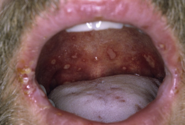
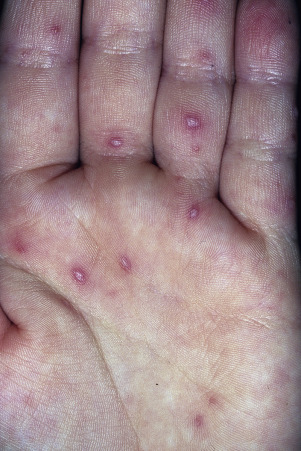
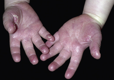
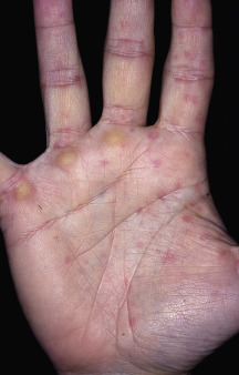
Oral lesions, present in 90% of cases, are generally the initial sign ( Fig. 14.4 ). Aphthae-like erosions varying from a few to 10 or more appear anywhere in the oral cavity and are most frequently small and asymptomatic. The cutaneous lesions, which occur in approximately two thirds of patients, appear less than 24 hours after the enanthem. They begin as 3- to 7-mm, red macules that rapidly become pale, white, oval vesicles with a red areola. There may be a few inconspicuous lesions, or there may be dozens. The vesicles occur on the palms, soles ( Figs. 14.5 to 14.7 ), dorsal aspects of the fingers and toes, and occasionally on the face, buttocks, and legs. They heal in approximately 7 days, usually without crusting or scarring. Nail matrix arrest was reported in a small group of children after HFMD. Beau lines (transverse ridging) and/or onychomadesis (nail shedding) followed HFMD by 3 to 8 weeks.
Serious and Fatal Cases.
Although most cases of HFMD do not result in serious complications, outbreaks of HFMD caused by EV71 can present with a high rate of neurologic complications, including meningoencephalitis, pulmonary complications, and possibly death. HFMD caused by EV71 has become a major emerging infectious disease in Asia.
Differential Diagnosis.
When cutaneous lesions are absent, the disease may be confused with aphthous stomatitis. The oral erosions of HFMD are usually smaller and more uniform. The vesicles of herpes appear in clusters, and those of varicella endure longer and always crust. Both varicella and herpes have multinucleated, giant cells in smears taken from the moist skin exposed when a vesicle is removed (Tzanck smear). Giant cells are not present in lesions of HFMD. Eczema herpeticum, due to herpes simplex virus, may be differentiated from eczema coxsackium with a herpes simplex viral culture or PCR.
Treatment.
Symptomatic relief and reassurance are all that are required.
Scarlet Fever
Scarlet fever (scarlatina) is caused by pyrogenic exotoxins (streptococcal pyrogenic exotoxins) produced by group A streptococcus. Multiple erythrogenic toxins (SpeA, SpeC, and SSA) have been isolated in scarlet fever outbreaks and alone or in combination are thought to be responsible for scarlet fever. Scarlet fever is more common in the winter and spring and less common in the autumn. Some parts of the world, such as England and Asia (Vietnam, South Korea, Hong Kong and mainland China) are seeing a rise in the incidence of scarlet fever. The reason for the increase in scarlet fever is unknown, but the rise may represent the natural cyclical pattern of scarlet fever (once a feared disease of the 19th and early 20th centuries) or an expansion of a single clone of group A streptococcus to which there is no population immunity. The infection may originate in the pharynx or skin and is most common in children (ages 5 to 15 years).
Incubation Period.
The incubation period of scarlet fever ( Fig. 14.8 ) is 2 to 4 days.
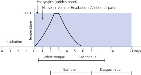
Prodromal and Eruptive Phase.
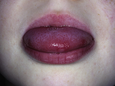
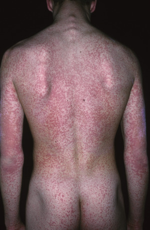
The sudden onset of fever and pharyngitis is followed shortly by nausea, vomiting, headache, and abdominal pain. The entire oral cavity may be red, and the tongue is covered with a yellowish white coat through which red papillae protrude. Diffuse lymphadenopathy may appear just before the onset of the eruption. The systemic symptoms continue until the fever subsides. The rash begins around the neck and face and spreads in 48 hours to the trunk and extremities; the palms and soles are spared ( Fig. 14.9 ). The face is flushed except for circumoral pallor, whereas all other involved areas exhibit a vivid scarlet hue with innumerable pinpoint papules that give a sandpaper quality to the skin ( Fig. 14.10 ). The rash is more limited and less dramatic in milder cases. Linear petechiae (Pastia sign) are characteristic; they are found in skin folds, particularly the antecubital fossa and inguinal area. The tongue sheds the white coat to reveal a red, raw, glazed surface with engorged papillae ( Fig. 14.11 ).
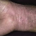
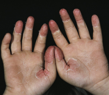
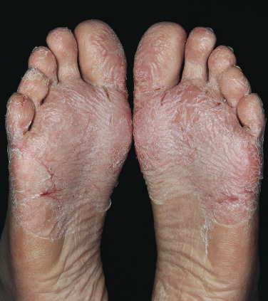
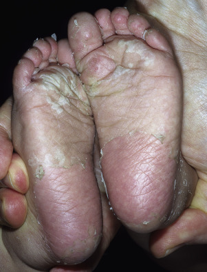
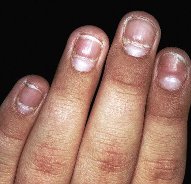
The fever and rash subside and desquamation appears, more pronounced than in any of the eruptive fevers. It begins on the face, where it is sparse and superficial; progresses to the trunk, often with a circular, punched-out appearance; and finally spreads to the hands ( Fig. 14.12 ) and feet ( Figs. 14.13 and 14.14 ), where the epidermis is the thickest. Clinically, the hands and feet appear normal during the initial stages of the disease. Large sheaths of epidermis may be shed from the palms and soles in a glove-like cast, exposing new and often tender epidermis beneath. A transverse groove may be produced in all of the nails (Beau lines) ( Fig. 14.15 ). The pattern of desquamation of the palms and soles and grooving of the nails is such a distinct characteristic of scarlet fever that it is helpful in making a retrospective diagnosis in cases where the eruption is minimal. A rising antistreptolysin-O titer constitutes additional supporting evidence for a recent infection. Desquamation is generally complete in 4 weeks, but it may last for 8 weeks. Recurrence rates of scarlet fever as high as 18% have been reported.
Treatment.
Treat with penicillin, amoxicillin, cephalosporin such as cephalexin or cefadroxil, clindamycin, azithromycin and clarithromycin ( Table 14.2 ).
| Drug, Route | Dose or Dosage | Duration or Quantity |
|---|---|---|
| FOR INDIVIDUALS WITHOUT PENICILLIN ALLERGIES | ||
| Penicillin V, oral | Children: 250 mg twice daily or 3 times daily; adolescents and adults: 250 mg 4 times daily or 500 mg twice daily | 10 days |
| Amoxicillin, oral | 50 mg/kg once daily (max: 1000 mg); alternate: 25 mg/kg (max: 500 mg) twice daily | 10 days |
| Benzathine penicillin G, intramuscular | <27 kg: 600,000 U; ≥27 kg: 1,200,000 U | 1 dose |
| FOR INDIVIDUALS WITH PENICILLIN ALLERGIES | ||
| Cephalexin, * oral | 20 mg/kg/dose, twice daily (max: 500 mg/dose) | 10 days |
| Cefadroxil, * oral | 30 mg/kg once daily (max: 1 g) | 10 days |
| Clindamycin, oral | 7 mg/kg/dose 3 times daily (max: 300 mg/dose) | 10 days |
| Azithromycin, † oral | 12 mg/kg once daily (max: 500 mg) | 5 days |
| Clarithromycin, † oral | 7.5 mg/kg/dose twice daily (max: 250 mg/dose) | 10 days |
* Avoid in individuals with immediate-type hypersensitivity to penicillin.
† Resistance of S. pyogenes to these agents is well known and varies both geographically and temporally.
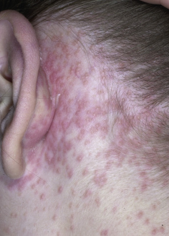
Rubella
Rubella (German measles, 3-day measles) is a contagious exanthematous viral infection characterized by nonspecific signs and symptoms including transient erythematous and sometimes pruritic rash, postauricular or suboccipital lymphadenopathy, arthralgia, and low-grade fever. Clinically similar exanthematous illnesses are caused by parvovirus, adenoviruses, and enteroviruses. Most individuals experience a mild illness and 25% to 50% of rubella infections are subclinical. Rubella contracted during pregnancy (congenital rubella syndrome) can cause severe birth defects and fetal loss, which are preventable with vaccination. In 2004 and 2015 rubella and congenital rubella syndrome were eliminated in the United States and the Americas region, respectively. However in many parts of the world, especially Africa, the Middle East, and South and Southeast Asia, rubella continues to be a public health concern with over 100,000 infants born with congenital rubella syndrome.
Congenital Rubella Syndrome.
Women who become infected with rubella early in the first trimester of pregnancy may transmit the virus to the fetus, resulting in a number of congenital defects (congenital rubella syndrome).
Incubation Period.
The incubation period of rubella ( Fig. 14.16 ) is 18 days, with a range of 14 to 21 days.
Prodromal Phase.
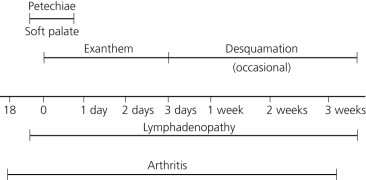
Mild symptoms of malaise, headache, and moderate temperature elevation may precede the eruption by a few hours or a day. Children are usually asymptomatic. Lymphadenopathy, characteristically postauricular, suboccipital, and cervical, may appear 4 to 7 days before the rash and be maximal at the onset of the exanthem. In 2% of cases, petechiae on the soft palate occur late in the prodromal phase or early in the eruptive phase.
Eruptive Phase.
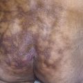
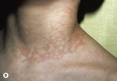
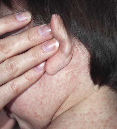
The eruption begins on the neck or face and spreads in hours to the trunk and extremities ( Fig. 14.17 ). The lesions are pinpoint to 1 cm, round or oval, pinkish or rosy red macules or maculopapules. The color is less vivid than that of scarlet fever and lacks the blue or violaceous tinge seen in measles. The lesions are usually discrete but may be grouped or coalesced on the face or trunk. The rash fades in 24 to 48 hours in the same order in which it appeared and may be followed by a fine desquamation.
Among adults infected with rubella, transient polyarthralgia or polyarthritis occurs frequently. These manifestations are particularly common among women.
Arthritis, affecting primarily the phalangeal joints of women, may occur in the prodromal period and may last for 2 to 3 weeks after the rash has disappeared. No treatment is required.
Central nervous system (CNS) complications (e.g., encephalitis) occur at a ratio of 1 : 6000 cases and are more likely to affect adults. Thrombocytopenia occurs at a ratio of 1 : 3000 cases and is more likely to affect children.
Serologic Testing.
Rubella IgG is often used as a marker of past infection or response to vaccination. This test is especially important in women of child-bearing age and is included in the recommended prenatal blood tests. The presence of IgG typically indicates immunity to acute rubella infection. Diagnosis is made on clinical findings as well as specific rubella IgM or a significant rise in rubella IgG titers in acute and convalescent serum specimens. RT-PCR and viral culture are other methods to detect rubella infection. Rubella is a reportable disease in the United States.
Vaccine.
The rubella vaccine is a live attenuated virus. Although it is available as a single preparation, it is recommended that in most cases rubella vaccine be given as part of the MMR vaccine. MMR is recommended at 12 to 15 months (not earlier) and a second dose when the child is 4 to 6 years old (before kindergarten or first grade). Rubella vaccination is important for nonimmune women who may become pregnant because of the risk for serious birth defects if they acquire the disease during pregnancy.
Erythema Infectiosum (Parvovirus B19 Infection)
Parvovirus B19 is a common viral infection. Over 50% of adults have been exposed. Most infections are asymptomatic. Parvovirus B19 is associated with many disease manifestations that vary with the immunologic and hematologic status of the patient. The main target of B19 infection is the red cell receptor globoside (blood group P antigen) of erythroid progenitor cells of the bone marrow. People who do not have the virus receptor (erythrocyte P antigen) are naturally resistant to infection with this virus. Parvovirus B19 infection induces various host responses, including DNA damage response, cell cycle arrest, and erythroid cell death via apoptosis (erythroid progenitors in bone marrow and fetal tissues). Parvovirus B19 may stay in tissues for years and not produce infection (unproductive infection) and has been associated with acute and chronic inflammatory cardiomyopathy, rheumatoid arthritis, vasculitis, meningoencephalitis, hepatitis, and thyroid disease. Other cutaneous eruptions such as peripheral purpuric papules (gloves and socks eruption), Gianotti–Crosti syndrome, and vesicular and morbilliform eruptions have been attributed to parvovirus B19 infections.
Erythema Infectiosum (Acute B19 Viral Infection).
Erythema infectiosum (fifth disease) is caused by the B19 parvovirus. It is relatively common and mildly contagious and appears sporadically or in epidemics, especially in the winter and spring. Peak attack rates occur in children between 5 and 14 years of age. Asymptomatic infection is common.
Infection causes erythema infectiosum in immunocompetent patients. It is the primary cause of transient aplastic crisis in patients with underlying hemolytic disorders. Persistent infection in immunosuppressed patients may present as red cell aplasia and chronic anemia. In utero infection may result in hydrops fetalis or congenital anemia. There is no evidence of reinfection in immunocompetent individuals.
Seroprevalence is 2% to 10% in children younger than 5 years, 40% to 60% in adults older than 20 years, and 85% or more in those older than 70 years. The average annual seroconversion rate of women of child-bearing age is 1.5%.
The virus may be transmitted via the respiratory route and via the transfusion of infected blood and blood products. Nosocomial transmission has been well documented. Individuals are not viremic or infectious at the rash or arthropathy stage of disease.
Incubation Period.
The incubation period of erythema infectiosum ( Fig. 14.18 ) is 13 to 18 days. Viremia occurs after the incubation period and reticulocyte numbers fall, resulting in a temporary drop in hemoglobin concentration of 1 g/dL in a normal person.
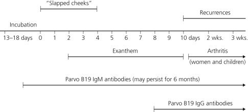
There is a nonspecific prodromal illness, followed by a three-stage erythematous disease.
Prodromal Symptoms.
Symptoms are usually mild or absent. Pruritus, low-grade fever, malaise, and sore throat precede the eruption in approximately 10% of cases. Lymphadenopathy is absent. Older individuals may complain of joint pain.
Eruptive Phase.
There are three distinct, overlapping stages.
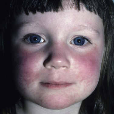
Facial Erythema (“Slapped Cheek”).
Red papules on the cheeks rapidly coalesce in hours, forming fiery red, slightly edematous, warm, erysipelas-like plaques that are symmetric on both cheeks and spare the nasolabial fold and the circumoral region ( Fig. 14.19 ). The “slapped cheek” appearance fades in 4 days. The classic “slapped cheek” is much more common in children than in adults.
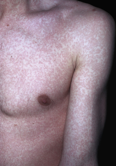
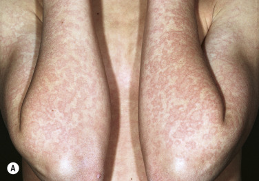
Net Pattern Erythema.
This unique characteristic eruption – erythema in a fishnet-like pattern – begins on the extremities approximately 2 days after the onset of facial erythema and extends to the trunk and buttocks, fading in 6 to 14 days ( Fig. 14.20 ). At times, the exanthem begins with erythema and does not become characteristic until irregular clearing takes place. This second-stage rash may vary from very faint erythema to a florid exanthem. Livedo reticularis has a similar net-like pattern, but it does not fade quickly.
Recurrent Phase.
The eruption may fade and then reappear in previously affected sites on the face and body during the next 2 to 3 weeks. Temperature changes, emotional upsets, and sunlight exposure may stimulate recurrences. The rash fades without scaling or pigmentation. There may be a slight lymphocytosis or eosinophilia.
Polyarthropathy Syndrome and Pruritus
Adults.
Adults exposed to the parvovirus during outbreaks may develop itching and arthralgia or arthritis. Women tend to be affected much more than men. The itching varies from mild to intense and is localized or generalized. In most cases a nonspecific macular eruption occurs without the appearance of the typical net-like pattern before the arthritis.
Affected individuals develop moderate to severe, symmetric polyarthritis that may be difficult to distinguish from rheumatoid arthritis (peripheral symmetric polyarthropathy involving the small joints of the hands, wrists, knees and feet). B19-induced arthralgia and arthritis typically resolves in 3 weeks and does not result in articular destruction. In the minority, joint symptoms may persist for years. Rheumatoid factor and other autoantibodies may be detected, although usually they are transient.
There are striking similarities between B19 infection and systemic lupus erythematosus (SLE): both may present with malar rash, fever, arthropathy, myalgia, cytopenia, hypocomplementemia, and anti-DNA and antinuclear antibodies (ANAs).
The differential diagnosis includes acute rheumatoid arthritis, seronegative arthritis, Lyme disease, and SLE. Parvovirus infection should be considered when an adult woman has acute polyarthropathy associated with pruritus, especially if she has been exposed to children with erythema infectiosum.
The demonstration of anti–parvovirus B19 IgM and a 4-fold increase in anti–parvovirus B19 IgG (acute and convalescent samples) are standard diagnostic tests. In certain clinical circumstances (e.g., chronic aplasia), measurement of B19 DNA by PCR may be considered. Measurement of IgM level must be done within the first months, since it disappears later in the course of the disease. Immunosuppressive agents used to treat rheumatoid arthritis may prolong the persistence of the virus and disease in parvovirus B19–induced arthritis/arthropathy.
B19 infection is not associated with the joint destruction seen in rheumatoid arthritis. Prolonged symptoms do not correlate with serologic studies, such as the duration of B19 IgM response, or persistent viremia.
Adult flu-like symptoms and arthropathies begin coincidentally with IgG antibody production in 18 to 24 days after exposure and are probably immune complex mediated.
Children.
Both male and female children may develop joint symptoms. Most cases have acute arthritis of brief duration; a few have arthralgias. Two patterns are seen: polyarticular, affecting five or more joints; and pauciarticular, affecting four or fewer joints. Large joints are affected more often than small joints. The knee is the most common joint affected (82%). Laboratory findings are normal. The duration of joint symptoms is usually less than 4 months, but some patients have persistent arthritis for 2 to 13 months, which fulfills the criteria for the diagnosis of juvenile rheumatoid arthritis.
Infection in the Pregnant Woman (Intrauterine Infection and Spontaneous Abortion).
In pregnant women, infection can, but usually does not, lead to fetal infection ( Box 14.2 ). Fetal infection sometimes causes severe anemia, congestive heart failure, generalized edema (fetal hydrops), and death. Many fetuses dying because of this infection are not noticeably hydropic.
Risk of Maternal Infection During an Epidemic: 30% to 65%
- •
Infected women may be asymptomatic.
Nurses and School Teachers: High Rate of Infection if Exposed
- •
People with fifth disease are contagious before they develop the rash; therefore exposure at the onset of an outbreak cannot be avoided.
- •
Pregnant personnel should remain at home until 2 to 3 weeks after the last identified case.
Laboratory Tests
- •
Immunoglobulin M (IgM) tests for symptomatic exposed women
- •
Fetal ultrasonography for confirmed or suspected cases
- •
Polymerase chain reaction testing of amniotic fluid and fetal serum – a sensitive and rapid method for diagnosis of intrauterine infection
- •
Monitor alpha-fetoprotein *
* Maternal serum alpha-fetoprotein level is a marker for fetal aplastic crisis during intrauterine human parvovirus infection.
levels in exposed and infected women
- •
Ultrasonography when alpha-fetoprotein levels are increased
- •
Abnormal ultrasound: fetal blood sampling as a guide for possible fetal transfusion
- •
Teratogenic effects not demonstrated
- •
Therapeutic abortion not indicated
Parvovirus B19 probably causes 10% to 15% of all cases of nonimmune hydrops. If the fetus survives fetal hydrops caused by B19, there usually are no long-term sequelae.
The fetus has a high rate of red cell production; its immature immune system may not be able to mount an adequate immune response. Parvovirus has been implicated as a cause of spontaneous abortion (from severe fetal anemia and hydrops fetalis).
Maternal infection in the first half of pregnancy is associated with 10% excess fetal loss and hydrops fetalis in 3% of cases (of which up to 60% resolve spontaneously or with appropriate management). B19-associated congenital abnormalities have not been reported among several hundred live-born infants of B19-infected mothers. The overall risk of serious adverse outcome from occupational exposure to parvovirus B19 infection during pregnancy is low (excess early fetal loss in 2 to 6 of 1000 pregnancies and fetal death from hydrops in 2 to 5 of 10,000 pregnancies). It is not recommended that susceptible pregnant women be excluded routinely from working with children during epidemics.
The polymerase chain reaction is a sensitive and rapid method for the diagnosis of intrauterine infection.
Anemia.
The virus has the propensity to infect and lyse erythroid precursor cells and interrupt normal red cell production. In a person with normal hematopoiesis, B19 infection produces a self-limited red cell aplasia that is clinically unapparent. In patients who have increased rates of red cell destruction or loss and who depend on compensatory increases in red cell production to maintain stable red cell indices, B19 infection may lead to transient aplastic crisis. Patients at risk for transient aplastic crisis include those with hemoglobinopathies (sickle cell disease, thalassemia, hereditary spherocytosis, and pyruvate kinase deficiency) and those with anemias associated with acute or chronic blood loss. B19 infection accounts for most, if not all, aplastic crises in sickle cell disease, but at least 20% of infections do not result in aplasia. Up to 30% of hospital staff members may be infected when exposed to infected sickle cell patients. In immunodeficient persons and those with AIDS, B19 may persist, causing chronic red cell aplasia, which results in chronic anemia. Some of these patients may be cured with immune globulin therapy.
Laboratory Evaluation.
Most acute infections with B19 are diagnosed in the laboratory by serologically detecting IgG and IgM class antibodies with enzyme immunoassay. IgM antibodies are the most sensitive indicator of acute B19 infection in immunologically normal persons. Measuring IgM level is useful for diagnosing recent parvovirus infection. Both IgG and IgM may be present at or soon after onset of illness and reach peak titers within 30 days. Because IgG antibody may persist for years, diagnosis of acute infection is made by the detection of IgM antibodies, unless one can demonstrate a 4-fold rise in IgG in acute and convalescent titers. The prevalence of parvovirus B19 IgG antibodies increases with age. The age-specific prevalence of antibodies to parvovirus is 2% to 9% of children younger than 5 years, 15% to 35% in children 5 to 18 years of age, and 30% to 60% in adults (19 years or older). IgM is detectable for as long as 3 months after exposure. Parvovirus B19 can be detected in blood, amniotic fluid, and synovial fluid by rapid PCR.
Management.
Most health departments do not recommend exclusion from school for children with fifth disease. Many infections are unapparent, and exposure may occur in the community, as well as in school.
Evaluate the immune status of exposed pregnant women. The risks are nil if the women is IgG positive. If she is not immune (although the risk of the fetus being affected is very low), fetal surveillance by repeated ultrasonographic examination and immune status reevaluation is recommended.
Because the major immune response appears to be humoral, patients with chronic infection have been treated with immune globulin. These patients often respond with a marked reduction in the level of B19 viremia and with reticulocytosis, followed within a few weeks by resolution of the anemia. Patients with persistent infection should be monitored for evidence of relapse by observation of the reticulocyte counts and by assays for B19 viremia when indicated.
Roseola Infantum (Human Herpesvirus 6 and 7 Infection)
Roseola infantum (exanthema subitum, “sudden rash,” sixth disease, rose rash of infants, 3-day fever) is caused primarily by human herpesvirus 6 (HHV-6), which is epidemiologically and biologically similar to cytomegalovirus. As with other herpesviruses, HHV-6 shows persistent and intermittent or chronic shedding in the normal population, making the unusually early infection of children (seroconversion in the first year of life in up to 80% of all children) understandable. The virus remains latent in monocytes and macrophages and probably in the salivary glands. Virus may infect infants through the saliva mainly from mother to child. A severe, infectious, mononucleosis-like syndrome in adults may be caused by a primary infection with HHV-6. HHV-6 has also been implicated in idiopathic pneumonitis in immunocompromised hosts.
Most cases are asymptomatic or present with fever of unknown origin and occur without a rash. The disease is sporadic, and the majority of cases occur between the ages of 6 months and 4 years. Primary HHV-7 infection occurs later in childhood (at approximately 3 years of age) and also causes exanthema subitum, although less often than HHV-6. HHV-6 antibody is present in 90% to 100% of the population older than age 2 years. The development of high fever, as is seen in roseola, is worrisome, but the onset of the characteristic rash is reassuring.
One study showed that HHV-6 was responsible for 10% of hospital visits for acute illness in infants younger than 25 months of age and 33% of the febrile seizures occurring in children younger than 2 years.
In infants and young children HHV-6 is a major cause of visits to the emergency department, febrile seizures, and hospitalizations.
Incubation Period.
The incubation period of roseola infantum ( Fig. 14.21 ) is 12 days, with a range of 5 to 15 days.
Prodromal Symptoms.
There is a sudden onset of high fever of 103° F to 106° F, with few or minor symptoms. Most children appear inappropriately well for the degree of temperature elevation, but they may experience slight anorexia or one or two episodes of vomiting, running nose, cough, and hepatomegaly. Seizures (but more frequently general cerebral irritability) may occur before the eruptive phase. Most recover without sequelae. Cases of encephalitis/encephalopathy with abnormal electroencephalograms and cerebral computed tomograms have been reported; epilepsy developed in one case and in another case the patient died. HHV-6 DNA has been detected in the cerebrospinal fluid (CSF); this suggests that HHV-6 may invade the brain during the acute phase. HHV-6 infection should be suspected in infants with febrile convulsions, even those without the exanthem. Mild to moderate lymphadenopathy, usually in the occipital regions, begins at the onset of the febrile period and persists until after the eruption has subsided.
Eruptive Phase.
The rash begins as the fever subsides. The term exanthema subitum indicates the sudden “surprise” of the blossoming rash after the fall of the fever. Numerous pale pink, almond-shaped macules appear on the trunk and neck, become confluent, and then fade in a few hours to 2 days without scaling or pigmentation ( Figs. 14.22 and 14.23 ). The exanthem may resemble rubella or measles, but the pattern of development, distribution, and associated symptoms of these other exanthematous diseases are different.
Laboratory Evaluation.
Leukocytosis develops at the onset of fever, but leukopenia with a neutropenia and relative lymphocytosis appears as the temperature increases and persists until the eruption fades. Seroconversion during the convalescent phase can be detected with immunofluorescence or enzyme immunoassays. HHV-6 can be detected by rapid PCR.
Treatment.
Control the patient’s temperature with aspirin and provide reassurance. HHV-6 is inhibited by several antiviral drugs in the laboratory, including ganciclovir and foscarnet. Treatment may be considered for patients with serious HHV-6-associated disease confirmed with virologic tests.
Enterovirus (Echovirus and Coxsackievirus) and Parechovirus Exanthems
The previously described diseases characteristically display a predictable set of signs and symptoms. Roseola and erythema infectiosum are relatively common. Many physicians never see cases of measles, German measles, or scarlet fever. The most common exanthematous eruptions are caused by the enteroviruses, echovirus and coxsackievirus. Parechovirus is a new genus (formally echovirus 22 and echovirus 23) that causes viral illnesses (respiratory, gastrointestinal, and CNS) worldwide, especially in young infants. A large number of these viruses may begin with a skin eruption. Some of these eruptions are characteristic of the virus type, but in most cases one must be satisfied with the diagnosis of “viral rash.” In many cases, drug eruptions cannot be distinguished from the nonspecific exanthems of these enteroviruses.
Systemic Symptoms.
Many are possible, such as fever, nausea, vomiting, and diarrhea, along with typical viral symptoms of photophobia, lymphadenopathy, sore throat, and possibly encephalitis.
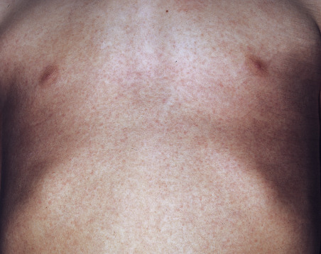
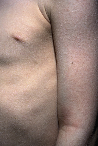
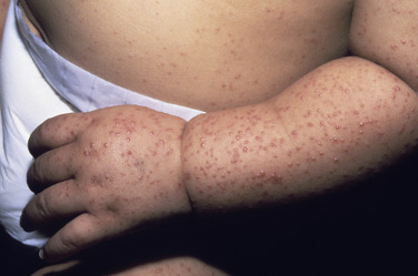
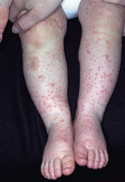
Exanthem.
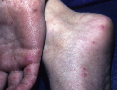
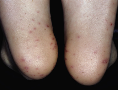
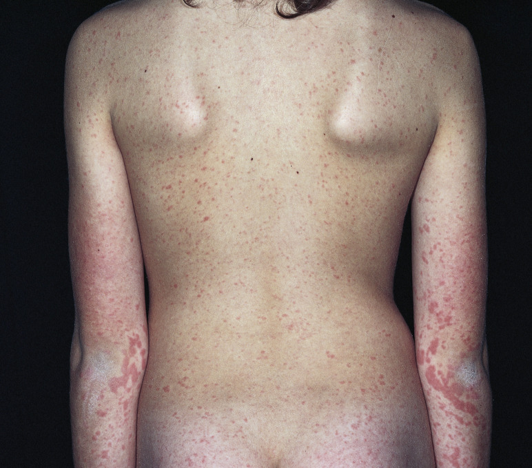
The rash may appear at any time during the course of the illness, and it is usually generalized. Lesions are erythematous maculopapules with areas of confluence, but they may be urticarial, vesicular, or sometimes petechial ( Figs. 14.24 to 14.32 ). The palms and soles may be involved. The eruptions are more common in children than in adults. In most cases, the rash fades without pigmentation or scaling.
Treatment.
Treatment consists of relieving symptoms.
Kawasaki Disease
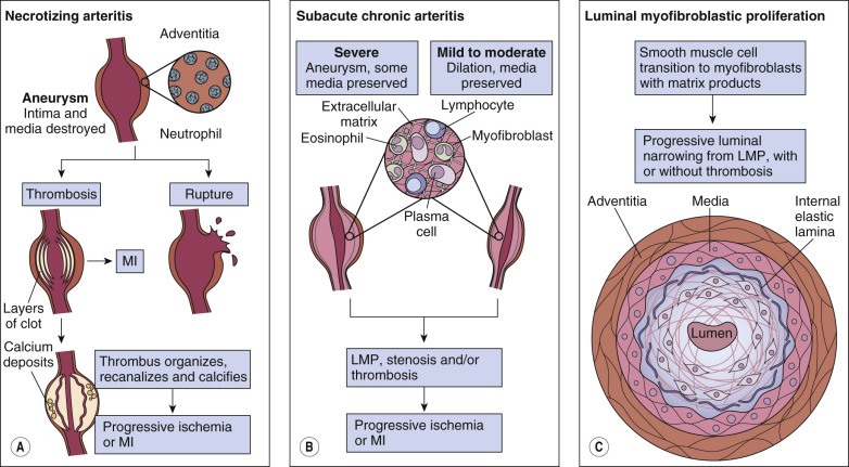
Kawasaki disease (KD) is an acute febrile multisystem disorder, primarily affecting children under 5 years of age. Originally described in Japan in 1967 as mucocutaneous lymph node syndrome, KD occurs worldwide ( Fig. 14.33 ![]() ), but especially in those of Asian descent (most common in those with Japanese ancestry) and now is the most common cause of acquired heart disease in children in developed countries. Genetic studies have identified susceptibility genes and genes influencing disease outcomes. Such studies have led to clinical trials utilizing medications targeting specific pathways (e.g., cyclosporine to block calcineurin-NFAT (nuclear factor of activated T cells) and statin drugs to block effects of TGF-β signaling pathway) Despite large-scale studies, the cause of KD remains unknown. Evidence suggests an antigen is inhaled that triggers an immune response in genetically susceptible hosts. Adaptive immune responses involving proinflammatory and regulatory T lymphocytes produce damage in multiple organs and tissues ( Fig. 14.34 ). Poor clinical outcomes are primarily attributed to coronary artery vasculopathy, characterized by necrotizing arteritis, subacute and chronic vasculitis and luminal myofibroblastic proliferation. Large coronary artery aneurysms occur and are an important distinguishing feature of KD.
), but especially in those of Asian descent (most common in those with Japanese ancestry) and now is the most common cause of acquired heart disease in children in developed countries. Genetic studies have identified susceptibility genes and genes influencing disease outcomes. Such studies have led to clinical trials utilizing medications targeting specific pathways (e.g., cyclosporine to block calcineurin-NFAT (nuclear factor of activated T cells) and statin drugs to block effects of TGF-β signaling pathway) Despite large-scale studies, the cause of KD remains unknown. Evidence suggests an antigen is inhaled that triggers an immune response in genetically susceptible hosts. Adaptive immune responses involving proinflammatory and regulatory T lymphocytes produce damage in multiple organs and tissues ( Fig. 14.34 ). Poor clinical outcomes are primarily attributed to coronary artery vasculopathy, characterized by necrotizing arteritis, subacute and chronic vasculitis and luminal myofibroblastic proliferation. Large coronary artery aneurysms occur and are an important distinguishing feature of KD.