Vulvar edema is associated with a variety of conditions. The edema can result from inflammatory conditions, infections, infestations, trauma, pregnancy, tumors and iatrogenic causes. At times, it is difficult to determine the cause of the vulvar edema. Treatment consists of determining the origin of the edema and giving the appropriate therapy for that diagnosis as well as the use of compression and, at times, lymphatic massage.
Vulvar edema is associated with a variety of medical conditions. There are also several other vulvar conditions that closely mimic edema making it difficult at times to clearly differentiate these conditions from edema. Vulvar edema can be a diagnostic dilemma and a treatment challenge for health care providers as well a significant frustration for affected individuals.
Pathophysiology
Edema is defined as abnormal and excessive accumulation of fluid within the skin. The amount of interstitial fluid is a result of the balance of fluid hemostasis. Increased secretion or impaired removal of fluid results in edema. Edema is facilitated by any of the following factors: increased intravascular hydrostastic pressure, reduced plasma oncotic pressure, increased blood vessel wall permeability, obstructed lymphatic clearance of fluids, and, finally, changes in the water retention properties of tissue. Body areas with distensible and loose skin, including the genitalia, are common sites for edema formation. Depending on the underlying cause of edema, the resultant fluid accumulation is either of plasma or lymphatic origin and at times both. Fluid of plasma origin often results in pitting edema (soft on palpation producing an indentation of the skin) whereas fluid of lymphatic origin is nonpitting (firm to palpation and nonindenting). Edema can develop acutely on exposure to allergens, including topical medications, infections, and toxins, and in certain medical conditions, such as lupus, leukemia, and lymphoma. The resulting edema in these situations is termed, angioedema . This is a result of increased vascular permeability in response to mediators of inflammation. Angioedema, a rare condition, may be hereditary (C1 esterase inhibitor deficiency). Generally, when acute angioedema occurs, onset of symptoms is often within minutes to hours and effects may last up to several days. Edema may be generalized or occasionally restricted to specific body sites, such as the vulva.
Any inflammatory or obstructive process can produce edema. At times, remarkable vulvar edema can occur without obvious inflammatory disease or trauma ( Box 1 ).
Contact dermatitis
Crohn disease
Hidradenitis suppurativa
Primary herpes simplex virus (HSV) infection
Recurrent vulvovaginal candidiasis (RVVC)
Pelvic obstruction due to tumor, radiation, or surgery
Edema associated with pregnancy/prolonged delivery
Subcutanous tumors that mimic edema (lipomas, cysts, and so forth)
Inflammatory conditions
Contact Dermatitis
Contact dermatitis of the vulva is inflammation occurring as a result of contact of the skin with a variety of substances and can manifest as either an acute dermatitis or a chronic dermatitis after prolonged exposure ( Fig. 1 ). The presentation of contact dermatitis is variable and this impairs diagnosis. Often the cause is multifactorial. There are two types of contact dermatitis, irritant and allergic contact dermatitis. Strong or caustic irritants cause an immediate irritant contact dermatitis that corresponds to the area of contact, as in the use of trichloroacetic acid for anogenital warts or an azole cream used in already inflamed vulvovaginal candidiasis (VVC). This is characterized by immediate pain on contact and well-circumscribed skin changes, which make diagnosis easy. More common is a delayed, chronic irritant contact dermatitis due to prolonged or frequent exposure to milder irritants. Common contact irritants include overwashing, laundry detergent, fabric softener, dyes in clothing, soaps, body washes, hygiene sprays, bubble bath, bath oils, tampons, panty liners, menstrual and incontinence pads, urine, sweat, semen, feces, douches, spermicides (nonoxynol-9 and benzalkonium chloride), and condoms. The clinical presentation consists of irritation and burning in a setting of poorly demarcated erythema, and more severe disease manifests edema, and, sometimes, superficial erosions, within glazed erythema.
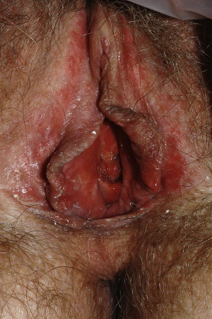
Allergic contact dermatitis is usually a type IV delayed hypersensitivity reaction and only occurs in those patients previously sensitized to the antigen. Vulvar allergens include anesthetics (benzocaine and diphenhydramine), antibiotics (neomycin sulfate and sulfa), antimycotics (clotrimazole), nonsteroidal anti-inflammatory drugs (bufexamac), antivirals (acyclovir), corticosteroids, chlorhexidine, fragrances, preservatives (parabens and propylene glycol), and lanolin. It may be difficult to differentiate chronic irritant contact dermatitis from allergic contact dermatitis; however, allergic contact dermatitis is extremely pruritic, and skin changes follow exposure by 1 to 2 days. The appearance of affected skin ranges from normal to erythema, vesicles, and, in severe cases, edema. Treatment of contact vulvitis includes eliminating the offending agent and symptomatic relief with cool gel packs applied to the vulvar skin for comfort. Nighttime scratching should be prevented with sedation, because there are no medications with inherently anti-itch properties. Elimination of the itch-scratch cycle is essential, and sedation with medications, such as amitriptyline (25 to 50 mg at bedtime) or hydroxyzine (10 to 50 mg) usually is helpful. Corticosteroids ointment of high to mid potency are used in a taper to reverse inflammation and subsequently the vulvar edema. At times, oral steroid tapers are required; prednisone (40 mg for small adults to 60 mg for large adults) each morning for 7 to 10 days can be discontinued without a taper other than transition to a topical corticosteroid. Allergic contact dermatitis requires up to a month of corticosteroid therapy, and irritant contact requires less.
Crohn Disease
Crohn disease, a chronic relapsing and remitting inflammatory bowel condition, can affect the entire length of the gastrointestinal tract from the oral cavity to the anus. Extraintestinal disease with or without continuity to the intestine sometimes occurs. Fifteen percent of patients have disease involving the mucocutaneous tissues. Extraintestinal Crohn disease, also called metastatic Crohn disease, may present with gynecologic manifestations, which often are not recognized and are difficult to treat. In 85% of cases, the diagnosis of gastrointestinal Crohn disease precedes vulvar involvement. Vulvar lesions occur in approximately 2% of women with Crohn disease. More common is perianal disease, with fistulae, tags, and edema. Fairly often, the only manifestation of vulvar Crohn disease is asymmetric labial edema, without other skin signs that suggest this diagnosis. More severe disease is characterized by labial edema with linear knife-like ulcerations, abscesses, or fissures located in the interlabial sulcus, perineum, or crural folds ( Fig. 2 ). Rectovaginal, anocutaneous, and recto-Bartholin fistulae can occur. Development of enterovaginal fistulas leads to vaginal scarring. Vulvar fistulae and abscess formation may precede active intestinal disease by years, making diagnosis of anorectal Crohn difficult. At times, repeat gastrointestinal screening for Crohn disease may be required for diagnosis. The diagnosis also is suggested by the presence of noncaseating granulomas on skin biopsy, but granulomatous inflammation is characteristic of hidradenitis suppurativa (HS) as well, another cause of anogential edema (discussed later).
The first-line treatment of vulvar Crohn disease can vary from the usual gastrointestinal treatment regimen. Generally, for initial treatment, oral metronidazole is used. Ciprofloxacin, both for its antimicrobial and its nonspecific anti-inflammatory properties, may also be used. Metronidazole and ciprofloxacin may be combined if needed for better response. For resistant disease or for the treatment of open, draining fistulas, infliximab (Remicade), etanercept (Enbrel), or adalimumab (Humira) may be used. These are anti–tumor necrosis factor (TNF) substances. TNF is a protein produced by the immune system that may cause the inflammation associated with Crohn disease. Corticosteroids and the 5-aminosalicylic acid drugs are generally effective only for bowel disease rather than for perineal disease; however, an occasional patient with vulvar Crohn disease has responded well to corticosteroids. Immunosuppressants, such as 6-mercaptopurine and azathioprine, have also been used at times.
Hidradenitis Suppurativa (Inverse Acne)
HS is a chronic, noninfectious, follicular occlusive condition resulting in painful nodules and abscesses in characteristic locations of the axillae and groin ( Fig. 3 ). After occlusion of hair follicles with desquamated keratin debris, the distending follicle enlarges with accumulating trapped keratin, until there is rupture of the distended follicle, with a resulting foreign body reaction to the keratin producing folliculitis. This results in noninfectious abscess formation. The abscesses drain, forming chronic sinus tracts and hypertrophic scarring with decreased mobility, lymphedema, and deformity. HS occurs on a spectrum of severity, from an occasional dermal nodule to severe, foul-smelling, draining skin with deformity, pain, and dysfunction. Even extremely mild disease is sometimes manifested by edema that is far out of proportion to subtle scarring noted on examination. Affected body areas include one or more of the apocrine gland-containing areas; the axillae, groin, and inframammary, periareola, perianal, and perineal regions. Aprocrine glands are not found on the inner aspects of the labia minora and vestibule. The presence of abscess formation in these areas should raise suspicion of other conditions, such as anorectal Crohn disease.
The prevalence of HS is described as from 1% to 4% of the population. It often begins after menarche and improves after menopause with peak incidence at ages 20 to 30 years. Women are more commonly affected than men, with female/male ratios ranging from 2:1 to 5:1. Predisposing factors include smoking, diabetes mellitus, and obesity. There are several reports of familial involvement with a possible single gene transmission. Chronic HS with draining sinuses and abscesses is difficult to differentiate from Crohn disease, lymphogranuloma venereum (LGV), pyoderma gangrenosum, and squamous cell carcinoma. Biopsy specimens looking for follicular hyperkeratosis, active folliculitis, sinus tracts, apocrine, and eccrine stasis may help clarify the diagnosis. Biopsies generally are not required for the diagnoses of hidradenitis suppurativa, however. There have been reports of squamous cell carcinoma in up to 3.2% of patients with perineal hidradenitis suppurativa. HS is staged by Hurley’s criteria (I–III) based on the severity of tissue involvement with abscesses and scarring. Generally, 75% remain in stage I, 24% progress to stage II, and 1% progress to stage III.
The goal of treatment is to prevent the progression of the disease and decrease existing disease to the mildest form. General treatment measures include education, support, reduction of friction heat and sweating in the area, and the use of antiseptic washes and cleansers. Weight loss and smoking cessation should be advised. Medical treatment uses antiandrogens, such as spirinolactone- or aldactone-containing oral contraceptive pills, in an extended cycle regimen to decrease end organ sensitivity. Chronic administration of antibiotics with anti-inflammatory effects minimizes the activity of this disease; these include doxycycline (100 mg twice daily), clindamycin (150 mg twice daily), and trimethoprim-sulfamethoxyzole (double strength twice daily). Measures to reduce the duration of flares and secondary infection include clindamycin 1% lotion twice daily and a 7- to 10-day course of tetracycline (250–500 mg orally 4 times daily), doxycycline (100 mg orally twice daily), amoxicillin/clavulinic acid (875/125 mg twice daily), or clindamycin (300 mg orally twice daily). Zinc gluconate tablets (50 mg orally twice daily) have anti-inflammatory and wound-healing purposes and are used as an adjunct to treatment. Intralesional triamcinolone acetonide (0.5 to 1 mL of a 10 mg/mL concentration) injected into the center of painful early papules and nodules is also helpful in suppressing inflammation. Dapsone (50–100 mg orally once a day chronically) can also be used. Monitoring parameters for dapsone include a weekly complete blood cell count ×4, then monthly ×6, then every 6 months. Liver function tests should be obtained as a baseline, then periodically. Long-term suppression of lesions is maintained with tetracyclines, clindamycin, or trimethoprim-sulfamethoxyzole, dapsone, and/or high-dose zinc. A therapy for resistant cases, requiring vigorous monitoring, is cyclosporine (4–5 mg/kg per day). Anti–TNF-α agents, such as infliximab, adalimumab, and etanercept, have been used with good responses, but these are expensive and do not have Food and Drug Administration (FDA) indication approval. Invariably, severe HS characterized by multiple interconnected tracts, sinuses, and scarring requires surgical removal to achieve long-term relief. Even with surgery, it is essential that concurrent medical therapy be used for prophylaxis.
Inflammatory conditions
Contact Dermatitis
Contact dermatitis of the vulva is inflammation occurring as a result of contact of the skin with a variety of substances and can manifest as either an acute dermatitis or a chronic dermatitis after prolonged exposure ( Fig. 1 ). The presentation of contact dermatitis is variable and this impairs diagnosis. Often the cause is multifactorial. There are two types of contact dermatitis, irritant and allergic contact dermatitis. Strong or caustic irritants cause an immediate irritant contact dermatitis that corresponds to the area of contact, as in the use of trichloroacetic acid for anogenital warts or an azole cream used in already inflamed vulvovaginal candidiasis (VVC). This is characterized by immediate pain on contact and well-circumscribed skin changes, which make diagnosis easy. More common is a delayed, chronic irritant contact dermatitis due to prolonged or frequent exposure to milder irritants. Common contact irritants include overwashing, laundry detergent, fabric softener, dyes in clothing, soaps, body washes, hygiene sprays, bubble bath, bath oils, tampons, panty liners, menstrual and incontinence pads, urine, sweat, semen, feces, douches, spermicides (nonoxynol-9 and benzalkonium chloride), and condoms. The clinical presentation consists of irritation and burning in a setting of poorly demarcated erythema, and more severe disease manifests edema, and, sometimes, superficial erosions, within glazed erythema.
Allergic contact dermatitis is usually a type IV delayed hypersensitivity reaction and only occurs in those patients previously sensitized to the antigen. Vulvar allergens include anesthetics (benzocaine and diphenhydramine), antibiotics (neomycin sulfate and sulfa), antimycotics (clotrimazole), nonsteroidal anti-inflammatory drugs (bufexamac), antivirals (acyclovir), corticosteroids, chlorhexidine, fragrances, preservatives (parabens and propylene glycol), and lanolin. It may be difficult to differentiate chronic irritant contact dermatitis from allergic contact dermatitis; however, allergic contact dermatitis is extremely pruritic, and skin changes follow exposure by 1 to 2 days. The appearance of affected skin ranges from normal to erythema, vesicles, and, in severe cases, edema. Treatment of contact vulvitis includes eliminating the offending agent and symptomatic relief with cool gel packs applied to the vulvar skin for comfort. Nighttime scratching should be prevented with sedation, because there are no medications with inherently anti-itch properties. Elimination of the itch-scratch cycle is essential, and sedation with medications, such as amitriptyline (25 to 50 mg at bedtime) or hydroxyzine (10 to 50 mg) usually is helpful. Corticosteroids ointment of high to mid potency are used in a taper to reverse inflammation and subsequently the vulvar edema. At times, oral steroid tapers are required; prednisone (40 mg for small adults to 60 mg for large adults) each morning for 7 to 10 days can be discontinued without a taper other than transition to a topical corticosteroid. Allergic contact dermatitis requires up to a month of corticosteroid therapy, and irritant contact requires less.
Crohn Disease
Crohn disease, a chronic relapsing and remitting inflammatory bowel condition, can affect the entire length of the gastrointestinal tract from the oral cavity to the anus. Extraintestinal disease with or without continuity to the intestine sometimes occurs. Fifteen percent of patients have disease involving the mucocutaneous tissues. Extraintestinal Crohn disease, also called metastatic Crohn disease, may present with gynecologic manifestations, which often are not recognized and are difficult to treat. In 85% of cases, the diagnosis of gastrointestinal Crohn disease precedes vulvar involvement. Vulvar lesions occur in approximately 2% of women with Crohn disease. More common is perianal disease, with fistulae, tags, and edema. Fairly often, the only manifestation of vulvar Crohn disease is asymmetric labial edema, without other skin signs that suggest this diagnosis. More severe disease is characterized by labial edema with linear knife-like ulcerations, abscesses, or fissures located in the interlabial sulcus, perineum, or crural folds ( Fig. 2 ). Rectovaginal, anocutaneous, and recto-Bartholin fistulae can occur. Development of enterovaginal fistulas leads to vaginal scarring. Vulvar fistulae and abscess formation may precede active intestinal disease by years, making diagnosis of anorectal Crohn difficult. At times, repeat gastrointestinal screening for Crohn disease may be required for diagnosis. The diagnosis also is suggested by the presence of noncaseating granulomas on skin biopsy, but granulomatous inflammation is characteristic of hidradenitis suppurativa (HS) as well, another cause of anogential edema (discussed later).
The first-line treatment of vulvar Crohn disease can vary from the usual gastrointestinal treatment regimen. Generally, for initial treatment, oral metronidazole is used. Ciprofloxacin, both for its antimicrobial and its nonspecific anti-inflammatory properties, may also be used. Metronidazole and ciprofloxacin may be combined if needed for better response. For resistant disease or for the treatment of open, draining fistulas, infliximab (Remicade), etanercept (Enbrel), or adalimumab (Humira) may be used. These are anti–tumor necrosis factor (TNF) substances. TNF is a protein produced by the immune system that may cause the inflammation associated with Crohn disease. Corticosteroids and the 5-aminosalicylic acid drugs are generally effective only for bowel disease rather than for perineal disease; however, an occasional patient with vulvar Crohn disease has responded well to corticosteroids. Immunosuppressants, such as 6-mercaptopurine and azathioprine, have also been used at times.
Hidradenitis Suppurativa (Inverse Acne)
HS is a chronic, noninfectious, follicular occlusive condition resulting in painful nodules and abscesses in characteristic locations of the axillae and groin ( Fig. 3 ). After occlusion of hair follicles with desquamated keratin debris, the distending follicle enlarges with accumulating trapped keratin, until there is rupture of the distended follicle, with a resulting foreign body reaction to the keratin producing folliculitis. This results in noninfectious abscess formation. The abscesses drain, forming chronic sinus tracts and hypertrophic scarring with decreased mobility, lymphedema, and deformity. HS occurs on a spectrum of severity, from an occasional dermal nodule to severe, foul-smelling, draining skin with deformity, pain, and dysfunction. Even extremely mild disease is sometimes manifested by edema that is far out of proportion to subtle scarring noted on examination. Affected body areas include one or more of the apocrine gland-containing areas; the axillae, groin, and inframammary, periareola, perianal, and perineal regions. Aprocrine glands are not found on the inner aspects of the labia minora and vestibule. The presence of abscess formation in these areas should raise suspicion of other conditions, such as anorectal Crohn disease.
The prevalence of HS is described as from 1% to 4% of the population. It often begins after menarche and improves after menopause with peak incidence at ages 20 to 30 years. Women are more commonly affected than men, with female/male ratios ranging from 2:1 to 5:1. Predisposing factors include smoking, diabetes mellitus, and obesity. There are several reports of familial involvement with a possible single gene transmission. Chronic HS with draining sinuses and abscesses is difficult to differentiate from Crohn disease, lymphogranuloma venereum (LGV), pyoderma gangrenosum, and squamous cell carcinoma. Biopsy specimens looking for follicular hyperkeratosis, active folliculitis, sinus tracts, apocrine, and eccrine stasis may help clarify the diagnosis. Biopsies generally are not required for the diagnoses of hidradenitis suppurativa, however. There have been reports of squamous cell carcinoma in up to 3.2% of patients with perineal hidradenitis suppurativa. HS is staged by Hurley’s criteria (I–III) based on the severity of tissue involvement with abscesses and scarring. Generally, 75% remain in stage I, 24% progress to stage II, and 1% progress to stage III.
The goal of treatment is to prevent the progression of the disease and decrease existing disease to the mildest form. General treatment measures include education, support, reduction of friction heat and sweating in the area, and the use of antiseptic washes and cleansers. Weight loss and smoking cessation should be advised. Medical treatment uses antiandrogens, such as spirinolactone- or aldactone-containing oral contraceptive pills, in an extended cycle regimen to decrease end organ sensitivity. Chronic administration of antibiotics with anti-inflammatory effects minimizes the activity of this disease; these include doxycycline (100 mg twice daily), clindamycin (150 mg twice daily), and trimethoprim-sulfamethoxyzole (double strength twice daily). Measures to reduce the duration of flares and secondary infection include clindamycin 1% lotion twice daily and a 7- to 10-day course of tetracycline (250–500 mg orally 4 times daily), doxycycline (100 mg orally twice daily), amoxicillin/clavulinic acid (875/125 mg twice daily), or clindamycin (300 mg orally twice daily). Zinc gluconate tablets (50 mg orally twice daily) have anti-inflammatory and wound-healing purposes and are used as an adjunct to treatment. Intralesional triamcinolone acetonide (0.5 to 1 mL of a 10 mg/mL concentration) injected into the center of painful early papules and nodules is also helpful in suppressing inflammation. Dapsone (50–100 mg orally once a day chronically) can also be used. Monitoring parameters for dapsone include a weekly complete blood cell count ×4, then monthly ×6, then every 6 months. Liver function tests should be obtained as a baseline, then periodically. Long-term suppression of lesions is maintained with tetracyclines, clindamycin, or trimethoprim-sulfamethoxyzole, dapsone, and/or high-dose zinc. A therapy for resistant cases, requiring vigorous monitoring, is cyclosporine (4–5 mg/kg per day). Anti–TNF-α agents, such as infliximab, adalimumab, and etanercept, have been used with good responses, but these are expensive and do not have Food and Drug Administration (FDA) indication approval. Invariably, severe HS characterized by multiple interconnected tracts, sinuses, and scarring requires surgical removal to achieve long-term relief. Even with surgery, it is essential that concurrent medical therapy be used for prophylaxis.
Related posts:


Stay updated, free articles. Join our Telegram channel

Full access? Get Clinical Tree


