Approximately one of every 10 women has a pigmented vulvar lesion. Given the risk of melanomas and pigmented vulvar intraepithelial neoplasia (squamous cell carcinoma in situ), proper evaluation of vulvar pigmented lesions is critical. Most vulvar lesions are benign; however, vulvar lesions grossly, dermoscopically, and histologically can appear atypical compared with pigmented lesions on the rest of the body. Thus, it is imperative to use not only a keen eye but also a low threshold for biopsy.
Roughly 1 of every 10 women will have a pigmented vulvar lesion in her lifetime. Given the risk associated with melanomas and vulvar intraepithelial neoplasia (VIN, squamous cell carcinoma in situ), careful evaluation is critical. In contrast to melanocytic lesions on fully keratinized skin, pigmented lesions on the vulva can appear quite different, both grossly and histologically. Pigmented lesions encompass lesions containing melanin as well as lesions that appear pigmented, but do not contain melanin, such as purpura, vascular lesions, and debris-filled comedones.
This review article outlines an approach to the undifferentiated pigmented vulvar lesion and includes descriptions of the most clinically significant pigmented lesions of the vulva.
Evaluating the pigmented vulvar lesion
Relevant History
Women rarely examine the vulva, so history is of minimal importance. A lesion should be evaluated on its appearance and, when atypical, a biopsy taken, rather than the patient’s report of duration and change.
Relevant Clinical Features
Clinical morphology is the primary, initial indicator of risk and abnormality. In addition, some pigmented lesions are more likely to be single, whereas others are more likely to be multifocal.
Diagnostic Tools
As with keratinized skin, skin biopsies are sometimes essential to distinguish benign from malignant growths. Because pigmented lesions on genital skin are less typical morphologically than on extragenital skin, the threshold to biopsy a genital pigmented lesion should be lower. A punch or excisional biopsy, rather than a shave biopsy, should be performed for any potential melanoma, to facilitate diagnosis and measurement of thickness. Borderline histologic results may require consultation with other dermatopathologists with an expertise in the diagnosis of melanocytic lesions.
Dermoscopy can be a useful diagnostic tool in the evaluation of pigmented lesions on nonmodified mucous membranes, but this is a tool not available to nondermatologists, and also this is not used routinely in many dermatology practices. This can be useful for vulvar lesions as well and is discussed in context with the relevant diseases.
Differential diagnosis
Physiologic Hyperpigmentation
Different skin types show different degrees of pigmentation on the vulva. Physiologic hyperpigmentation in the vulvar region sometimes is concerning for a disease process when it is outside the range of normal for that skin type ( Fig. 1 ). Physiologic hyperpigmentation is normal hyperpigmentation that is most common in individuals who are darkly complexioned; this is accentuated at the posterior introitus, the tips of the labia minora, and the perianal skin. Often there is hyperpigmentation of the hair-bearing labia majora as well. The proximal, medial thighs sometimes exhibit uniform hyperpigmentation that fades to the color of the nonmodified mucous membrane. These lesions are macular and symmetric, with no scale or change in texture from normal skin, and this is asymptomatic. Genital tissue has a higher density of melanocytes than the rest of the body, magnifying the effect of physiologic hyperpigmentation. The degree of hyperpigmentation can change with different hormonal stages, such as adolescence and menopause, as well as pregnancy and contraceptive use. If a biopsy is performed, the melanin content and number of melanosomes of the melanocytes and keratinocytes of the basal layer is higher than normal. Congenital adrenal hyperplasia, Addison’s disease, or Cushing’s disease can result in hyperpigmentation that is similar in appearance.
Postinflammatory hyperpigmentation
Postinflammatory hyperpigmentation (PIH) occurs in patients of all skin types, including on the vulva, but this is most striking in patients of color ( Fig. 2 ). This presents as macules and patches of varying shades of brown, which are usually less symmetric than physiologic hyperpigmentation, and occur most often in a distribution of previous injury or dermatosis. The postinflammatory pigment change in and of itself is asymptomatic, but pruritus can be present with concomitant active inflammation from an underlying skin disease. Causes of PIH include inflammatory skin disease, trauma, dermatologic treatments, and fixed drug eruption.
Signs of associated inflammatory conditions may be, but are not always, present. Lichen sclerosus and lichen planus are relatively common inflammatory dermatoses that sometimes produce striking postinflammatory hyperpigmentation. The basement membrane disruption of these diseases, as well as of fixed drug eruptions and erythema multiforme, are especially likely to create postinflammatory hyperpigmentation because of the characteristic basement membrane damage that allows melanin release into the dermis. If the pattern of the hyperpigmentation is atypical, the diagnosis should be confirmed by biopsy, especially because there may be an association of lichen sclerosus with melanoma. Patients with darker skin types sometimes exhibit dark color not only from postinflammatory hyperpigmentation, but also from active inflammation that appears pigmented only because of their skin type. Treatment of any underlying active inflammation minimizes patient discomfort and further worsening of postinflammatory hyperpigmentation. PIH is usually not treated. Hydroquinone can be tried, but it removes epidermal pigment rather than dermal pigment and it can be irritating to genital skin. The hyperpigmentation of lichen sclerosus and lichen planus resolves slowly if at all.
Acanthosis nigricans
Practitioners are familiar with acanthosis nigricans on the folds of the neck, axilla, and groin. Acanthosis nigricans can also be found on the vulva and consists of the same velvety, leathery, wrinkly texture with a darkened appearance as on other parts of the body, and is often associated with skin tags within skin folds ( Fig. 3 ). Acanthosis nigricans is most commonly associated with diabetes mellitus, obesity, and some medications such as niacin and prednisone. Rarely, this is a marker of cancer, most commonly adenocarcinoma. In the latter case, this is most likely in thin patients with marked changes of acanthosis nigricans, and can occur in atypical areas rather than skin folds. Histologically, acanthosis nigricans is papillomatous, producing its thickened and darkened appearance clinically. In a cross-sectional study of nondiabetic women being evaluated for hirsutism with documented hyperandrogenism at a university teaching hospital, 56% of the women were found to have acanthosis nigricans. Although different body sites were found to be involved, such as the axilla, nape of neck, breasts, and inner thighs, interestingly, vulvar acanthosis nigricans was always present in the women who displayed at least one lesion. Acanthosis nigricans can usually be distinguished from physiologic hyperpigmentation by its velvety texture and usual sparing of mucous membranes and modified mucous membranes.
Seborrheic keratoses
Seborrheic keratoses (SKs) are common benign tumors of epidermal cells that are most prevalent in older adults and found on all keratinized skin surfaces, although there is no predisposition for the genitalia ( Fig. 4 ). They are a proliferation of keratinocytes of unknown cause, occurring in all white patients older than 40 years, with a familial predilection for large numbers. These can be seen in other races and in younger patients as well. Typical SKs are keratotic, flat-topped, sharply demarcated, brown lesions with a stuck-on appearance, showing keratotic and follicular plugging, uniform color and shape, and a slightly verrucous surface. They can lose their coloration and uniformity when irritated, and the moisture and heat of the genital area can make vulvar seborrheic keratoses appear less keratotic. These lesions often mimic nevi, pigmented condylomata acuminata, and vulvar intraepithelial neoplasia (VIN, squamous cell carcinoma in situ, Bowenoid papulosis) and, less often, melanoma or dysplastic nevi. These atypical lesions should be biopsied. In general, HPV (human papillomavirus)-related lesions tend to be multiple, whereas SKs are more likely to be single within this area. SKs are unlikely to occur on the vulva if there are not typical SKs noted on other skin surfaces.
Atypical-appearing SKs may benefit from the use of dermoscopy. At least one melanomalike SK was analyzed with dermoscopy and found to demonstrate numerous milialike cysts, a “pseudonetwork” of numerous gland openings, and an absence of pigment network, globules, and streaks. The comedolike openings (ie, keratin-filled invaginations in the epidermis) appreciated in cutaneous SKs are not seen in the vulva, possibly because of friction preventing their formation.
In difficult-to-diagnose, suspected SKs, biopsy is recommended, as roughly 0.5% may be melanomas.
SKs are usually not removed. If the diagnosis is in doubt, they should be removed and submitted for histology. Typical but symptomatic lesions can be removed by cryotherapy, curettage, liquid nitrogen, electrocautery, or shave biopsy.
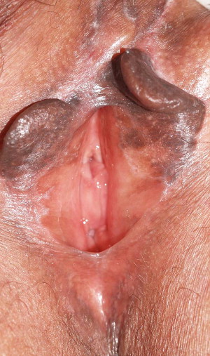
Pigmented condylomata acuminata (anogenital warts)
Condylomata acuminata, or anogenital warts, are HPV-related growths usually found as multiple lesions on the genitalia ( Fig. 5 ). They have variable morphology, to include papillomatous, verrucous, fleshy papules, and flat-topped. Although most warts are skin colored, some are hyperpigmented, as regularly occurs in patients of darker skin types. Flat-topped, brown, anogenital warts in individuals of light skin types should be biopsied to evaluate for VIN, also known as squamous cell in situ and Bowenoid papulosis. Anogential warts are often indistinguishable from SKs. Dermoscopically, anogenital warts exhibit exophytic papillary structures with variation in pigment that includes jet-black color (ie, hemorrhage), red dots, and whitish halo (ie, keratinization).
Vulvar melanosis/lentiginosis
Roughly 68% of pigmented vulvar lesions in reproductive-age women are lentigines. Vulvar melanosis or lentiginosis presents as multiple asymptomatic, asymmetric macules that have the following features: tan to black coloration, irregular borders, color variation within single lesions, and are of varying size ( Fig. 6 ). Their macular, nontextured nature is an important distinguishing factor from other vulvar lesions. Melanosis can occur throughout the vulva including the labia minora and medial labia majora, introitus, and perineum. Vulvar melanosis can occur as a postinflammatory hyperpigmentation, such as in patients with lichen sclerosus.
Although the diagnosis of vulvar melanosis is largely made by inspection, and biopsy confirmation, dermoscopy may play a role as well. Vulvar melanosis demonstrates different patterns, including structureless, parallel, and reticularlike or ringlike pattern, differing from dermoscopic features of melanoma. More studies are needed to differentiate melanomas from melanosis using these patterns. Features that indicate a diagnosis other than vulvar lentiginosis include the presence of a papular component, erosions, or symptoms such as pruritus or pain. A biopsy is nearly always indicated to rule out melanoma and pigmented VIN.
Histologically, these lesions contain melanin pigment confined to the basal layer of squamous epithelium. Lentigines show basal layer hyperpigmentation, mild melanocytic hyperplasia arranged as solitary units at the dermoepidermal junction rather than nests, epithelial hyperplasia, and stromal melanophages without cytologic atypia. If biopsies are negative, reassurance and observation is the appropriate management. Removal is solely a cosmetic issue and can be attempted via destructive or excisional modalities. The association of vulvar melanosis with malignancy is unlikely but has not been disproven at this point. Patients with vulvar melanosis appear to have no increase in melanomas or squamous cell carcinomas, although a single case of genital melanosis in a patient with melanoma of the urinary bladder has been reported. Also, vulvar melanosis is associated with lichen sclerosus, and there has been reported a possible association between lichen sclerosus and melanoma. Thus, a reasonable approach would be to follow these patients at some sort of regular interval, especially if they have concomitant lichen sclerosus.
Genodermatoses characterized by lentigines
There are several genodermatoses that exhibit genital and, often, extragenital mucosal pigmentation showing histologic changes of lentigines as a characteristic feature. These lentigines generally consist of multiple small, dark macules that variably occur on the mucous membranes, modified mucous membranes, and nearby keratinized skin. Bannayan-Riley-Ruvalcaba syndrome carries autosomal dominant inheritance and involves the triad of macrocephaly, genital lentiginosis, and intestinal polyposis. The genital lentigines have been shown to histologically demonstrate hyperplasia of the epidermis with increased basal layer pigmentation and a slight increase in number of melanocytes compared with normal. LAMB syndrome is a cardiocutaneous syndrome that includes atrial myxomas, black and blue nevi in the skin and genital mucosa, and papules and dermal nodules on the skin and tongue. Laugier-Hunziker syndrome involves acquired hyperpigmentation of the oral and genital mucosa (benign to malignant) that can include longitudinal melanonychia. Beare-Stevenson cutis gyrate syndrome is a rare genetic disorder characterized by craniosynostosis, cutis gyrate (furrowed, wrinkled skin), and acanthosis nigricans that can involve the genital skin. Dowling-Degos disease is a rare inherited disease that is characterized by flexoral reticular hyperpigmentation where pigmented macules may be present on the genitalia. LEOPARD, or multiple lentigines syndrome, is an autosomal dominant trait characterized by hypertelorism, sensoneurial deafness, and cardiac abnormalities. Carney complex or NAME syndrome is characterized by hypercortisolism, mucocutaneous lentigines, and nonendocrine and endocrine tumors such as myxomas. Peutz-Jeghers syndrome is an autosomal dominant disorder characterized by intestinal hamartomatous polyps and mucocutaneous melanocytic macules, with an increased risk of a variety of cancers.
Melanocytic nevi (pigmented nevi, nevocellular nevi, common nevi)
A melanocytic nevus is a benign proliferation of melanocytes. Roughly 23% of pigmented vulvar lesions in reproductive-aged women are melanocytic nevi, and roughly 2% of women have vulvar nevi. Clinically, nevi can be flat (junctional) or domed (compound or intradermal). Common nevi range from tan to dark brown in color; rarely these can be blue because of dermal pigment. Individual nevi are generally symmetric, with sharp and regular borders, even color throughout the lesion, and size smaller than 7 mm ( Fig. 7 ). Nevi that stray from this characterization warrant biopsy, and may be dysplastic nevi (atypical moles) whose presence increases an individual’s risk of cutaneous melanoma. The gross and microscopic appearance of dysplastic nevi falls between those of common nevi and melanoma. Surgical removal of atypical nevi on the vulva is warranted. Nevi on the vulva can at times be difficult to distinguish from seborrheic keratoses, pigmented warts, and pigmented VIN, for which dermoscopy and/or biopsy can be helpful to differentiate these lesions. Virtually all nevi are acquired, but uncommon congenital nevi can be larger and carry a greater risk of malignancy.
The early medical literature described concern over the premalignant potential of vulvar nevi as compared with nevi on other parts of the body. However, a large majority of vulvar nevi are identical histologically to extragenital nevi, with most being junctional. A minority subgroup of unusual vulvar nevi occurs in premenopausal women, with the distinguishing feature of enlarged junctional melanocytic nests. Distinct from dysplastic nevi found on all skin surfaces, these vulvar nevi sometimes demonstrate cytologic atypia, focal pagetoid spread, with a dermal nevus component, adnexal spread, and dense eosinophilic fibrosis in the superficial dermis. The Gleason and colleagues study reported recurrence of a single nevus from 45 removed by excision after 3.5 years, and no recurrences after 11.5 years, suggesting that these are not malignant. Such lesions have been termed “nevi with site-related atypia,” of which the genitalia is one of these sites, or “atypical melanocytic nevus of the genital type” (AMNGT). It is important to note that these nevi can be misdiagnosed as melanoma even among experienced pathologists.
Some clinicians suggest removal of any nevi found within lichen sclerosus. These nevi can appear histologically similar to melanoma despite being of benign character, with features such as confluence of junctional nests, nests within dermal fibrosis, lymphocytic inflammation, and pagetoid upward spread of melanocytes. Ideally, lichen sclerosus should be controlled before benign-appearing nevi are excised.
Melanoma
Melanoma is a malignancy of the pigment-producing cells of the epidermis. Only about 3% of all melanomas involve the genital tract ; however, 8% to 10% of genital malignancies are melanomas, making it the second most common malignancy of the vulva behind squamous cell carcinoma. Patients with lighter skin types are most at risk; however, it should be noted that the incidence ratio of genital melanomas between different ethnicities is less marked than that observed for other cutaneous melanomas. The Surveillance, Epidemiology, and End Results (SEER) database of vulvar and vaginal melanomas from 1992 to 2005 found the overall white-to-black incidence ratio in vulvar melanomas was 3.14 to 1.00 and in vaginal melanomas was 1.02 to 1.00, which is much less than compared with cutaneous, nongenital melanomas (13 to 1 and 17 to 1, respectively). Genetic factors, such as family history of melanoma or inherited dysplastic nevus syndrome, appear to be more important than UV exposure in the development of vulvar melanomas. Vulvar melanomas at time of diagnosis tend to be large, with red, white, blue, or black hues, irregular coloration, asymmetric and indistinct borders, and size larger than 7 mm ( Fig. 8 ). They most commonly occur on the labia majora in women of age 50 or older, and can be flat or nodular. At least one quarter of cases in lighter skin types are amelanotic, which distinguishes vulvar melanomas from those on other parts of the body. Because vulvar melanomas usually present at a more advanced stage than melanoma on more visible surfaces, patients are often symptomatic with a palpable mass, pain, pruritus, or bleeding, or they have noticed an enlarging lesion. Most vulvar melanomas are of the mucosal lentiginous subtype on the mucosal surface of the genitalia, but these can also be superficial spreading or nodular, especially on the keratinized surfaces. Vulvar melanomas of the mucosal lentiginous subtype appear to emerge de novo rather than being associated with prior nevi as is often observed in cutaneous, nongenital melanomas.
If melanoma is suspected, an excisional biopsy with 1- to 2-mm margins is ideal, but punch biopsy of a subsection of a large lesion is reasonable. Shave biopsies should not be performed to preserve diagnostic and prognostic accuracy. Pathologists experienced in reading large volumes of vulvar pigmented lesions should be consulted when the initial biopsy is inconclusive but identifies atypia. Histologically, melanoma shows a significant increase in the number of atypical melanocytes at all epidermal levels, arranged as solitary units and as nests, with striking dendrites and some melanocytes being mitotic.
Once diagnosed, melanomas are staged with Breslow thickness being the most important predictor of survival. Because vulvar melanomas tend to be more advanced on presentation, the typical Breslow thickness at presentation is 2 to 3 mm. Staging is performed using the American Joint Committee on Cancer classification system. In a large review of SEER cases, most vulvar melanomas were found in white women (90%), of which 61% represented localized disease, 9% had nodal metastases, and only 6% had distant disease. Five-year disease-specific survival rates were 75.5% in patients with local disease, 38.7% with regional disease, and 22.1% in distant disease. Younger women and those with fewer positive lymph nodes had the best chance at survival.
Treatment is largely surgical. Local excision with 1- to 2-cm margins carries similar survival as a radical vulvectomy, with groin dissection or sentinel lymph node biopsy performed for tumors thicker than 1 mm. Sentinel lymph node biopsies sometimes have a useful place in practice. A recent small study of patients with vulvar melanoma without palpable groin nodes demonstrated that Breslow thickness alone does not predict the presence of lymph node metastases, and that sentinel lymph node biopsy could be useful to help guide which patients should be recommended radical lymph node dissection. Dacarbazine chemotherapy has been shown to have response rates in the 15% to 25% range and interferon has been used with melanoma in general as an adjuvant therapy. Radiation and chemotherapy are often used for palliation. Routine skin checks should be performed as follow-up in patients with a history of melanoma.
Vulvar intraepithelial neoplasia
There are 2 major clinical types of vulvar intraepithelial neoplasia (VIN): those that are largely multifocal HPV-related disease occurring in younger women, and those that are largely unifocal HPV-unrelated disease occurring in older women. HPV-related VIN is linked to high-risk HPV types, most commonly 16, 18, and 31. Risk factors include smoking, multiple sexual partners, and immunosuppression, such as HIV. Multifocal VIN, or Bowenoid papulosis, is HPV-related and often appears as hyperpigmented, flat-topped papules or plaques with distinct margins ( Fig. 9 ). The diagnosis is based clinically with histologic confirmation, as the lesions can grossly be mistaken for banal genital warts, SKs, and melanocytic nevi. They most commonly occur on the vestibule and lateral labia minora. Clinicians should avoid biopsying lesions recently treated with podophyllin, as its effect on the skin may be interpreted histologically as VIN.
VIN generally is treated because of the risk of progression to invasive vulvar carcinoma without treatment. This can be as high as 87.5% in those, usually older, women with untreated unifocal disease often associated with lichen sclerosus or lichen planus. However, a small study of almost completely non-white Pacific Islanders showed spontaneous regression of warty/basaloid VIN in women younger than 30 with multifocal pigmented lesions, many of whom were smokers and symptomatic. Thus, multifocal lesions can sometimes be observed over a 12-month period in woman younger than 30 before treating them, assuming patients have regular clinical follow-up.
Typical treatment modalities include surgery, cryotherapy, and CO2 laser excision or vaporization. For larger lesions, imiquimod has become an often-used therapy, but this does not have Food and Drug Administration approval. Less often, 5-fluorouracil (5-FU) cream is considered, and topical 5-aminolevulinic acid–based photodynamic therapy is a new modality. It involves application of a photosensitizing compound that collects in neoplastic tissue, followed by nonthermal light of a wavelength matching the photosensitizer’s absorption characteristics, thereby producing reactive oxidants than can kill the local cells. It has been studied to treat high-grade VIN, but pigmented and multifocal lesions appear to be less responsive compared with unifocal lesions.
Squamous cell carcinoma
Squamous cell carcinoma (SCC) constitutes the great majority of vulvar malignancies. From 1999 to 2004, the rate of vulvar cancer in the United States among all women was 1.7 per 100,000. Cancer rates rose about 3% per year among black women but were stable among other racial and ethnic groups. White women older than 50 carry the highest rates of SCC. Risk factors include long-term inflammation (eg, lichen sclerosus and lichen planus) in older woman and HPV-related disease in younger women. It appears that roughly 60% of vulvar SCC cases are associated with lichen sclerosus, and that untreated lichen sclerosus carries a 3% to 5% risk of SCC. Risk factors for HPV-positive SCCs include tobacco and alcohol use and cervical dysplasia. Immunosuppression by virtue of disease or medication significantly increases the risk of invasive SCC in all types of VIN.
At presentation, about half of invasive SCC cases are symptomatic, notably pruritic. Multiple biopsies of lesions of different morphologies increase the yield of detecting SCC. There are 3 main types of SCC. Two are associated with HPV: the classic Bowenoid type, which most often presents as keratotic, verrucous nodules or masses, and verrucous carcinoma (Buschke-Lowenstein tumor), which appears as large, cauliflowerlike lesions that rarely metastasize but are locally invasive. The third type of SCC is the non-HPV–associated keratinizing or differentiated SCC, which appears as crustlike lesions that on biopsy show groups of keratinocytes with cornified material in the lamina propria. This type most often occurs in a setting of long-term inflammation, such as lichen sclerosus or lichen planus.
Staging is dictated by the International Federation of Gynecology and Obstetrics. Five-year survival ranges from 100% for localized, stage I disease, to 20% for stage IVB disease. Unlike melanoma, tumor depth rather than thickness is the primary histologic feature that predicts prognosis and directs therapy. The primary treatment modality is surgery, reserving radical vulvectomies for only advanced cases. Alternatively, chemotherapy and radiation are used in those who are not surgical candidates. Sentinel lymph node biopsy compared with inguinofemoral lymph node dissection for staging carries a reduction in morbidity and has a false negative rate of about 2% ; however, both approaches are still used in practice.
Pigmented basal cell carcinoma
Basal cell carcinoma (BCC) is a low-grade neoplasm often classified as an epithelioma rather than a carcinoma because of its very rare metastatic potential. This tumor usually is pink or flesh-colored, with a pearly, translucent sheen; however, it occasionally is pigmented, sometimes exhibiting irregular browns and black. BCC is uncommon in the vulva, constituting approximately 5% of primary vulvar cancers. BCCs can appear as nodules, polyps, ulcers, or flat areas of hyperpigmentation or hypopigmentation. These eventually enlarge and ultimately ulcerate. BCCs of the genitalia usually require a biopsy to diagnose. Although metastasis is not a concern, up to 10% recurred in a study of vulvar BCC in the United Kingdom, and were virtually never fatal. Basal cell nevus syndrome is a rare disorder that can also affect the perineum. Thus, patients with this disease are recommended to have routine anogenital examinations.
Others
There are a variety of other vulvar lesions that may appear pigmented to the naked eye but do not contain melanin. Using a lens magnifier can help differentiate these from true pigmented lesions.
Angiokeratomas are benign, common hemangiomas that are usually multiple and familial ( Fig. 10 ) and present as small, smooth papules usually found on the hair-bearing vulvar skin, increasing with age. They vary in color from red to purple that is so dark as to appear black, and, when solitary, these can mimic nodular melanoma, thus warranting biopsy. Treatment is cosmetic. Electrocautery can be done, but new lesions occur.