Hidradenitis suppurativa (HS) is a chronic, inflammatory, scarring condition involving the intertriginous skin of the axillary, inguinal, inframammary, genital, and perineal areas of the body. It is also referred to as acne inversa and Verneuil disease. Follicular occlusion is the primary event in HS. It is now accepted that the first pathogenetic change is in the pilosebaceous follicular ducts, like acne, and so there has been a move to rename this disorder acne inversa. Despite the legitimate argument that hidradenitis suppurativa is a misnomer, the term has become generally accepted.
Hidradenitis suppurativa (HS) is a chronic, inflammatory, scarring condition involving the intertriginous skin of the axillary, inguinal, inframammary, genital, and perineal areas of the body. It is also referred to as acne inversa and, in the old literature, as Verneuil disease.
HS has been linked to the apocrine sweat (hidros) glands (aden) that were first described in 1921. This link became merely historical in 1990, when Yu and Cook showed follicular occlusion to be the primary event in HS. It is now accepted that the first pathogenetic change is in the pilosebaceous follicular ducts, like acne, and so there has been a move to rename this disorder acne inversa. Despite the legitimate argument that HS is a misnomer, the term has become generally accepted.
Definition
HS is defined clinically by its various features and by its chronicity. It is a recurrent disease, classically, but not exclusively, in the apocrine gland–bearing areas of the skin, manifesting as painful, recurrent, deep-seated, inflamed nodules that can result in abscesses, sinuses, and varying degrees of chronic draining sinus tracts with scarring, disfigurement, and disability.
The Second International HS Research Symposium (San Francisco, March 2009) adopted the following consensus definition : “HS is a chronic, inflammatory, recurrent, debilitating, skin follicular disease that usually presents after puberty with painful deep seated, inflamed lesions in the apocrine gland-bearing areas of the body, most commonly the axilla, inguinal and anogenital region.”
HS is frequently misdiagnosed as boils. This results in delayed diagnosis, fragmented care, and progression to a chronic, disabling condition that has a profoundly negative effect on quality of life.
Diagnostic criteria
The Second International HS Research Symposium also adopted the following diagnostic criteria:
- 1.
Typical lesions: either deep-seated painful nodules (blind boils) in early primary lesions or abscesses, draining sinuses, bridged scars, and tombstone open comedones in secondary lesions
- 2.
Typical topography: axillae, groin, genitals, perineal, and perianal region, buttocks, infra- and intermammary folds
- 3.
Chronicity and recurrences.
These 3 criteria must be met to establish the diagnosis.
HS is recognized by the characteristic skin lesions appearing in the typical locations. The pattern in one area of recurrent boils that do not respond to standard antibiotics is a good clue. Normal boils caused by bacteria respond well to antibiotics and seldom recur after treatment. Normal boils point vertically to, and discharge onto, the surface, unlike typical HS lesions, which are rounded and tend not to burst. HS is characterized by deep, painful, subcutaneous nodules that rupture horizontally under the skin and then tend to track subcutaneously. HS is chronic; 90% of patients in one study had the disease for an average of 19 years.
There are questions that can help diagnose HS and differentiate it from other disorders ( Box 1 ).
- 1.
Does anyone in your family have the same symptoms?
- 2.
Do the boils recur in the same spots?
- 3.
Do you smoke or use tobacco products?
- 4.
Do your boils flare before your menstrual period?
- 5.
Have the treatments received been helpful?
- 6.
Do you get a fever with these boils?
- 7.
Do you have infections elsewhere?
Patients with HS normally respond “Yes” to questions 1 to 4 and “No” to 5 to 7.
Data from Poli F, Jemec GB, Revuz J. Clinical presentation. In: Jemec GB, Revuz J, Leyden J, editors. Hidradenitis suppurativa. Berlin: Springer; 2006. p. 22.
Differential diagnosis
HS has an extensive differential diagnosis ( Box 2 ). The appearance, age of onset, typical locations, poor response to antibiotics, and lack of signs of systemic sepsis can all help distinguish this condition, so the diagnosis should be obvious. There are few conditions that cause recurrent abscesses and sinus tract formation in the intertriginous skin areas, such as Crohn disease, ulcerative colitis, and granuloma inguinale. The most common differential diagnoses are the follicular pyodermas: folliculitis, furuncles, and carbuncles. Infected Bartholin glands and epidermal cysts can also present with HS-like lesions. Atypical infections from any organism, from deep fungi to mycobacteria, can mimic HS, although this is rare. Tumors such as epidermoid cysts, even steatocystoma multiplex, can be confused with HS. The swelling and lymphedema of Crohn disease can cause confusion. Acne produces many lesions similar to HS but the distribution is different.
- •
Infections
Bacterial
Carbuncles, furuncles, abscesses, ischiorectal/perirectal abscess, Bartholin duct abscess, erysipelas
Mycobacteria: tuberculous abscess
Sexually transmitted infections: granuloma inguinale, lymphogranuloma venereum, noduloulcerative syphilis
Deep fungi: blastomyces, nocardia
- •
Tumors
Cysts: epidermoid, Bartholin, pilonidal
Other: steatocystoma multiplex
Trichoepithelioma
- •
Miscellaneous
Crohn disease
Anal or vulvovaginal fistulae
Because the sites of involvement of HS are so varied and the lesions are sometimes nonspecific, patients present the problem to many specialists. They may see surgeons, gynecologists, urologists, plastic surgeons, dermatologists, infectious disease specialists, proctologists, and even gastroenterologists. Patients seen in emergency departments are often treated with simple incision and drainage. Too often, a short course of antibiotic is given or the lesions are just incised and drained. This treatment is generally ineffective in controlling the disorder and is discouraging for patients. Delay in diagnosis is common, averaging about 7 years, and can be decades.
Prevalence and epidemiology
HS is a common, forgotten, and orphaned disease. It is often not recognized by physicians, even dermatologists, and the results are devastating for patients. HS is mistakenly referred to as a rare disease. The global prevalence has been reported as between 1% and 4%, depending on the definitions used. Jemec reported a prevalence of 4% in a series of self-reported cases, with a high prevalence in young adults that was confirmed in another study. Revuz reported the prevalence in persons 55 years and older as 0.5% in contrast to 1.4% for younger people. There may be an increased incidence in black people but most investigators report no racial differences. It is more common in women than men, at a ratio of 3.3:1.14. HS affects the genders differently, being more common found under the breasts (22%) and in the groin (93%) in women and on the buttocks (40%) and perianal area (51%) in men. The average age of onset is 23 years, with a range of 11 to 50 years. It rarely occurs before puberty, occurs earlier in those with a family history of HS, and is unusual after menopause. In men, HS can continue into old age, it can be more severe, and associated squamous cell carcinoma, although infrequent, is more common in men.
Etiology
Several factors are related to the development of HS.
Genetic Factors
Patients with HS have a 35% to 40% positive family history. An autosomal dominant inheritance pattern has been noted. Studies on 4 generations in a Chinese family indicate linkage to a locus at chromosome 1p21.1 to 1q25.3 but no specific gene was defined. Despite the dominant inheritance pattern, a positive history is usually found in fewer than 50% of family members, probably because of lack of adequate family reporting, poor recognition or hiding of HS, or development of HS in family members after the survey. These findings were supported by von der Werth and Williams who also suggested that HS is most likely a heterogeneous disease, probably with several genes involved. Revuz pointed out that patients with a family history of HS usually have milder disease with earlier onset. More genetic studies are needed.
Infection
Bacteria have long been considered in the pathogenesis of HS. Various strains have been cultured but all are considered to be secondary invaders. It is generally agreed that bacteria do not have a major direct role in the cause of HS but may share in the pathogenesis of the chronic relapsing lesions of deep Hurley stage III, in which they may be responsible for some of the destructive processes that are seen.
Hormonal Factors
Although there has been controversy for years about the role of androgens in HS, there is a strong relationship between sex hormones and HS. It has been suggested that the preponderance in women argues against the role of androgens, a suggestion that ignores the greater sensitivity of women to androgens. HS is more common in women; it occurs around menarche, flares premenstrually, improves with pregnancy, fades after menopause, and does not occur in eunuchs or eunuchoids. Even the studies of HS in children are associated with premature adrenarche or early puberty, when androgens are dominant. As in acne, there are no increases in serum androgens in most patients with HS and the effect is assumed to be caused by end organ sensitivity. The ultimate support for the role of androgens is the effectiveness of antiandrogen therapy, as discussed later.
Immune Factors
Even in its most aggressive Hurley stage III form, the disease does not usually produce acute systemic inflammatory effects. There is no fever, no lymphadenopathy, no septicemia, no local cellulitis, cultures are often sterile, and, if the offending material beneath the surface is removed, the disease heals without further difficulty and without antibiotics, which is strongly suggestive of inflammation mediated on the local level by the innate immune system. The simple model of the disorder is the inflammatory reaction around a simple ingrown hair; flick out the foreign hair so it is no longer in contact with dermal toll-like receptors (as an unproven example) and the inflammation fades immediately. Adaptive immunity therefore seems to play no significant part in the cause of HS.
Other Factors
HS can be triggered by, or flared because of, lithium, which can enhance neutrophil migration, increase epithelial cell proliferation, and cause follicular plugging by a direct effect on follicular keratinocytes, as in acne. Sirolimus has been related to the new onset of HS, and medroxyprogesterone acetate acting as an androgen has precipitated or aggravated HS in personal cases. Although HS can be found in both obese and thin patients, shearing forces and hormone-related factors in the obese can worsen HS, and obesity seems to result in more severe disease. Smoking is common in patients with HS, 70% to 89% of patients with HS have been smokers, and nicotine has been shown to activate nonneuronal acetylcholine receptors causing increased keratinization of the pilosebaceous duct, suggesting a role for nicotine in the cause of HS.
Pathogenesis
The pathogenesis of the disease consists of follicular plugging, ductal rupture, and secondary inflammation leading to numerous downstream changes. HS is subject to genetic, mechanical, hormonal, and other influences. The sequential story is likely as follows, although some links remain to be proven.
When hormonal overstimulation of the production of ductal keratinocytes results in failure of terminal differentiation of the keratinocytes lining the ducts, they fail to separate from each other, leading to the accumulation of keratinocytes known as a comedo. The comedo causes a tight plug in the acroinfundibulum of the duct. It seems that a genetic weakness or deficiency of the PAS-positive glassy membrane glycoprotein material that supports the duct (Danby FW. The glassy membrane in hidradenitis suppurativa, unpublished work, 2009) under the centrifugal pressure of a bulging follicular canal, permits the wall of the duct to lose its structural integrity. Molecule-sized follicular contents leak out, stimulating the innate immune system, leading to the rupture of the congenitally weak wall of the duct. Healing processes attempt to repair the normal anatomy of the pilosebaceous unit, and sometimes succeed, but the failure of the inflammatory contents to simply discharge to the surface and then heal is what differentiates HS from acne, folliculitis, or a simple boil. After rupturing beneath the surface, the follicular fragments and their growth cause the extensive lateral spread and the characteristic inflammatory reaction.
The epithelial material caught in the dermis is not simply sitting there as isolated, benign pieces of damaged tissue, waiting to dissolve. These fragments are alive and are still exposed to the hormones and growth factors that nourished the initial overproduction of keratinocytes. Continuous growth of these hormonally stimulated remnants beneath the surface produces the communicating sinuses ( Fig. 1 ) and provides increasing volumes of irritating material, some almost certainly derived from the pluripotential stem cells in the bulge area of the exploded follicle. This aberrant epithelial repair response drives the inflammation in the dermis and subcutis.
The innate immune system is the prime mover in HS (–discussed earlier), just as in acne vulgaris and even acne rosacea. The ongoing reaction to an enlarging and extending mass of epithelial and ductal elements produces continuous and increasing amounts of innate inflammatory reaction, which explains the relentless course of the disorder. This mass of interactive material forms the central core of HS, and the disorder will not settle until this active mass is eliminated. These and numerous other pathogenetic considerations were recently discussed by a panel of experts.
Clinical Description
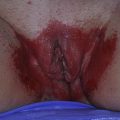
HS classically occurs in the early 20s. The onset can be insidious, with apparently random small lesions that develop as a red, indurated papule, pustule, or nodule that may resolve without leaving any mark ( Box 3 ). The lesions may develop in only 1 area, or several areas over weeks or months. The discomfort varies from vague itching to mild to moderate pain. In some cases, the onset can be severe, even frightening, with large, deep, painful lesions that result in restriction of activities. Too often they are diagnosed as recurrent boils or furunculosis. Lesions are generally intertriginous and involve, in order of frequency, the axillae, inguinal areas, inner thigh, perianal and perineal areas, mammary and inframammary area, buttocks, pubic region, scrotum, vulva, chest, scalp, and retroauricular region. Women typically have involvement in the groin, axilla, and under the breasts. In men, it is found in the axillae and groin and is more common perianally than in women. Involvement of the perineal and perianal areas is more debilitating than axillary involvement because of frequent recurrence.
HS Lesions
Primary lesions
Recurrent or persistent/painful
- •
Red papules less than 1 cm in diameter
- •
Red nodules larger than 1 cm in diameter
- •
Pustules or abscesses
- •
Secondary lesions
Persistent red, painful, draining sinus(es)
Ulcerations with or without granulation tissue
Tertiary lesions
Scars
- •
Pitted or cribriform
- •
Dense fibrotic plaques or linear, ropelike scars
- •
Epidermoid cysts
Single or multiple tombstone comedones
Lymphedema with or without lymphangiectasia
Primary lesions of HS are individual, painful nodules, 0.5 to 2 cm in diameter that persist for weeks or months with a varying degree of inflammation. The diagnosis is frequently delayed. These lesions can be deep and patients complain of pain although all that is visible is redness with barely any swelling, despite major discomfort. A prodrome has been reported in 50% of all patients, consisting of burning, stinging, pain, and pruritus, with or without hyperhidrosis, occurring 12 to 48 hours before the onset of nodules. The first lesion may be a small (<1 cm), red papule that may resolve and recur or rupture with purulent drainage. Deep, red, round, painful nodules (>1 cm) are typical. They may be individual or coalesce into groups. The HS nodules do not point centrally but are deep, round, red, and sore, without the spontaneous drainage seen in furunculosis. Nodules can last from 7 to 15 days. They then may progress to resolution, or persist, or drain. With more severe disease, and more time, there is eventual drainage with spontaneous resolution of the pain. Patients often squeeze or pinch these lesions to get relief (some even attempt home surgery to open lesions). Lesions may be grouped or come and go in various areas, depending on the individual. Sheets of small folliculopapules and folliculopustules and acneform lesions may be scattered over the buttocks, mons pubis, and around the breasts. These lesions occur both in early and late HS and may occur in an area separate from the main HS activity. Patients may present, for example, active papules, nodules, and draining sinuses in one area (the groin) and sheets of perifollicular papule pustules elsewhere (around the breasts or buttocks). All these lesions wax and wane.
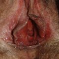
Secondary lesions develop as a result of persistence of the process in an area. The subcutaneous coalescence of several neighboring cysts, or the lateral extension of actively proliferating pilosebaceous material with rupture to the surface, results in formation of chronic interlinked sinuses. Drainage from these lesions may be serous, purulent, bloody, or a mixture, with or without odor. Uncommonly there may be persistent ulcerations and even the formation of red granulation tissue around a sinus opening. With healing, hypertrophic scars develop and eventually dense ropelike linear fibrotic bands develop that may crisscross an area of involvement. Sinus tracts can be single or multiple; they may be hardly visible, with intermittent serous drainage, or they may be swollen, painful, and inflamed with 1 or more draining sinuses forming a honeycombed pattern of draining nodules or tracts.
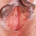
Tertiary lesions occur as a result of aberrant healing. Small HS lesions typically heal with small, acneform, pitted, and, less commonly, cribriform scars. Occasionally, indolent epidermal cysts of 1 to 6 cm can develop on the face, behind the ears, at the nape of the neck, on the trunk, and in the genital area. These cysts are more common in men than in women. Sinus tracks can coalesce forming hypertrophic, fibrous, fistular tracts involving entire zones. These subcutaneous networks of sinuses can form a solid plaque or thick, bridged, ropelike scars. Both can result in contractures and may decrease range of motion so that patients cannot lift arms or separate thighs. Scarring can result in lymphatic obstruction with lymphedema and eventual lymphangiectasia. One of the classic features of HS is the tombstone comedones that form in a burned out area because of permanently dilated pores. They are often associated with multiple small, superficial sinuses and are present in up to 50% of patients.
Clinical course
The mean age of onset is 22.1 years and HS lasts about 19 years. It can remit or partially remit with pregnancy and breastfeeding. The course for patients can be variable. It may be intermittent and benign, with mild but chronic painful disease, acute exacerbations, premenstrual flares, and usually resolution after menopause. It may remit for weeks to months, lesions may flare continuously or only intermittently, and marked involvement may occur in only 1 area with a long deep-draining sinus and discharge typical of Hurley stage III. Revuz describes 2 types of severe disease. The first shows typical solid plaques of coalescent nodules, fistulae, and scars with smelly discharge, pain, and debility. The other type shows new lesions; deep, painful, separate nodules, and draining lesions, continually appearing and each lasting 10 to 30 days. Patients do not necessarily progress from mild to moderate to severe disease but may instead present with a certain degree of severity and remain in that range. Those with severe disease may have marked severity from the beginning and the lesions often develop rapidly, sometimes with the predictive prodrome of itch, pain, and/or malaise.
Patients have varied difficulty with mobility, ranging from minor soreness and discomfort to being unable to walk or sit without pain. With large draining areas, odor may be significant. Patients may need to wear diapers for the drainage. With such pain, debility, and disruption in their daily lives it is common for patients with HS to withdraw and become depressed and, eventually, dysfunctional.
The severity of HS can be classified using Hurley clinical staging and this can also be used to help direct future management.
Hurley stages
In the recent report by Canoui-Poitrine and colleagues, 68.2% of patients are Hurley stage I, 27.6% Hurley stage II, and 3.9% Hurley stage III ( Box 4 ). To more accurately assess outcome variables related to HS treatment, the Sartorius score has been developed to monitor patients. It is to act as a complimentary system to the Hurley classification, and is presently undergoing evaluation as a research tool.
Stage I: abscess formation, single or multiple without sinus tracts and cicatrization
Stage II: recurrent abscesses with tract formation and cicatrization, single or multiple widely separated lesions
Stage III: diffuse or near-diffuse involvement, or multiple interconnecting tracts and abscesses across entire area
Morbidity/quality of life
It is usual for patients with severe disease to be unemployable and socially isolated because of the painful, draining, and malodorous lesions. HS has a profoundly negative effect on patients, not only at a physical level but socially and economically. von der Werth and Jemec studied 144 patients with HS with a dermatology life quality index and found a higher morbidity index than in mild to moderate psoriasis or alopecia. The high score was related to pain, malodorous discharge, intimate sites of eruptions, and lack of medical care because of incorrect diagnosis.
In a study of the quality of life of 61 patients with HS, impairment was higher than that for urticaria, neurofibromatosis, psoriasis, and atopic dermatitis. The effect on quality of life is correlated with the number of active lesions and, in particular, severity of pain. Pain is the most limiting factor. Long, continuous duration, and pelvic area involvement, especially with a malodorous discharge, are other major factors.
Patients with HS lose an average of 2 to 7 days of work per year, and this number tends to be greater in women. They are often unemployed, poor, socially isolated, or reclusive.
Associated diseases
HS has been associated with severe acne (acne conglobata), dissecting cellulitis of the scalp, and pilonidal cysts ( Box 5 ). Pilonidal cysts have been associated with HS in up to 30% of cases. Acne vulgaris is only found with HS in about 10% of women and 30% of men but a history of long-lasting, scarring acne was found in 23% of women and 44% of men. Dissecting cellulitis of the scalp is rare (1%); Revuz has never seen the follicular triad in 500 patients he has followed.
Related posts:

Stay updated, free articles. Join our Telegram channel

Full access? Get Clinical Tree


