28 Velopharyngeal dysfunction
Synopsis


Anatomy and physiology of the velopharynx
Anatomy
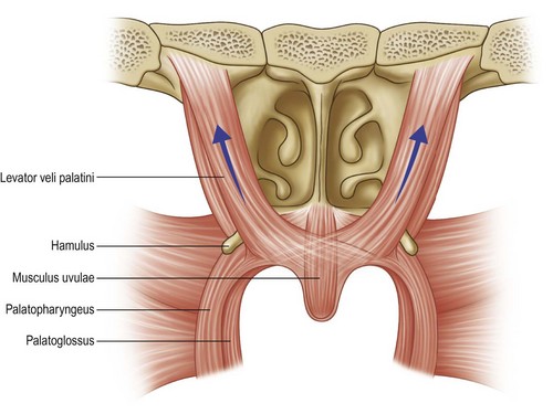
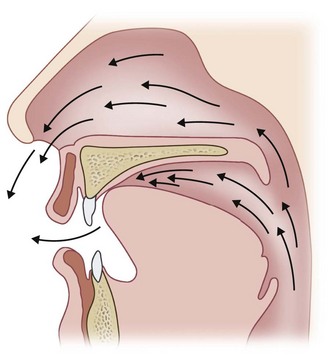
The velopharyngeal port is defined anteriorly by the soft palate, or velum, laterally by the lateral pharyngeal walls, and posteriorly by the posterior pharyngeal wall. Closure of the velopharynx during speech is a voluntary action that is mediated by the motor cortex and that requires the coordinated action of the velopharyngeal musculature. The muscles of the soft palate include the levator veli palatini, the tensor veli palatini, the palatoglossus, the palatopharyngeus, and the musculus uvulae (Fig. 28.1). The levator takes its origin from the petrous portion of the temporal bone and from the medial aspect of the eustachian tube. Its fibers course anteriorly, inferiorly, and medially, inserting into the palatal aponeurosis and decussating with the levator fibers from the opposite side (Fig. 28.2). Contraction of the muscular sling formed by the paired levators is the primary mechanism for velar elevation and closure of the velopharyngeal port, although evidence suggests that the palatoglossus and palatopharyngeus muscles may act as antagonists to the levators to provide fine motor control of velar position during speech.1,2 The musculus uvulae is a paired intrinsic muscle that likely contributes to velopharyngeal closure both by adding bulk to the dorsal surface of the velum and by contributing to velar stretch.3–5 It is usually absent in patients with overt and submucosal clefts of the palate.6
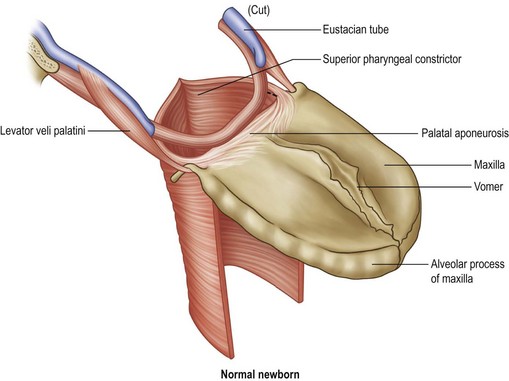
The superior pharyngeal constrictor is a broad, thin muscle that takes origin from the velum, the medial pterygoid, and the pterygomandibular raphe, inserting into the median pharyngeal raphe along with the constrictor muscle fibers from the opposite side. Contraction of the superior constrictor may contribute to velopharyngeal closure by effecting medial movement of the lateral walls and anterior movement of the posterior wall of the velopharynx.7,8 The anatomy of the superior constrictor and its contribution to velopharyngeal closure, however, are highly variable.
With the exception of the tensor veli palatini, which is innervated by the third division of the trigeminal nerve (V3), all of the muscles of the velopharynx receive motor innervation from the pharyngeal plexus, which is composed of fibers from the glossopharyngeal (IX), vagus (X), and accessory (XI) nerves.9 Studies have suggested that the facial nerve (VII) may also play a minor role in velopharyngeal motor function.10,11 It is important to note that, although the functional activity of the velopharyngeal valve (that is, uncoupling of the oropharynx and nasopharynx) during speech and swallowing may be similar, the neurological pathways for these activities are distinct. Velopharyngeal movements for speech are learned, voluntary activities that are controlled by the motor cortex, whereas similar movements for swallowing are primarily involuntary activities that originate from the brainstem.
Physiology
It is widely accepted that the levator veli palatini is the muscle that is primarily responsible for velar motion and, hence, for velopharyngeal closure.12 Fine motor control of velar position may also be governed by the palatoglossus and palatopharyngeus. As noted above, the paired musculus uvulae may play an important role in velar stretch and in filling the gap between the velum and the posterior pharynx during velopharyngeal closure. The relative contribution of the levator and of the superior pharyngeal constrictor to lateral pharyngeal wall movement has been the subject of some debate.
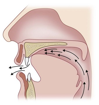
In normal individuals, the velum lifts posteriorly and superiorly during velopharyngeal closure. The normal point of contact with the posterior pharyngeal wall is located approximately three-quarters of the way back on the velum from the posterior nasal spine (Fig. 28.3). The site of velopharyngeal closure is usually at or just inferior to the palatal plane, but velar height, as well as the extent of velopharyngeal contact, varies systematically depending upon the phonetic context.13–15
The contribution of lateral pharyngeal wall movement to velopharyngeal closure varies amongst individuals with or without cleft palate and, as with velar movement, varies with the phonemic task. Maximal lateral pharyngeal wall displacement generally occurs at the level of velopharyngeal contact. Skolnick et al.16 and Croft et al.17 have described three basic patterns of velopharyngeal closure observed in normal subjects (Fig. 28.4): (1) coronal, in which closure is effected primarily by velar elevation; (2) circular (with or without Passavant’s ridge), in which medial movement of the lateral pharyngeal walls contributes to velopharyngeal closure in near-equal proportion to the velum; and (3) sagittal, in which closure is effected primarily by medial movement of the lateral pharyngeal walls and the velum contacts the lateral walls rather than the posterior wall. Of these, the coronal pattern of closure is observed most commonly in both normal individuals and in patients with VPD.
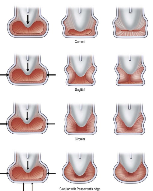
In some individuals, a localized transverse ridge of tissue may be seen to form on the posterior pharyngeal wall during speech. This anterior movement of the posterior pharyngeal wall during velopharyngeal closure was first described by Passavant in 186318 and is therefore frequently referred to as “Passavant’s ridge.” Although some have written that its appearance is always indicative of pathologic velopharyngeal function, Croft et al.17 have demonstrated that Passavant’s ridge may play a role in velopharyngeal closure in both normal speakers and in those with VPD.
Electromyographic studies support the notion that normal velopharyngeal function requires the central coordination of velopharyngeal muscle activity with other articulatory movements.19 Changes in velar position during sound production represent the end result of a complex interaction of several interrelated variables, including auditory and proprioceptive feedback. Moreover, for a single individual, there may be significant flexibility in the system of sound production such that there may be a limited but variable repertoire of velopharyngeal movements that may produce the same perceived sounds. Despite decades of speech science research, the precise neurophysiology of both normal and abnormal velopharyngeal function remains incompletely understood.
Basic science/disease process of velopharyngeal dysfunction
Velopharyngeal insufficiency
The first major diagnostic category of VPD is velopharyngeal insufficiency, a term used to denote an anatomic, or structural, defect responsible for inadequate closure of the velopharyngeal valve. Such defects may be congenital, as in cases of cleft palate or congenital velopharyngeal disproportion (i.e., a short soft palate relative to the depth of the pharynx) (Fig. 28.5), or they may be secondary to surgical procedures that alter velopharyngeal anatomy, as in cases of palatoplasty, tumor resection, or adenoidectomy. The most common congenital structural defects associated with VPD are cleft palate and submucosal cleft palate. The reported incidence of persistent VPD after cleft palate repair varies widely and is influenced by a large number of variables. In the absence of an oronasal fistula, however, VPD after palatoplasty is most commonly the result of impaired velar mobility, velopharyngeal disproportion, or a combination of both.
Since adequacy of velopharyngeal closure is largely a function of the ratio of pharyngeal depth to palatal length, patients with a proportionally short palate or deep pharynx may demonstrate incomplete velopharyngeal closure (Fig. 28.5). Each of these conditions may be the result of either a congenital anomaly or an iatrogenic alteration in velopharyngeal architecture. Cicatricial changes following palatoplasty, for example, may lead to velopharyngeal insufficiency secondary to velar shortening. Congenital differences in skeletal architecture may also play a role in postpalatoplasty VPD. Patients with clefts have been demonstrated to have a broader nasopharynx than controls, likely the result of alterations in cranial base dimensions.20–22 Osborne et al.23 and Ross and Lindsay24 have shown that a higher prevalence of upper cervical spine abnormalities in patients with clefts may result in increased pharyngeal depth. Likewise, platybasia, or flattening of the cranial base angle, may contribute to VPD by increasing pharyngeal depth, and thus the depth-to-length ratio. Ruotolo et al.25 have shown that patients with 22q11.2 deletion syndrome, a condition associated with a high frequency of severe noncleft VPD, demonstrate several predisposing skeletal and soft-tissue anomalies, including increased pharyngeal depth, platybasia, and cervical spine anomalies.
Velopharyngeal insufficiency may also be caused by postsurgical changes in velopharyngeal anatomy. In young children, velopharyngeal closure is most often velar-adenoidal. Removal of hyperplastic adenoids for the management of nasopharyngeal airway obstruction or chronic otitis media results in an acute increase in pharyngeal depth. In the majority of noncleft patients, the capacity for velar stretch allows the palate to accommodate for this change. In most cases, postadenoidectomy velopharyngeal insufficiency is transient, and resonance returns to normal within 6–12 months. VPD may persist, however, in a small number of patients, some of whom may have predisposing factors for VPD, including submucosal cleft palate, a short velum, a deep pharynx, or neuromuscular disorders. For patients with these conditions, the adenoids may play a critical role in velopharyngeal closure and even their normal involution may result in velopharyngeal insufficiency.12 Careful assessment of velopharyngeal anatomy is therefore essential in all patients prior to adenoidectomy, and should be avoided if possible when anatomical factors that predispose to VPD are identified.
In some patients, irregularity of the surface contour of the adenoid pad may interfere with the ability of the velum to achieve complete velopharyngeal closure.26 In others, enlarged tonsils may intrude between the velum and the posterior pharyngeal wall, resulting in incomplete closure.27,28 In these cases, the first step in management should be a selective adenoidectomy or tonsillectomy, respectively, as such may be sufficient to solve the problem, obviating the need for any of the surgical procedures later described.
Velopharyngeal incompetence
There are extensive possibilities for neurologic diagnoses that may be associated with congenital velopharyngeal incompetence including, but not limited to, cerebral palsy, myotonic dystrophy, muscular dystrophy, and congenital hypotonia. Asymmetrical velopharyngeal function, such as that typical of patients with hemifacial microsomia, is also a common cause of velopharyngeal incompetency. Acquired or “late” causes of velopharyngeal incompetence may include traumatic brain injury, cerebrovascular accident or brainstem stroke, and progressive diseases such as Parkinson’s disease, amyotrophic lateral sclerosis, muscular dystrophies, multiple sclerosis, and other demyelinating diseases.29–33
Children and adults with motor speech disorders have also been shown to demonstrate velopharyngeal incompetence of varying degrees of severity. Apraxia of speech (also referred to as developmental apraxia of speech or childhood apraxia of speech, in children) is a neurologic condition resulting in difficulties with speech motor programming and control.34 Apraxia may be characterized by inconsistent symptoms of VPD such as inconsistent nasalization of vowels or consonants and inconsistent nasal emission. In addition, many individuals with apraxia may exhibit a combination of both inconsistent hypernasality and hyponasality, providing additional evidence of the abnormal coordination of the palate during speech sound production. Younger children with apraxia may demonstrate some overlapping speech features often seen in children with congenital velopharyngeal incompetence of other causes. For example, children with a history of VPD may have a limited inventory of sounds and demonstrate difficulties learning to produce oral consonants in the first few years of life. These children may produce a pattern of compensatory articulation errors (i.e., glottal stop substitutions) or omit sounds completely. Children with apraxia have difficulty with generating the appropriate “motor program” (blueprint) for producing the sequence of motor movements to produce a sound, which is not typical of children with isolated VPD or clefting. The importance of obtaining a thorough speech pathology evaluation to diagnose these conditions differentially is critical for appropriate treatment decision-making.
Lastly, stress velopharyngeal incompetence is a special case of inadequate velopharyngeal closure for nonspeech behaviors. It is most commonly observed in wind musicians given the high pressure demands.35,36 There may or may not be comorbid hypernasality or nasal emission in speech. In some cases, stress velopharyngeal incompetence may be an indicator of an underlying physical cause of velopharyngeal incompetence, which may have been masked, or very mild, in the past. In some cases, neurologic or structural causes of VPD (e.g., submucous cleft palate) are diagnosed following the presentation of stress velopharyngeal incompetence, further emphasizing the importance of a thorough clinical evaluation of all patients with any form of VPD.37 Treatment for stress velopharyngeal incompetence may follow a similar course as that for speech disorders, although there have been reported cases of spontaneous recovery after a period of rest.
Velopharyngeal mislearning
The third, and lesser known, type of VPD involves velopharyngeal mislearning.38 In this category, the velopharyngeal mechanism appears to be anatomically and physiologically capable of consistent and complete velopharyngeal closure for speech, despite the observation of inconsistent velopharyngeal closure for speech. In this type of VPD, the patient has mislearned how to produce certain speech sounds accurately. The most common example is that of phoneme-specific nasal emission, in which nasal airflow is produced as a complete substitution for an oral consonant, despite adequate velopharyngeal closure ability for other consonants.39 Clinically, this will often be observed in a child with nasal emission heard on a selected set of sounds, most commonly S, Z, SH, or CH, but not on other sounds such as P, B, T, D, K, G. Another example of velopharyngeal mislearning is the case in which a child produces compensatory articulation errors (e.g., glottal stops) which may prevent or interfere with the achievement of adequate velopharyngeal closure for speech. The velopharyngeal movements during the production of these aberrant speech errors have been shown to be counterproductive (distal versus medial movement of the lateral pharyngeal walls) to the achievement of velopharyngeal closure for speech. A related example is that seen in children with congenital hearing loss with an inability to self-monitor their own speech production, resulting in nasalized speech errors with an otherwise physiologically intact velopharyngeal mechanism.
Combined types
In some cases, individuals with craniofacial anomalies and/or clefting may exhibit a combined disorder with evidence of both velopharyngeal insufficiency and velopharyngeal incompetency, resulting in a challenge for surgeons. Some patients with 22q11.2 deletion syndrome have been shown to demonstrate evidence of a combined type of VPD due to the combination of structural clefting disorders, increased pharyngeal depth, and hypotonia of the velopharynx.40 Regardless of the type of VPD which is present, a complete and thorough physical exam, clinical speech evaluation, and any necessary instrumental or imaging studies should be completed to confirm the etiology and identify the most appropriate treatment plan.
Diagnosis/patient presentation
Patient history and physical exam
• Current patient/family concerns with speech
• Pregnancy history, complications, medication use, and any exposure to teratogens
• Birth and delivery history and complications
• Primary medical diagnoses (e.g., cleft palate, syndromes, cardiac defects, neuromuscular disease)
• History of any feeding or swallowing difficulties during infancy and any current swallowing concerns, including nasal regurgitation and difficulty with breastfeeding or bottlefeeding during infancy
• History of hearing loss or ear disease, including history of frequent ear infections or effusions
• History of snoring or symptoms of sleep apnea
• Surgical history, including prior tonsillectomy, adenoidectomy, and, if appropriate, cleft-related surgical history and timing
• History of any genetic testing and results
• Family history of cleft lip/palate, nasal speech, speech delay, or articulation/pronunciation difficulties; hearing loss, learning disabilities, and medical conditions
• Oral–facial movement and symmetry
• Presence and location of any fistulae
• Presence of signs of submucous cleft palate, including bifid uvula, zona pellucida, and palpate for notch
• Soft palate length, symmetry, and degree of elevation and symmetry during phonation
Perceptual speech evaluation
Perceptual speech assessment is considered the gold standard in the diagnosis of speech disorders of persons with cleft palate and VPD.41 Additional instrumental assessment and imaging are considered adjunct to the perceptual speech findings, which are the ultimate arbiter of a patient’s need for treatment. Perceptual speech evaluation of this patient population should be completed by a speech pathologist with coursework, training, and continuing education in the area of cleft palate and craniofacial anomalies, whenever possible.
During the speech evaluation, the speech pathologist obtains the necessary clinical information regarding the presence and perceived severity of VPD, suspected etiology, and makes preliminary decisions regarding treatment recommendations to discuss with the team. In addition, the speech pathologist is making diagnostic decisions regarding the presence of comorbid conditions such as articulation disorders, voice disorders, and language difficulties. Box 28.1 provides a list of common speech pathology terminology used for describing the speech characteristics associated with VPD. The most common speech sequelae of VPD include reduced speech intelligibility; articulation disorders ranging from severe compensatory articulation disorders (e.g., pervasive use of glottal stop substitutions) to mild articulation errors and distortions secondary to malocclusion; reduced intraoral pressure of oral pressure consonants; audible nasal emission or nasal turbulence on oral pressure consonants; hypernasal resonance; possible hoarseness and decreased loudness.41–43
BOX 28.1
Common speech pathology terminology
Intelligibility: perceived amount of speech (i.e., number of words) understood
Nasal substitutions: the active replacement of oral sounds P, B, T, D with nasal sounds M, N
The components of a standard speech evaluation for the assessment of velopharyngeal closure for speech should include an assessment of intelligibility, resonance, voice, and articulation during a spontaneous speech sample, conversation, and/or picture description tasks.44 A standard reading passage is suggested for use with adolescent and adult patients. Articulation skills should be assessed with standardized measures (i.e., a standardized articulation test), as well as word and sentence repetition tasks (standard lists available44). Oral-only or nasal-only stimuli (e.g., Buy baby a bib, Pet the puppy, Mama made muffins, etc.) are also used to assess resonance, nasal emission (audible and inaudible), and pressure for consonants. Special mirrors or listening tubes may also be utilized to evaluate the presence of inaudible nasal emission. The speech parameters are typically rated with five- or seven-point equal-appearing interval scales or other ratio-based scaling methods (e.g., visual analog scales).41 In some centers, after the clinician rates each speech characteristic individually, a composite decision is made regarding the overall perceived adequacy of velopharyngeal closure for speech.
Indirect measures of velopharyngeal closure for speech
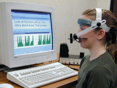
Nasalance is an acoustic index of nasality which has been shown to correlate with perceptual judgments of resonance.45 Nasalance can be measured using commercially available products such as the Nasometer (Kay Pentax) (Fig. 28.6), Nasality Visualization System (Glottal Enterprises), Nasalview (Tiger DRS), and other similar systems. Nasalance is a ratio of the nasal sound energy divided by the sum of the oral plus nasal sound energy in the speech signal.45 The patient wears a specialized headpiece with nasal and oral microphones that capture the speech signal while the patient reads or repeats a standardized speech sample (Fig. 28.6). Automated analysis provides a nasalance score (expressed as a percentage), which is then interpreted against the perceptual speech observations. Nasalance can range from 0 to 100%; higher numbers represent a higher degree of nasality in speech. A variety of normative and “cutoff” scores have been suggested, which are dependent upon the type of speech stimuli used for the nasalance score calculation.46–49 Surgeons and clinicians should exercise caution in reliance on nasalance scores, however, due to the variety of potential confounding variables that can artificially inflate or reduce nasalance scores. These include nasal turbulence, articulation errors, vocal hoarseness, mixed resonance, and equipment placement variations, which may reduce with the validity of this measure.49–51 Nasalance should be considered a supplement to the perceptual speech evaluation, not a substitute for it.
Pioneered by Warren and colleagues,52,53 pressure–flow testing was developed to obtain quantitative measurement of intraoral and nasal pressure and airflow, velopharyngeal orifice size, and velopharyngeal closure timing for speech. The schematic in Figure 28.7 illustrates the type of instrumentation and set-up often utilized for aerodynamic assessment of velopharyngeal closure for speech. Custom-designed and commercially available aerodynamic systems, e.g., PERCI-SARS (Microtronics), offer both clinical and research applications for the assessment of velopharyngeal closure for speech. Warren and others52–


Stay updated, free articles. Join our Telegram channel

Full access? Get Clinical Tree


