, Julie Karen1 and Perry Robins1
(1)
New York University School of Medicine, New York, NY, USA
Leg Vein Treatment Overview
Superficial telangiectasia (pink, red, <1 mm), reticular veins (blue, green, 1–3 mm), and varicose veins (blue, green, colorless, >3 mm) of the lower extremities are interconnected and commonly develop after impairment of venous return due to valvular incompetence.
Prior to treatment, a comprehensive history should be taken with particular attention to family history of varicose veins, prior venous procedures, coagulopathy, anticipated immobility or travel, pregnancy/lactation, concurrent minocycline therapy (due to risk of long-lasting pigmentary alteration), arterial insufficiency, migraine with aura, patent foramen ovale (PFO), and presence of venous symptoms (see below).
Consider duplex ultrasound if bulging varicose veins within zones of influence of saphenous systems or presence of signs/symptoms of superficial venous insufficiency, including swelling, aching, burning, restless legs, cramping, stasis dermatitis, ulceration, or lipodermatosclerosis.
Sclerotherapy remains the gold standard for treatment of non-varicose leg veins.
Detergent solutions (see Table 6.2) can be combined with air (1:3–4) and agitated via a two- or three-way stop-cock in order to create foam—Tessari method.
Foam sclerotherapy (which is not FDA approved) can be used for treatment of large networks of vessels, and is particularly useful for duplex guided sclerotherapy due to its echogenicity. There is an increased risk of adverse side effects, including visual disturbances and transient CNS events. Use should be avoided in patients with a known patent foramen ovale (PFO).
Proximal reflux/larger veins should be treated before related small veins.
Treatments should be spaced approximately 6 weeks apart.
Most common side effects are ecchymosis, postsclerotherapy pigmentation (more common with higher concentrations and after foam sclerotherapy), matting, and ulceration.
Postoperative compression improves efficacy of vein treatments, reduces risk of thromboembolism, reduces risk of pigmentation (due to intravascular coagulum formation), and increases the rate of recovery, although optimal duration of compression is unclear.
Laser treatment is an option for patients with severe needle phobia or recalcitrant vessels (see Table 6.1).
Leg Vein Treatment Algorithm
1.
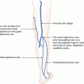
Saphenous incompetence (see Figs. 1.15 and 1.16)
(a)
Ablation (endovenous laser/radiofrequency), ligation +/− short stripping
(b)
Duplex-guided foam sclerotherapy
2.
Get Clinical Tree app for offline access

Saphenous branches
(a)


Ambulatory phlebectomy
Related posts:

Stay updated, free articles. Join our Telegram channel
Full access? Get Clinical Tree