Urticarial eruptions yield, as a primary lesion, a wheal (hivelike) or something that resembles a wheal, creating a fixed indurated papule or plaque, without scale. This pattern may be produced by internal and external causes. Most lesions are erythematous (red).
Important History Questions for Urticarial Eruptions
How long have you had these lesions?
Attempt to establish the duration of illness. Some urticarial processes are acute (e.g., acute urticaria, Sweet syndrome), whereas others are present for weeks, months, or even years (e.g., chronic urticaria).
How long do individual lesions last?
In simple urticaria (hives), the individual lesions lasts less than 24 hours, although the rash may last longer. This differs from urticarial vasculitis, in which individual lesions last longer than 24 hours. As a separate question, it is important to query the patient about how long individual lesions last, distinct from how long the overall condition has been present. This information is often overlooked in emergency department (ED) and urgent care settings.
What medications do you take?
Most urticarial eruptions represent a hypersensitivity process. These conditions, including urticaria, angioedema, serum sickness, and Sweet syndrome, may be induced by drugs.
Do the lesions itch or are they painful?
Simple urticaria (hives) is usually pruritic, whereas urticarial vasculitis, Sweet syndrome, cellulitis, and especially necrotizing fasciitis may be described as burning or painful.
Have you had any difficulty breathing?
This is a critical question to ask and document in the medical record. Sinopulmonary involvement distinguishes angioedema and anaphylaxis from simple urticaria, which is without mucosal or sinopulmonary involvement.
Important Physical Findings for Urticarial Eruptions
Are any of the lesions annular?
Some urticarial eruptions may be annular, or vaguely annular, and this is an important pattern to observe. Simple urticaria (hives) may present with annular and nonannular lesions.
Are any of the lesions linear?
Linear lesions suggest dermatographism, which can be tested by stroking the skin with a firm object and waiting 3 to 6 minutes to identify an erythematous line. The line should disappear in 15 to 30 minutes.
Do any of the lesions demonstrate focal hemorrhage?
Simple urticaria does not demonstrate associated hemorrhage unless the hemorrhage is simply due to increased hydrostatic pressure in the lower extremities. Urticarial vasculitis and hemorrhagic edema of infancy often demonstrate associated hemorrhage.
How large are the lesions?
Small papular wheals (1–4 mm) often suggests cholinergic urticaria, which is related to heat or sweating.
What is the distribution of the lesions?
Some forms of physical urticaria may demonstrate a distinct distribution. For example, solar urticaria is photoaccentuated, whereas pressure urticaria occurs in areas of pressure, such as the hands, feet, trunk, buttocks, and legs.
Is any fever present?
A fever should prompt consideration of an infectious cause of urticaria (e.g., reaction to streptococcal pharyngitis or a viral infection). Also, some mimics of urticaria may be caused by infection, such as erythema migrans, the cutaneous manifestation of Lyme disease.
Urticaria
ICD10 Code L50.0 (allergic) and L50.1 (idiopathic)
INTERNAL ETIOLOGY
- •
Ampicillin
- •
Amoxicillin
- •
Penicillin
- •
Sulfonamides
Pathogenesis
Urticaria (hives) is ubiquitous, with about 20% of the world’s population experiencing at least one episode of urticaria in a lifetime. A cause is identified in about 50% of cases of acute urticaria (<6 weeks’ duration). Common instigators include food (e.g., seafood, nuts), wasp and bee stings, blood products, viral infections, radiocontrast media, and drugs. It is believed that many cases of chronic urticaria (>6 weeks) are due to autoantibodies directed against the high-affinity immunoglobulin E (IgE) receptor and/or IgE itself. Hence, even extensive and detailed patient workups in chronic urticaria often fail to demonstrate a cause.
Clinical Features
- •
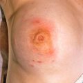
Acute urticaria (<6 weeks’ duration)—pruritic wheals, of variable size, which may be annular. Although the condition may last for a prolonged duration, individual lesions last less than 24 hours ( Figs. 5.1–5.3 ).
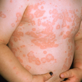
Fig. 5.1
Acute urticaria in a child from an unknown cause. Some lesions maintain an annular appearance.
(From the Fitzsimons Army Medical Center Collection, Aurora, CO.)
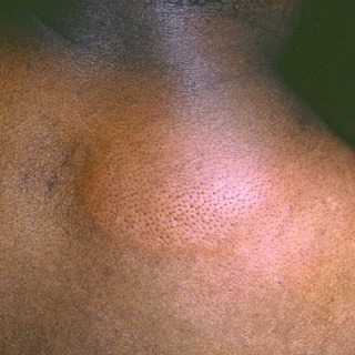
Fig. 5.2
Large lesion of urticaria demonstrating a dimpled appearance due to dermal edema (peau d’orange).
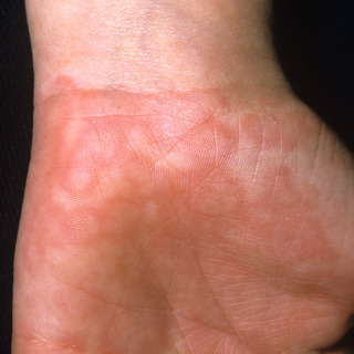
Fig. 5.3
Patient with acute urticaria of the palm from an unknown cause. The location suggests possible pressure urticaria.
(From the Fitzsimons Army Medical Center Collection, Aurora, CO.)
- •
Chronic urticaria (>6 weeks’ duration, with nearly daily eruptions)—a cause is less often identified.
Diagnosis
- •
A drug exposure history is critical. Most causative drugs were initiated in the last 8 to 14 days, but occasionally an eruption may be caused by a drug used for a longer period. The query should include also drugs used on an occasional basis, as well as any vitamins or supplements that the patient may have taken.
- •
Skin biopsies are not usually diagnostic but may exclude other disorders. The findings may suggest urticaria or another hypersensitivity process. Moreover, a biopsy does not discriminate among different causes of urticaria (e.g., drug-induced, pressure-induced, cholinergic urticaria).
- •
Sometimes, direct immunofluorescent studies may be useful to exclude urticarial vasculitis.
- •
Blind laboratory evaluations are usually not helpful. Laboratory studies are more significant when directed by a pertinent medical history, review of systems, or physical examination. Studies may include a complete blood count (CBC), erythrocyte sedimentation rate (ESR), thyroid antibodies (present in ~25% of patients with chronic urticaria), determination of cryoglobulin levels, and tests for hepatitis B, hepatitis C, and antinuclear antibody (ANA) and extractable nuclear antigen antibodies (ENA) (connective tissue disease can be associated with urticaria).
Treatment
- •
Acute urticaria
- •
Discontinue potentially offending medications when possible.
- •
Classic histamine-1 (H1) nonsedating antihistamines represent first-line therapy; the most common treatments are cetirizine, fexofenadine, loratadine, and desloratadine.
- •
Patients who fail nonsedating antihistamines might consider a sedating antihistamine in the evening (e.g., hydroxyzine, diphenhydramine).
- •
For patients failing H 1 antihistamines, an H2 antagonist (e.g., cimetidine, ranitidine) may be added.
- •
Oral prednisone may be used for severe cases. A typical adult dose is 40 mg/day for 3 days, tapering to 20 mg/day for 3 days and then 10 mg/day for 3 days.
- •
- •
Chronic urticaria
- •
Classic H1 antihistamines (as above)
- •
Doxepin (potent H1 and H2 receptor antagonist)—typical doses are 10 to 30 mg at night.
- •
Leukotriene inhibitors (zafirlukast, montelukast sodium)—anecdotal success
- •
Omalizumab is a monoclonal antibody that inhibits IgE binding to high-affinity IgE receptors on mast cells.
- •
Studies comparing inhibition of the wheal and flare response of injected histamine have demonstrated that of the nonsedating antihistamines such as cetirizine is more effective than terfenadine, epinastine, ebastine, fexofenadine, and loratadine. Despite the evidence provided by the experimental model, the clinical relevance of the findings was challenged.
Note that the term nonsedating is not always accurate, because some patients will note variable sedation with these drugs. The weaker sedation observed in this class of drugs is due to lowered crossing of the blood-brain barrier, at least relative to classic sedating antihistamines.
Physical Urticarias
ICD10 Code (dependent on subtype; see below)
EXTERNAL ETIOLOGY
- •
Aquagenic urticaria: L50.8
- •
Cholinergic urticaria: L50.5
- •
Cold urticaria: L50.2
- •
Dermatographism: L50.3
- •
Pressure urticaria: L50.8
- •
Solar urticaria: L50.8
- •
Vibratory urticaria: L50.6
Pathogenesis
In physical urticarias (see box), the wheal and flare response is a result of cutaneous physical stimuli (e.g., pressure, heat, cold). Physical urticaria accounts for about 15% to 20% of all cases. Dermatographism and cholinergic urticaria are the most common physical urticarias.
Clinical Features
- •
Clinical features depend on the type of physical urticaria:
- –
Aquagenic urticaria is a rare variant confined to sites of water exposure.
- –
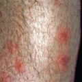
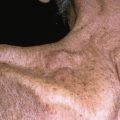
Cholinergic urticaria is relatively common and is caused by heat and sweating. It results in small (1- to 4-mm) papules on the trunk and proximal extremities ( Fig. 5.4 ). Patients often report that the condition is precipitated by warm showers, exercise, and emotional stress.
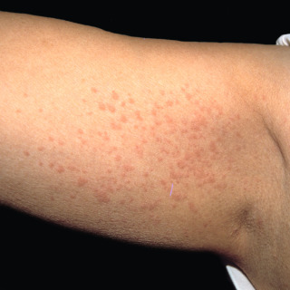
Fig. 5.4
Patient with small urticarial uniform papules characteristic of cholinergic urticaria.
(From the Fitzsimons Army Medical Center Collection, Aurora, CO.)
- –
Cold urticaria is an uncommon variant caused by cold exposure ( Fig. 5.5 ). In some cases, angioedema may be associated. In extreme circumstances, anaphylaxis may ensue. The condition is present in 1 in 2000 adults.
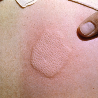
Fig. 5.5
Wheal produced by an ice cube in a patient with cold urticaria.
(From the Fitzsimons Army Medical Center Collection, Aurora, CO.)
- –
Dermatographism ( Fig. 5.6 ) is characterized by red linear indurated streaks with stroking of the skin.
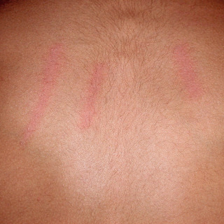
Fig. 5.6
Patient with dermatographism. Linear wheals of the back are created by stroking the skin with a pen.
- –
Solar urticaria occurs on photoexposed skin. It usually occurs within minutes to 1 hour of sun exposure, which differs from other photosensitive conditions, such as polymorphous light eruption, which is more delayed ( Fig. 5.7 ).

Fig. 5.7
Patient with solar urticaria on the back of the hand.
(From the Fitzsimons Army Medical Center Collection, Aurora, CO.)
- –
Diagnosis
- •
The diagnosis of cholinergic urticaria, the most common form of physical urticaria, requires clinical correlation about precipitating factors with the small size of the lesions. In addition to pruritus, cholinergic urticaria typically produces a subtle stinging sensation.
- •
Cold urticaria is diagnosed by the ice cube test, in which an ice cube is applied to the skin for 2 to 5 minutes, with re-examination at 10-minute intervals ( Fig. 5.5 ). Test sensitivity is about 85%, but the specificity is nearly 100%.
- •
Dermatographism is easily diagnosed by wheals produced by firmly stroking the skin with an object such as the blunt end of a pen (see Fig. 5.6 ).
Treatment
- •
Cold urticaria is treated with avoidance of exposure. Remember that angioedema and anaphylaxis can occur and can even be fatal.
- •
Cholinergic urticaria is treated with avoidance of activities that cause undue overheating.
- •
All forms of physical urticaria may be treated with antihistamines, as discussed for simple urticaria.
 Angioedema
ICD10 code T78.3
GENETIC AND EXTERNAL ETIOLOGIES
Hereditary
Acquired
- •
Acquired C1 esterase inhibitor (C1-INH) deficiency
- •
Allergic
- •
Drug-induced
- •
Angiotensin-converting enzyme (ACE) inhibitors
- •
Penicillin
- •
Nonsteroidal antiinflammatory drugs (NSAIDs)
- •
- •
Malignancy
Pathogenesis
Angioedema is characterized by deeply situated edema of the lower dermis and/or subcutaneous tissue (including the submucosa). Although some patients may present with both urticaria and angioedema, in many cases angioedema occurs in isolation.
Clinical Features
- •
Sudden localized swelling of skin and/or mucosal surfaces is seen, usually without a wheal and flare reaction ( Figs. 5.8 and 5.9 ).
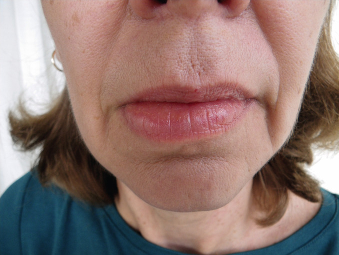
Fig. 5.8
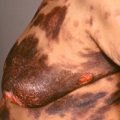
Patient with mild acquired angioedema of the lips that was unassociated with urticaria. The cause was never identified.
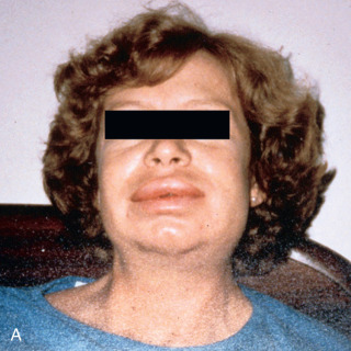
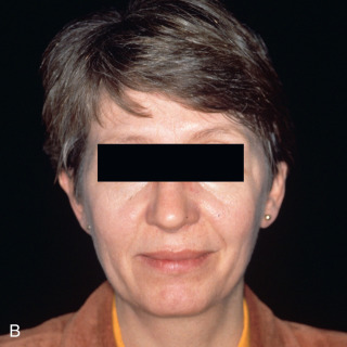
Fig. 5.9
A, Hereditary angioedema in a patient during an acute attack. The photograph was taken by the patient at home. B, Same patient as seen between attacks.
(From the Fitzsimons Army Medical Center Collection, Aurora, CO.)
- •
Typically, angioedema resolves in 1 to 3 days, without permanent sequelae.
- •
Involvement of the respiratory tract can be fatal, but this is more common in hereditary angioedema (see below).
- •
Abdominal pain due to edema of the bowel wall can be severe, but this is also more common in hereditary angioedema (see below).
Diagnosis
- •
The presence of coexisting urticaria is an important finding. Nearly all patients with hereditary angioedema have the condition in isolation. Patients with acquired disease often have both angioedema and urticaria at the same time.
- •
About 80% of patients with hereditary angioedema report similar attacks in family members.
- •
The serum C4 level is a sensitive but nonspecific screening tool. C4 levels are low in acquired angioedema and in hereditary forms of angioedema caused by C1-INH deficiency.
- •
Therefore, if a low level of C4 is detected, quantitative (type I) and functional (type II) C1-INH studies should be commissioned to refine the diagnosis further.
- •
Mast cell tryptase levels are usually normal in hereditary angioedema but may be elevated in acquired forms.
Treatment
- •
Intubation or tracheostomy may be necessary in patients with severe laryngeal edema and respiratory distress.
- •
Severe attacks need to be observed in the emergency room or hospital.
- •
Ecallantide is a subcutaneously administered recombinant plasma kallikrein inhibitor. However, it must be administered in a controlled environment because anaphylactic reactions to the drug have been reported.
- •
Icatibant is a bradykinin B2 receptor antagonist that is given subcutaneously and may even be self-administered by the patient during an acute attack.
- •
Danazol was used as a prophylactic treatment in the past, but it has now largely been replaced by more effective and safer treatments.
Related posts:
Stay updated, free articles. Join our Telegram channel

Full access? Get Clinical Tree


