Key Terms
Erythema Induratum
Nodular vasculitis
Lupus Panniculitis
Lupus erythematosus panniculitis
Lupus profundus
Subcutaneous Granuloma Annulare
Deep granuloma annulare
Pseudorheumatoid nodules
Rheumatoid Nodules
Rheumatoid nodulosis syndrome
Subcutaneous diseases include those conditions that are classically considered panniculitis (e.g., erythema nodosum, erythema induratum), in addition to diseases that may involve the subcutaneous fat (e.g., rheumatoid nodules, deep granuloma annulare), and even vasculitis, which may affect the vessels of the subcutaneous fat and resemble a panniculitis. It is important to note that later in the course of the disease, some forms of panniculitis may involve the dermis and/or epidermis, resulting in perforation or ulceration.
Important History Questions
How long have the lesions been present?
Some forms of panniculitis are usually acute and do not last long (e.g., pancreatic panniculitis), whereas others (e.g., lupus panniculitis) tend to be chronic.
Are the lesions painful, and, if so, how painful are they?
Some types of panniculitis are exquisitely painful (e.g., pancreatic panniculitis), whereas others are only tender (e.g., erythema nodosum) and some are typically not painful (e.g., subcutaneous granuloma annulare).
What medications do you take?
This is a very important question, because some forms of panniculitis can be drug induced (e.g., erythema nodosum, pancreatic panniculitis), which may mean that the condition is potentially curable.
Do you have any other medical conditions?
Tuberculosis is of particular interest; this specifically because it is associated with erythema induratum and erythema nodosum. Determine if there is a known history of hepatitis B infection, which is associated with polyarteritis nodosa; pancreatitis, which could indicate pancreatic fat necrosis; and sarcoidosis, which can present as subcutaneous nodules and plaques. The patient should also be asked about any known history of a connective tissue disorder, such as lupus erythematosus.
Have you ever had a positive tuberculosis skin test?
This is a most important evaluation of the patient with possible erythema induratum and, to a lesser extent, erythema nodosum.
How much alcohol do you consume in a week?
This question obviously needs to be used only when pancreatic fat necrosis is a clinical consideration. It needs to be stated with tact.
Important Physical Findings
What is the distribution of the lesions?
Some types of panniculitis are almost always located on the legs (e.g., erythema nodosum, erythema induratum), whereas others are frequently located in other areas. For example, subcutaneous granuloma annulare is not infrequently found on the head in infants and young children.
Are the lesions ulcerated, or is there any evidence of epidermal change?
Some forms of panniculitis are never ulcerated (e.g., erythema nodosum, subcutaneous fat necrosis of the newborn), whereas others, such as erythema induratum and pancreatic fat necrosis, are frequently ulcerated. Rheumatoid nodules can also demonstrate perforation of necrotic collagen.
Is there any physical evidence of active arthritis?
Rheumatoid nodules and pancreatic fat necrosis can both be associated with arthritis. It is not uncommon for erythema nodosum to be associated with arthralgias.
Are there any other types of skin lesions?
This is important, because some diseases, such as subcutaneous granuloma annulare (e.g., annular lesions) or sarcoidosis, may have different types of lesions present on the body, which is diagnostic.
Erythema Nodosum
ICD10 code L52
INTERNAL AND EXTERNAL ETIOLOGIES
Pathogenesis
The pathogenesis of erythema nodosum is poorly understood. Erythema nodosum, like urticaria, is not a primary disease but rather represents a reaction pattern to a variety of different antigenic insults, with the most common underlying factors being β-hemolytic streptococcal infection, drug therapy (particularly birth control pills), which includes herbal therapy (e.g., Echinacea), sarcoidosis, ulcerative colitis, tuberculosis, viral infections, Crohn disease, deep fungal infections, Behçet syndrome, pregnancy, and underlying malignancy. Patients with β-hemolytic streptococcal infection typically develop erythema nodosum 2 to 3 weeks after the infection, which is usually associated with an elevated antistreptolysin O (ASO) titer.
- •
Azathioprine
- •
Celecoxib
- •
Echinacea herbal therapy
- •
Gold
- •
Oral contraceptives
- •
Sulfonamides
Clinical Features
- •
This is the most common form of panniculitis; it may occur at any age, including children, but the peak incidence is in patients between 20 and 30 years of age.
- •
It is three to six times more common in women than men.
- •
Patients may demonstrate a prodrome of fever, malaise, and arthralgias.
- •
Primary lesions are tender, erythematous subcutaneous nodules of variable size (1–15 cm); they are never sharply demarcated.
- •
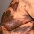
It is predominantly acral, with the classic location being the anterior shins ( Fig. 15.1 ).
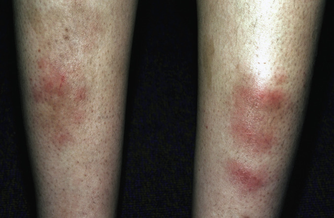
Fig. 15.1
Typical erythema nodosum of the anterior shins in a young woman. Note the discoloration of some of the lesions on the right leg.
(From the Fitzsimons Army Medical Center Collection, Aurora, CO.)
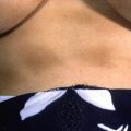
- •
Less common locations include other parts of the leg ( Fig. 15.2 ) and arms ( Fig. 15.3 ).
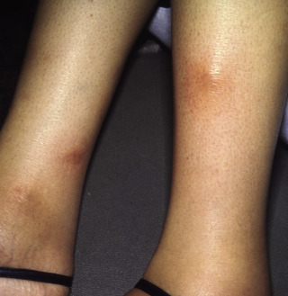
Fig. 15.2
Erythema nodosum on the lateral legs in a patient.
(From the Fitzsimons Army Medical Center Collection, Aurora, CO.)
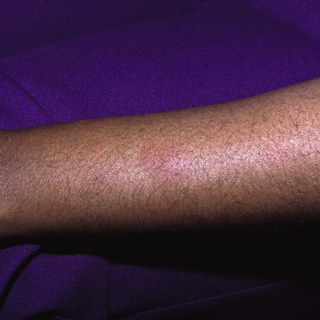
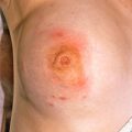
Fig. 15.3
Unusual location for erythema nodosum on the arm of a patient.
(From the Fitzsimons Army Medical Center Collection, Aurora, CO.)
- •
There are typically less than 10 lesions; however, more than 50 lesions are seen in rare patients.
- •
Ulceration does not occur.
Diagnosis
- •
Clinical presentation is usually characteristic, although the presentation may be mimicked by other forms of panniculitis, such as erythema induratum, or deep vasculitis, such as periarteritis nodosa.
- •
Routine laboratory tests that should be done include a throat culture to rule out active streptococcal infection and an ASO titer to rule out recent streptococcal infection.
- •
In select cases, consider testing stool for Yersinia infection and/or chest x-ray to rule out sarcoidosis or an underlying deep fungal infection.
- •
The histologic findings on biopsy—a 5-mm or larger punch biopsy or incisional biopsy that includes fat—is typically needed and is usually diagnostic.
Treatment
- •
No treatment is needed in mild cases but is always an option, because most cases are self-limited.
- •
Using nonsteroidal antiinflammatory drugs (NSAIDs) for pain relief and reduction of inflammation is recommended as initial treatment in more severe cases.
- •
Very painful or severe cases quickly respond to prednisone, 10 to 40 mg per day for 3 to 7 days. This therapy should be used with caution if an underlying infection is suspected.
- •
Oral potassium iodide (SSKI) is an alternate therapy, starting at two drops tid and increasing up to six drops tid.
Clinical Course
Individual lesions typically last 3 to 6 weeks, although new lesions may continue to develop. Rare patients may develop chronic erythema nodosum that is more likely to be unilateral.
Erythema Induratum
ICD10 code A18.4
INTERNAL ETIOLOGY
Pathogenesis
The term erythema induratum (of Bazin) is often used interchangeably with the term nodular vasculitis, although some dermatologists reserve the former for cases that are tuberculosis-associated and the latter term for those that are not. The pathogenesis is not entirely understood, although in cases associated with tuberculosis, polymerase chain reaction (PCR) studies have demonstrated Mycobacterium tuberculosis DNA in more than 75% of cases, suggesting that it is a hypersensitivity reaction. Less common associations have included Crohn disease, other infections, and, very rarely, medications (e.g., propylthiouracil).
Clinical Features
- •
It usually affects young and middle-aged women, although any age or gender can be affected.
- •
It is more common in clinical populations with a strong exposure to tuberculosis.
- •
It is usually located on the calves and shins, although the trunk, upper extremities, and even the face can be affected.
- •
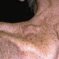
The primary lesion is comprised of one or more painful, erythematous, subcutaneous nodules ( Fig. 15.4 ).
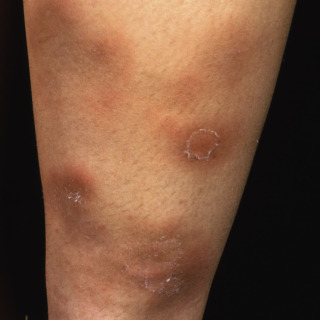
Fig. 15.4
Classic location of erythema induratum on the posterior aspect of the calf of a patient.
(From the Fitzsimons Army Medical Center Collection, Aurora, CO.)
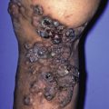
- •
Ulceration is frequently present in one or more lesions ( Figs. 15.5 and 15.6 ), a finding that is not found in erythema nodosum.
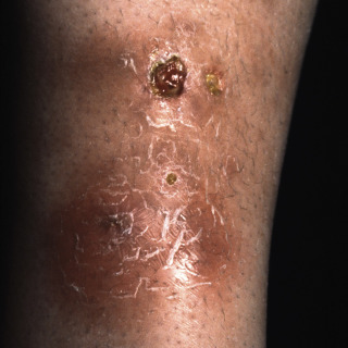
Fig. 15.5
Patient with indurated subcutaneous plaques of erythema induratum, with early ulceration.
(From the Fitzsimons Army Medical Center Collection, Aurora, CO.)
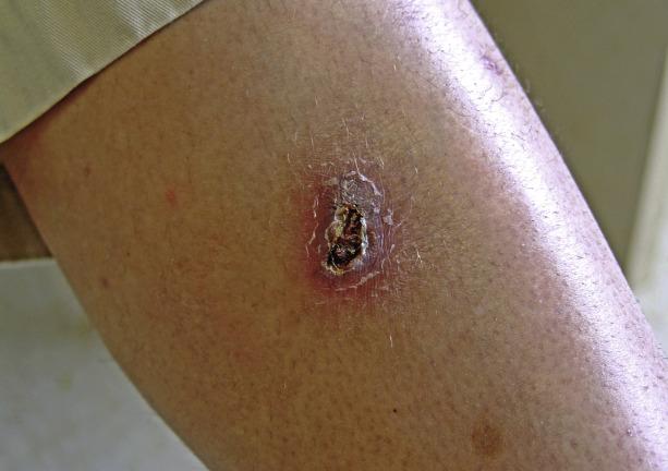
Fig. 15.6
Patient with ulcerated erythema induratum of the thigh that was secondary to undiagnosed tuberculosis.
Diagnosis
- •
In regard to the clinical presentation, be particularly suspicious in a patient with a known history of tuberculosis or exposure to individuals with active tuberculosis. The differential diagnosis includes erythema nodosum, lupus panniculitis, polyarteritis nodosa, and other rare forms of panniculitis.
- •
The diagnosis is typically established by a 5- to 8-mm punch biopsy or incisional biopsy. It is critical that the biopsy includes adequate subcutaneous fat. The histologic findings may be strongly suggestive or diagnostic. Cases associated with tuberculosis are culture negative and do not demonstrate organisms with special stains; they can only be demonstrated by PCR assay, which is not routinely available.
- •
Chest x-rays and a purified protein derivative (PPD) skin test are strongly recommended in all cases to exclude evidence of tuberculosis. If tuberculosis is strongly suspected, consider diluting the PPD to 1 : 10, because patients may demonstrate very exuberant reactions.
Treatment
- •
The treatment of choice is to identify any underlying cause (e.g., tuberculosis) and treat that disorder or withdraw any potentially offending drug.
- •
Oral potassium iodide (SSKI) is an alternate therapy, starting at two drops tid and increasing up to six drops tid.
- •
NSAIDs may be used to reduce pain and inflammation.
- •
Use oral prednisone, starting at a dose of 10 to 40 mg/day, that is tapered as quickly as possible as the patient responds to therapy. Prednisone is not recommended until active tuberculosis has been excluded.
Clinical Course
Untreated lesions tend to persist for months or even years and, when the lesions resolve, they may heal with atrophy and variable scarring.
Related posts:

Stay updated, free articles. Join our Telegram channel

Full access? Get Clinical Tree


