Key Terms
Seborrheic Dermatitis
Cradle cap
Dandruff
Sebopsoriasis
Seborrhea petaloides
Dermatitis, a term interchangeable with the term eczema , represents the most common cause of skin-related visits to health care providers in the United States. Histologically, dermatitis is characterized by variable epidermal edema (spongiosis), with inflammatory cells infiltrating the epidermis and dermis. Clinically, dermatitis presents with variable erythema and edema; it can appear to weep or even vesiculate. In many cases, a specific diagnosis cannot be made without clinical data, so the following questions are important to ask of all persons with possible dermatitis.
Important History Questions
How long has the dermatitis (rash) been present?
Attempt to ascertain if the condition is acute or chronic. Atopic dermatitis, nummular dermatitis, and seborrheic dermatitis are often chronic conditions, albeit with waxing and waning moments. Allergic contact dermatitis, id reactions (autoeczematous eruptions), and eczematous drug eruptions are more likely to be acute in nature.
Have you had a similar rash in the past?
An affirmative answer favors repeated exposures to an allergen in allergic contact dermatitis, an exacerbation of a chronic dermatitis (e.g., atopic dermatitis), or repeated exposure to a drug in a drug-induced eczematous process.
Where did the rash start?
Ascertaining the point of initial involvement may provide an important clue in allergic contact dermatitis (ACD) or irritant contact dermatitis (ICD). Be suspicious of a low-grade allergic contact dermatitis when the condition begins on the thin skin of the eyelids.
Do any of your immediate relatives have eczema, asthma, or hay fever?
When multiple persons in a family have these conditions, an atopic diathesis is suggested. It is important to remember that many laypersons refer to atopic dermatitis as childhood eczema.
Do you have allergies or sensitivity to things that come into contact with your skin?
An affirmative response should prompt additional questioning about a specific allergen and allergic contact dermatitis, or it may again suggest an atopic diathesis; persons with atopy often describe their skin as being sensitive.
What sort of work do you do?
Occupational exposure to an irritant or allergen is an important question. For example, a 2001 study of 1200 health care workers identified an irritant or allergic hand dermatitis in more than one-third of participants.
Have you started any new medications in the past month?
Although eczematous drug eruptions represent just 1% to 4% of drug eruptions, the condition with discontinuation of the offending drug.
How are you treating the rash?
Many patients use home remedies, over-the-counter medications, or borrowed or inappropriate prescription medications that can worsen (or improve) dermatitis.
What else do you put on your skin, and what type of soap do you use?
Many patients use lye-based soaps that are irritants or heavily fragranced soaps that can cause an allergic dermatitis. Dermatitis will improve if synthetic soaps are used instead of harsh, lye-based soaps.
Important Physical Findings
What is the distribution of the dermatitis?
Some types of dermatitis have characteristic patterns of involvement (e.g., flexural involvement in atopic dermatitis, involvement of the scalp, eyebrow, eyelid, and nasolabial folds in seborrheic dermatitis).
Is there a distinct pattern to the dermatitis?
Allergic contact dermatitis is initally sharply circumscribed and assumes linear configurations that begins after contact with the allergen. A sharply circumscribed edge, with central clearing, forming an annulus, should raise concern for a dermatophyte infection rather than a dermatitis.
Allergic Contact Dermatitis
ICD10 code L23.0 to L23.9
EXTERNAL ETIOLOGY
Pathogenesis
Allergic contact dermatitis (ACD) is a delayed type hypersensitivity process (type IV reaction) that occurs when a low-molecular-weight antigen (hapten) is processed by antigen-presenting cells of the skin. These antigen-presenting cells then travel to the lymph nodes and promote expansion of clonal effector T-cells. The T-cells traverse back to the skin to produce inflammation. The memory T-cells serve to facilitate a quicker immune response upon rechallenge. This process of initial sensitization requires 5 to 21 days to foment, but, upon rechallenge, the elicitation of a second response can occur in 24 to 72 hours.
Clinical Features
- •
ACD occurs about 5 to 21 days after exposure if the patient is not already sensitized to the antigen or 1 to 3 days after re-exposure if the patient is already sensitized.
- •
Acute ACD is intensely pruritic and causes erythema and variable edema. Allergic dermatitis is the most likely of all forms of dermatitis to present with blisters (see Chapter 11 ).
- •
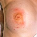
ACD often demonstrates peculiar configurations, reflecting the points of contact; hence, it may produce linear ( Figs. 10.1 and 10.2 ) or round dermatitis ( Figs. 10.3 and 10.4 ).
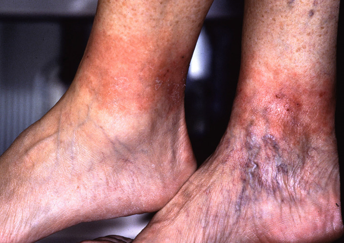
Fig. 10.1
Patient with allergic contact dermatitis to elastin in socks.
(From the Fitzsimons Army Medical Center Collection, Aurora, CO.)
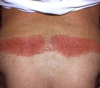
Fig. 10.2
Patient with allergic contact dermatitis to elastin in waistband of underwear.
(From the Fitzsimons Army Medical Center Collection, Aurora, CO.)
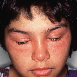
Fig. 10.3
Patient with eyelid dermatitis demonstrating a line of demarcation to the erythema.
(From the Fitzsimons Army Medical Center Collection, Aurora, CO.)
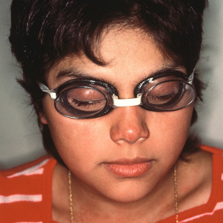
Fig. 10.4
Area of eyelid dermatitis corresponds to goggles worn during swimming. This patient was allergic to one of the rubber accelerators.
(From the Fitzsimons Army Medical Center Collection, Aurora, CO.)
- •
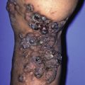
In severe ACD the dermatitis can spread past the point of contact ( Fig. 10.5 ).
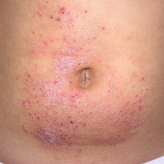
Fig. 10.5
Patient with allergic contact dermatitis due to the nickel found in the metal snap of the jeans. More than 14% of all adults are allergic to nickel. Note that the dermatitis has spread past the point of contact.
- •
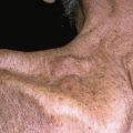
ACD often presents on the thin skin of the eyelids ( Fig. 10.6 ), neck ( Fig. 10.7 ), and web spaces between the fingers.
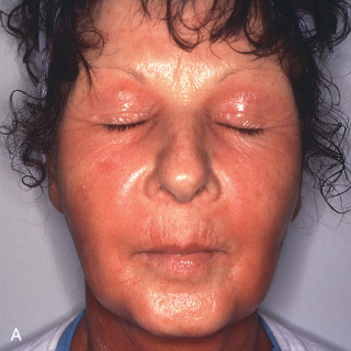
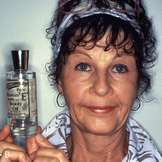
Fig. 10.6
A, Patient with acute onset of markedly pruritic dermatitis. B, The culprit turned out be topical vitamin E. Despite consumer perception, so-called natural products may produce allergic reactions.
(From the Fitzsimons Army Medical Center Collection, Aurora, CO.)
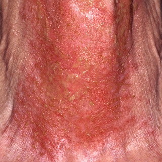
Fig. 10.7
Patient with oozing dermatitis of the thin skin of the anterior neck due to topical neomycin, one of the most common causes of allergic contact dermatitis.
(From the Joanna Burch Collection, Aurora, CO.)
Diagnosis
- •
Elicitation of a detailed exposure history is critical in ACD. The exposure period of interest is usually about 1 to 3 days but can be as long as 3 weeks with an initial episode.
- •
A skin biopsy is not diagnostic but may exclude other diseases that can mimic ACD.
- •
Patients with exposure to multiple potential allergens, such as a woman with facial dermatitis who uses facial soaps, creams, and other cosmetics, may require all topical preparations be discontinued. One single agent is then added back each week to identify the agent that produced the ACD.
- •
Some cases may require patch testing by a dermatologist to identify the offending agent or ingredients.
Treatment
For ACD caused by poison ivy, it is often useful to prescribe 78 5-mg tablets of prednisone. Then ask the patient to take 12 tablets the first day, 11 the second day, 10 the third day, and so on, until all the tablets have been taken.
- •
Mild cases of ACD may respond simply to withdrawal but can also be treated with a mild steroid (e.g., 1% hydrocortisone) or moderate steroid (e.g., 0.1% triamcinolone), applied bid.
- •
Ointments are a useful choice for ACD because these agents do not contain preservatives that could be involved in the process. Hydrocortisone, 1% ointment, is useful for eyelid dermatitis.
- •
Severe cases of ACD, especially those on thicker skin, may require potent topical corticosteroids applied bid (e.g., fluocinonide, clobetasol).
- •
Oral antihistamines (e.g., cetirizine, hydroxyzine) are used for relief of pruritus and provide sedation at night.
- •
Severe or generalized cases may require oral prednisone (40–100 mg PO qd) for 5 to 7 days. Some haptens, such as those in poison ivy, bind irreversibly to the skin, and new lesions may occur for up to 3 weeks. Prednisone may need to be continued for up to 3 weeks in these cases (see “Rule of 78” inset).
- •
Skin barrier creams (e.g., Stokogard, Hollister Moisture Barrier, Hydropel, Ivy Shield) are partially effective in preventing ACD to poison ivy.
As demonstrated by the two patients shown in Figs. 10.1 and 10.2 , ACD to elastic in clothing can occur. In some cases, it is due to compounds called rubber accelerators that are used during production of the elastic. In other cases, it is due to chemical changes from bleach. In patients with this type of reaction, switching to new clothing and not using bleach avoids the problem.
- •
Plant allergens—57% of all adults in the United States are allergic to rhus antigen, which is found in poison ivy, poison oak, and poison sumac. It is also found in a variety of related plants (e.g., mango, cashew nut trees)
- •
Nickel—14% of US adults are allergic to nickel. They can become allergic to other metals, but this is uncommon. It is worth noting that the incidence of ACD to gold has been increasing.
- •
Formaldehyde and formaldehyde releasers (quaternium-15)
- •
Neomycin—9% of US adults
- •
Rubber (thiuram, carba mix)
- •
Fragrances—8% of US adults
Figs. 10.3 and 10.4 demonstrate an important physical finding—namely, involvement of the eyelids. Because the eyelid skin is so thin, it is frequently involved in ACD. It is not uncommon for low-grade ACD to be confined to the eyelids. In a large study of 203 patients with eyelid dermatitis, ACD was the cause in 74% of cases. The remaining patients usually had atopic dermatitis, seborrheic dermatitis, psoriasis, dry eyes, dermatomyositis, and irritant contact dermatitis.
Guin JD: Eyelid dermatitis: experience in 203 patients, J Am Acad Dermatol 47:755-765, 2002.
Irritant Contact Dermatitis
ICD10 code L23.0 to L23.9
EXTERNAL ETIOLOGY
Pathogenesis
In contrast to ACD, irritant contact dermatitis (ICD) is not mediated by an immunologic cascade. Instead, ICD is produced by direct toxic injury to the skin. ICD is affected by the nature of the toxic substance, the degree and duration of exposure, and individual skin susceptibility. Some skin sites may be more susceptible, elderly skin is more susceptible, and repeated water exposure damages the barrier function of the skin, making it more susceptible to irritants. Most ICD is caused by soap (e.g., bath soap, dishwashing soap), cleaning products (e.g., window cleaner, bathtub cleaner), alcohols in medications and cosmetics, and glues, cements, and deodorants. ICD can also be caused by minor physical trauma such as exposure to fiberglass, sand, cement, and rough paper. ICD is four- to five-fold more common than ACD.
Clinical Features
- •
Strong irritants, such as strong acids, produce an immediate burning or stinging with exposure, followed by erythema, edema, and even blisters or ulcerations. Strong bases (e.g. lye) may not produce the same sensory changes but can be just as damaging to the skin.
- •
Mild irritants, such as soaps, may produce dermatitis through cumulative insult over days or weeks.
- •
In contrast to ACD, irritant dermatitis is limited to exposed skin, and new lesions do not continue to develop after exposure is terminated.
Diagnosis
- •
Acute ICD may be self-evident because the patient may observe an immediate and direct relationship to an exposure ( Fig. 10.8 ). Acute ICD follows points of contact with the skin ( Figs. 10.9 and 10.10 ).
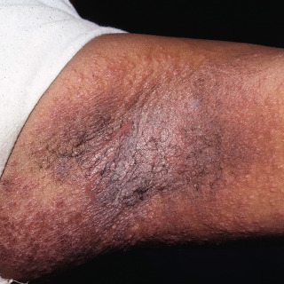
Fig. 10.8
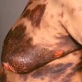
Patient with acute, severe, irritant dermatitis of the axillae due to a topical epilating agent.
(From the Fitzsimons Army Medical Center Collection, Aurora, CO.)
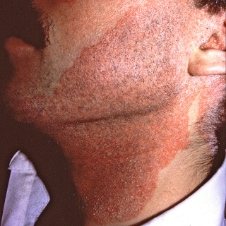
Fig. 10.9
Patient with acute severe irritant dermatitis of the face due to topical epilating agent.
(From the Fitzsimons Army Medical Center Collection, Aurora, CO.)
Fig. 10.10
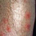
Patient with papular irritant dermatitis caused by exposure to fiberglass that followed a weekend of insulating an attic.
(From the Fitzsimons Army Medical Center Collection, Aurora, CO.)
- •
Low-grade and chronic ICD can be difficult to distinguish from other causes of dermatitis; the diagnosis may be established only with a careful exposure history and physical examination ( Fig. 10.11 ).
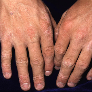
Fig. 10.11
Chronic, low-grade irritant dermatitis due to repeated exposure to soaps in a patient who worked as a car washer.
(From the Fitzsimons Army Medical Center Collection, Aurora, CO.)
- •
In general, biopsy of ICD is not helpful because only nonspecific features of spongiotic dermatitis are observed, but it may be useful in excluding other forms of dermatitis.
- •
Patch testing may be used to exclude ACD. Provocative use testing can be used, with application of the suspected irritant to a specific area of skin, only if the suspected substance is not excessively harmful. Do not use such testing with strong acids or strong bases.
- •
In many cases, the diagnosis of low-grade ICD depends on observed improvement with discontinuation of the suspected irritant.
Treatment
- •
The cornerstone of treatment for ICD is withdrawal of the offending agent, with protection from all irritants (e.g., gloves for irritant hand dermatitis). Because the effects of irritants are additive, all irritants should be avoided.
- •
The most common irritants are bath soaps, so instruction on proper bathing is important. Dove Sensitive Skin Body Wash, Olay Sensitive Body Wash, Aveeno Daily Moisturizing Body Wash, and Cetaphil skin-cleansing products are excellent choices when ICD is suspected.
- •
Severe irritant dermatitis (e.g., cantharone blister) is essentially a second-degree burn, and topical treatments are not helpful.
- •
Moisturizers should be recommended in cases of chronic irritant dermatitis, particularly those with ammonium lactate or sodium lactate, because these agents make the skin less susceptible to irritants. It may take 4 to 6 weeks of continued use to realize the effects of lactate-containing moisturizers.
- •
Mild to moderate potency topical corticosteroids may be of some benefit in chronic ICD.
- •
Patients should be cautioned that once the skin is irritated, normal barrier function may not return for 4 weeks and, during this time, the skin is more susceptible to all irritants.
Atopic Dermatitis (Childhood Eczema)
ICD10 code L20
GENETIC DISORDER WITH EXTERNAL EXACERBANTS
Pathogenesis
The precise incidence of atopy is unknown, but current evidence suggests that 9% to 20% of all individuals in the United States have an atopic diathesis. Atopy is inherited, and 70% of persons with atopic dermatitis (AD) have a family history of asthma, allergic rhinitis, and/or AD. Evidence suggests that an atopic diathesis is determined by the varied expression of 20 or more genes and is also affected by various environmental stimuli. The immunologic aberrations of atopy are poorly understood, but patients with AD manifest an increased release of histamine from mast cells and basophils, blood and tissue eosinophilia, and exaggerated immunoglobulin E (IgE) response mechanisms. Persons with atopy can be exquisitely sensitive to pruritic stimuli and have depressed cell-mediated immunity. Recent studies have linked some cases of AD to mutations in filaggrin, a protein important in the barrier function of the stratum corneum of the epidermis.
Clinical Features
- •
Most cases of AD present in childhood—60% present in the first year of life, and 90% present by the age of 5 years.
- •
During the infantile phase (2 months–2 years), patients demonstrate marked pruritus, excoriations, and diffuse dermatitis that usually involves the head, portions of the trunk, and diaper area ( Figs. 10.12–10.14 ). Half of these patients will clear by the age of 3 years.
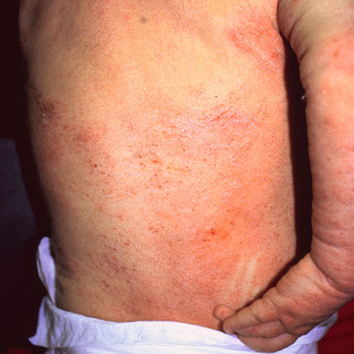
Fig. 10.12
Infant with atopic dermatitis demonstrating eczematoid dermatitis. Note the active excoriation and white dermatographism.
(From the Fitzsimons Army Medical Center Collection, Aurora, CO.)
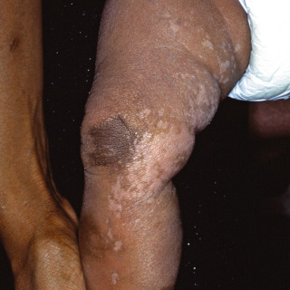
Fig. 10.13
Infant with chronic atopic dermatitis. Note the presence of hyperpigmented and hypopigmented areas.
(From the Fitzsimons Army Medical Center Collection, Aurora, CO.)

Fig. 10.14
Infant with atopic dermatitis demonstrating extensive scale, a common finding in atopic dermatitis.
(From the Fitzsimons Army Medical Center Collection, Aurora, CO.)
- •
During the childhood phase (3–11 years), lichenified plaques are common on the wrists, ankles, buttocks, and antecubital and popliteal fossae.
- •
During the adolescent phase (12–20 years), lichenified plaques are common on the face, neck, upper arms, back, and flexures ( Figs. 10.15 and 10.16 ).
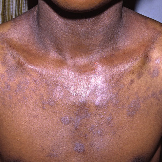
Fig. 10.15
Adolescent with atopic dermatitis of the flexural area of the neck. Some of the lesions demonstrate round configurations and resemble nummular dermatitis.
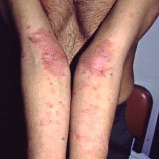
Fig. 10.16
Classic atopic dermatitis in an adolescent involving both antecubital fossae.
(From the Fitzsimons Army Medical Center Collection, Aurora, CO.)
- •
AD persists in adulthood in only 10% of patients, but, when this occurs, the disease presents in much the same way as in the adolescent phase ( Figs. 10.17–10.19 ).
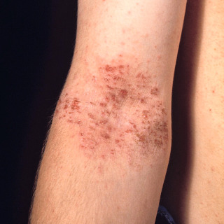
Fig. 10.17
Man with flexural atopic dermatitis demonstrating excoriations and yellow crust due to secondary staphylococcal infection.
(From the Fitzsimons Army Medical Center Collection, Aurora, CO.)

Fig. 10.18
Flexural atopic dermatitis in an adult demonstrating secondary linear excoriation.
(From the Fitzsimons Army Medical Center Collection, Aurora, CO.)
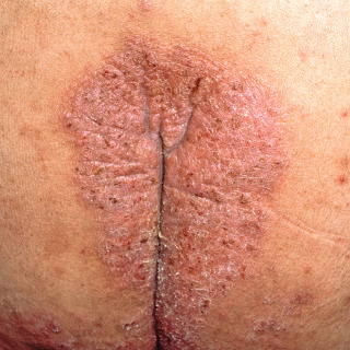
Fig. 10.19
Patient with atopic dermatitis of the sacral area demonstrating thick skin, with increased skin markings indicating secondary lichenification.
(From the Fitzsimons Army Medical Center Collection, Aurora, CO.)
- •
Adult patients with AD may experience only xerosis but are also more likely to develop chronic hand dermatitis.
- •
Other physical findings common in patients with AD include xerosis (see Chapter 9 ), keratosis pilaris, ichthyosis vulgaris, Dennie-Morgan lines (linear transverse folds below the lower eyelids), pityriasis alba (see Chapter 27 ), and transverse nasal creases (the so-called allergic salute).
- •
Patients with AD are more likely to harbor Staphylococcus aureus (~64% in one study) and are also susceptible to viral superinfection of inflamed skin (e.g., herpes simplex virus [HSV]—eczema herpeticum, vaccinia—eczema vaccinatum, or coxsackievirus A16—eczema coxsackium).
- •
In a large study of over 2500 children, AD was most often exacerbated by sweating from exercise, hot weather (possibly also because of sweating), and fabrics (especially wool).
Diagnosis
- •
A careful personal and family history and review of past skin disease are important.
- •
Unexplained pruritic dermatitis occurring in a child should prompt consideration of AD.
- •
AD cannot be diagnosed with any single laboratory test, but often patients with AD have an elevated serum IgE level and, possibly, peripheral eosinophilia. A raised total or allergen-specific IgE level was found in 74% of 1097 children with AD.
- •
A skin biopsy is usually nondiagnostic but may exclude other diseases that can mimic AD.
- •
A proposed diagnostic scheme for AD requires three of four major criteria to be present:
- 1.
Pruritus—some authorities believe that this is the principal problem (the itch that rashes). In one study, 52% of patients with AD reported pruritus, even without skin lesions, whereas only 6% of a matched control population reported pruritus.
- 2.
Typical morphology and distribution for age group (e.g., flexural areas)
- 3.
Chronic or chronically relapsing dermatitis
- 4.
Personal or family history of atopy (e.g., allergic rhinitis, asthma, AD)
- 1.
Treatment
- •
Removal of cutaneous irritants (soaps, wool) improves AD. Dove Sensitive Skin Body Wash, Olay Sensitive Body Wash, Aveeno Daily Moisturizing Body Wash, and Cetaphil skin-cleansing products are excellent choices for persons with AD. Fragrance-free laundry detergents and fabric softeners can be helpful but generally are not as important as gentle skin bathing choices.
- •
Food elimination diets are controversial. One study of 160 patients with AD has reported that 28% of individuals experienced exacerbations of AD when challenged with certain foods. The foods most often involved in exacerbations included milk, eggs, nuts, soy, wheat, and seafood. Note that food elimination diets need to proceed with extreme caution because there have been reports of such diets producing malnutrition in children (see discussion of kwashiorkor in Chapter 9 ). Consultation with a dietician is an option.
- •
Generous and liberal lubrication is critical. Ammonium lactate– or sodium lactate–containing moisturizers (e.g., AmLactin, Lac-Hydrin) are effective, if tolerated, but these agents may produce a burning sensation. Eucerine Smoothing Repair and Eucerin Intensive Repair, which also contain urea, are lotions that contain sodium lactate in weaker concentrations, and they are less likely to produce a burning sensation.
- •
Acutely inflamed or oozing skin may benefit from the application of open wet compresses. Some dermatologists use tap water, and others prefer a modified Burow solution (e.g., Domeboro).
- •
Topical corticosteroids are a mainstay of therapy for AD. The strength of the corticosteroid must to be tailored to the anatomic site involved and severity of disease. Mild disease may respond to 1% hydrocortisone cream, but more significant disease may require more potent topical corticosteroids, such as triamcinolone, fluocinonide, or even clobetasol for a short period. Severe AD may require corticosteroids under occlusion in conjunction with wet-dry wraps and/or with hospitalization.
- •
Oral antihistamines (e.g., cetirizine, hydroxyzine, diphenhydramine) are often used to decrease pruritus and provide sedation. At least two studies have demonstrated hydroxyzine to be effective in reducing pruritus in AD. One study has demonstrated that 0.7 mg/kg tid of hydroxyzine was as effective as 1.4 mg/kg tid in reducing the pruritus in children, with lesser sedation. Children or infants with severe AD not only keep themselves awake but drain the parent’s energy as well through familial sleep deprivation.
- •
Rare patients may require short-term oral corticosteroids for severe outbreaks. This should be avoided, if possible, due to the young ages of typical patients with AD and chronicity of the disease.
- •
Secondary infections, usually due to S. aureus, should be treated with appropriate oral antibiotics (e.g., cephalexin, dicloxacillin). In a study of 306 children with AD, 64% of patients were colonized with S. aureus . Clinical and experimental studies have supported the concept that topical steroids plus antistaphylococcal antibiotic therapy is more effective than topical steroids alone.
- •
Bleach baths may be effective in controlling staphylococcal infections and may even be antipruritic. However, the bleach bath must be dilute—
 cup of unscented household bleach in a full tub of water, approximately 1 tsp/gallon). Soaks should last 5 to 10 minutes. Explain to parents that the desired concentration is the same as that of pool water, and too much bleach can cause an irritant dermatitis.
- •
Topical tacrolimus and topical pimecrolimus can be used as alternatives for many patients, but both carry a black box warning with regard to use in patients younger than 2 years.
Related posts:

Stay updated, free articles. Join our Telegram channel

Full access? Get Clinical Tree


