Upper eyelid retraction
|
| History of thyroid-related orbitopathy |
| Prior facial surgery or trauma |
| Dry eye symptoms |
| Prior refractive surgery |
| Degree of dermatochalasis and fat prolapse |
| Presence of corneal exposure/lagophthalmos |
Introduction
Upper eyelid retraction repair is most commonly performed for thyroid-related orbitopathy (TRO). With upper eyelid retraction, there is often associated lagophthalmos and corneal exposure in addition to the startled appearance characteristic of this disfiguring disease. Upper eyelid retraction can also be seen after ptosis overcorrection as well as aggressive upper eyelid blepharoplasty and brow-lifting procedures.
When upper eyelid retraction is seen after ptosis repair, conservative measures such as downward eyelid massage should be performed, particularly early in the postoperative course. These maneuvers will frequently improve without surgical intervention. If the upper eyelid retraction is still present after 6 weeks, surgery can be considered.
The management of TRO-associated upper eyelid retraction is the final stage of surgical rehabilitation. Orbital decompression is performed first if indicated ( Chapter 64 ). Strabismus surgery is performed second for refractory diplopia followed by upper eyelid retraction repair. The surgical rehabilitation of the TRO patient should proceed after quiescence has been established unless vision-threatening corneal exposure or compressive optic neuropathy is present.
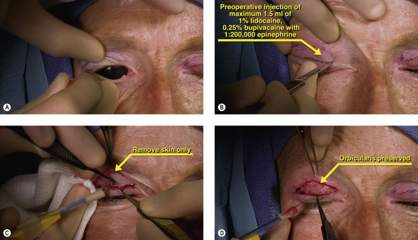
Several procedures have been described for repair of upper eyelid retraction associated with TRO, including full-thickness blepharotomy and posterior retractor recession techniques. Our preferred method is an anterior approach, single unit recession of the upper eyelid retractors (levator and Müller’s muscle). In this technique, the conjunctiva is kept intact and the amount of recession is titrated based on the degree of upper eyelid retraction. The surgery is performed conscious with minimal IV sedation so that an optimal eyelid height and contour can be achieved.
Surgical Technique
Related posts:


Stay updated, free articles. Join our Telegram channel

Full access? Get Clinical Tree


