Trunk Reconstruction and Pressure Sores


Chapter 11

Trunk Reconstruction and Pressure Sores


General Chest Wall Anatomy


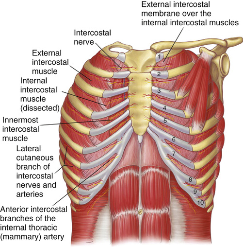
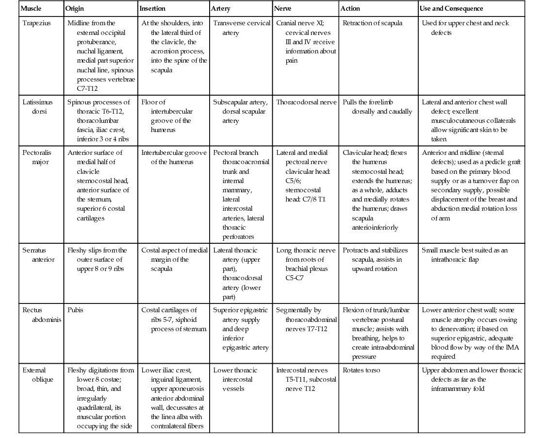
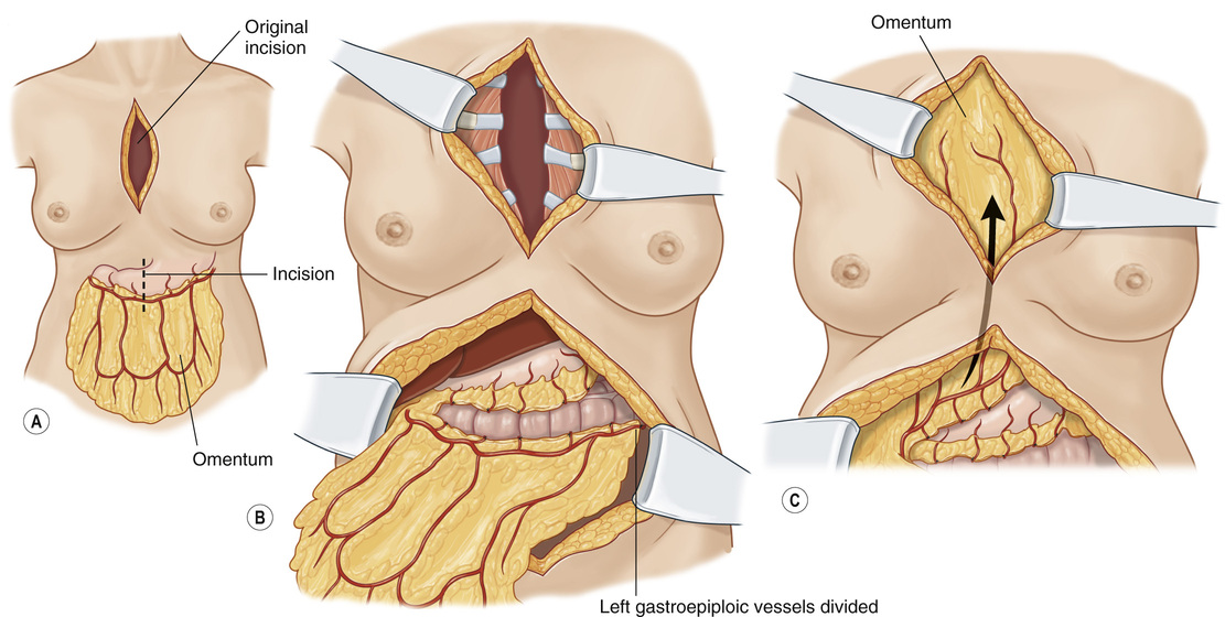
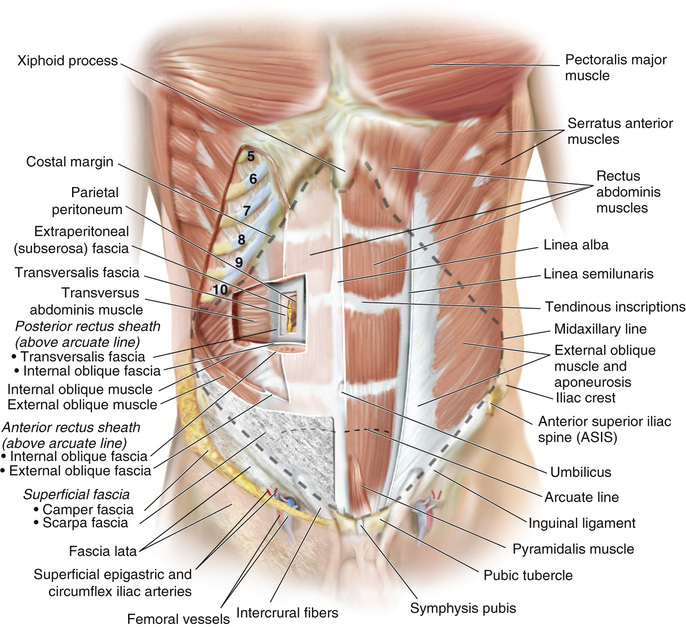
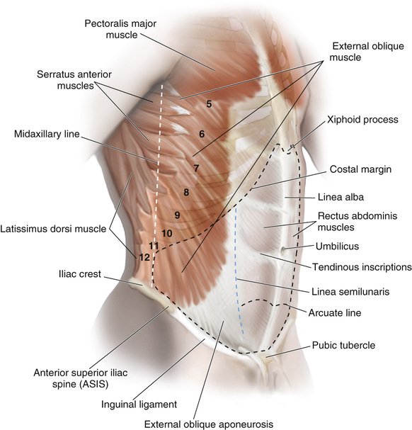
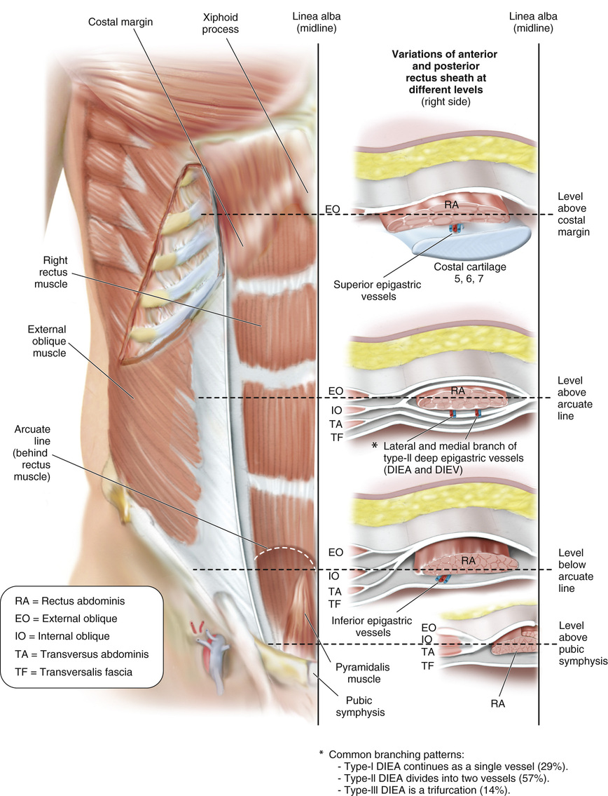
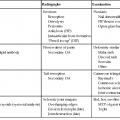
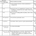
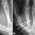
1. The chest wall is composed of a bony shell that protects critical structures (e.g., heart, lungs, liver, kidneys) with a muscular component that assists with respiration as well as stabilization and motion of the upper extremities (see Figure 11.1).


2. There are 12 paired ribs that make up the bony framework of the chest wall.


The first 7 ribs articulate posteriorly with the thoracic vertebrae and anteriorly with the sternum via a true sternocostal joint.


The last 5 ribs are often called “false ribs” because they articulate anteriorly with the 7th rib costal cartilage (ribs 8 to 10) or have no anterior articulation and are said to be “floating ribs” (ribs 11 and 12).


3. The sternum contains three bony parts.


The manubrium articulates with the first ribs and clavicles.


The body articulates with ribs 2 to 7.


The xiphoid process is insignificant.


4. Chest wall musculature


Intercostal muscles (external, internal, innermost m.) run between ribs, contain neurovascular bundles, and function to increase chest wall volume during inspiration.


The neurovascular bundles run between the internal intercostal muscles and the innermost intercostal muscles, just inferior to each rib (see Table 11.1).


Table 11.1


Extrathoracic Chest Wall Muscles


Muscle Origin Insertion Artery Nerve Action Use and Consequence
Trapezius Midline from the external occipital protuberance, nuchal ligament, medial part superior nuchal line, spinous processes vertebrae C7-T12 At the shoulders, into the lateral third of the clavicle, the acromion process, into the spine of the scapula Transverse cervical artery Cranial nerve XI; cervical nerves III and IV receive information about pain Retraction of scapula Used for upper chest and neck defects
Latissimus dorsi Spinous processes of thoracic T6-T12, thoracolumbar fascia, iliac crest, inferior 3 or 4 ribs Floor of intertubercular groove of the humerus Subscapular artery, dorsal scapular artery Thoracodorsal nerve Pulls the forelimb dorsally and caudally Lateral and anterior chest wall defect; excellent musculocutaneous collaterals allow significant skin to be taken
Pectoralis major Anterior surface of medial half of clavicle sternocostal head, anterior surface of the sternum, superior 6 costal cartilages Intertubercular groove of the humerus Pectoral branch thoracoacromial trunk and internal mammary, lateral intercostal arteries, lateral thoracic perforators Lateral and medial pectoral nerve clavicular head: C5/6; sternocostal head: C7/8 T1 Clavicular head; flexes the humerus sternocostal head; extends the humerus; as a whole, adducts and medially rotates the humerus; draws scapula anterioinferiorly Anterior and midline (sternal defects); used as a pedicle graft based on the primary blood supply or as a turnover flap on secondary supply, possible displacement of the breast and abduction medial rotation loss of arm
Serratus anterior Fleshy slips from the outer surface of upper 8 or 9 ribs Costal aspect of medial margin of the scapula Lateral thoracic artery (upper part), thoracodorsal artery (lower part) Long thoracic nerve from roots of brachial plexus C5-C7 Protracts and stabilizes scapula, assists in upward rotation Small muscle best suited as an intrathoracic flap
Rectus abdominis Pubis Costal cartilages of ribs 5-7, xiphoid process of sternum Superior epigastric artery supply and deep inferior epigastric artery Segmentally by thoracoabdominal nerves T7-T12 Flexion of trunk/lumbar vertebrae postural muscle; assists with breathing, helps to create intra-abdominal pressure Lower anterior chest wall; some muscle atrophy occurs owing to denervation; if based on superior epigastric, adequate blood flow by way of the IMA required
External oblique Fleshy digitations from lower 8 costae; broad, thin, and irregularly quadrilateral, its muscular portion occupying the side Lower iliac crest, inguinal ligament, upper aponeurosis anterior abdominal wall, decussates at the linea alba with contralateral fibers Lower thoracic intercostal vessels Intercostal nerves T5-T11, subcostal nerve T12 Rotates torso Upper abdomen and lower thoracic defects as far as the inframammary fold


image


IMA, Internal mammary artery.


Reprinted from Naidu, B.V., Rajesh, P.B., 2010. Relevant surgical anatomy of the chest wall. Thorac. Surg. Clin. 20, 453–463.


Chest Wall Reconstruction
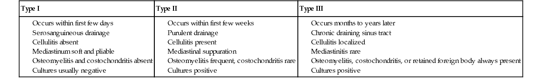


1. Chest wall defects are often the result of tumor resection, deep sternal wound infections, chronic empyemas, and/or trauma.


2. Chest wall defects following tumor extirpation often result in large segmental composite defects.


In general, stabilization of the chest wall, in addition to soft-tissue coverage, is required in defects >5 cm or more than 2 to 3 contiguous ribs.


Options for skeletal support include mesh (e.g., polypropylene, polytetrafluoroethylene [PTFE]), methylmethacrylate, and acellular dermal matrix.


Larger posterior rib defects may be tolerated because of the scapula.


3. Mediastinitis following sternotomy is reported to occur in 0.25% to 5% of cases.


Risk factors include diabetes, bilateral internal mammary artery (IMA) harvest, and obesity.


4. Pairolero and Arnold classified sternal wound infections into three types (see Table 11.2).


5. Reconstruction of sternal wounds largely depends on the type of infection, degree of purulence, and presence of bone and/or cartilage infection or necrosis.


For type-1 sternal wounds, if the wound is not purulent and the sternal bone is viable, sternal reconstruction with plates and/or wires, followed by flap coverage or closure, is possible.


In general, for type-2 and -3 sternal wound infections, staged reconstruction is required with serial washouts, debridement, and removal of hardware required to obtain a clean wound before flap reconstruction.


Vacuum-assisted closure (VAC) therapy is often used between serial debridement for assistance with wound control.


6. Common flaps for reconstruction of sternal wounds include


Pectoralis major


Vascular supply: Thoracoacromial artery (major), IMA perforators (minor, segmental)


Can be used as an advancement flap or turnover flap based off of the IMA perforators (requires intact IMA system)


Limited coverage of inferior sternal wounds


Muscle can be disinserted to allow for additional reach.


Rectus abdominis


Vascular supply: Superior and inferior epigastric a.


Useful for coverage of inferior sternal wounds


With ipsilateral IMA harvest sacrificing the superior epigastric artery, the flap can survive off of the 8th intercostal pedicle.


Latissimus dorsi


Vascular supply: Thoracodorsal a. (major), lumbar perforators (minor, segmental)


Useful for lateral chest wall defects


May require skin grafts


Omentum


Vascular supply: Gastroepiploic artery (major), short gastrics (minor)


Useful for large sternal wounds


Requires laparotomy and tunneling of flap through the diaphragm or extrathoracic


Requires skin graft (see Figure 11.2)


Abdominal Wall Anatomy


1. The abdominal wall is a hexagonal-shaped structure that functions to


Protect critical intra-abdominal structures


Stabilize the pelvis during walking, running, and jumping


Flex the vertebral column


Increase intra-abdominal pressure (e.g., to assist with expiration and defecation)


2. Vascular supply: Deep inferior epigastric a., superior epigastric a., superficial inferior epigastric a., intercostal and lumbar a.


3. Innervation: Nerves travel with the intercostal and lumbar arteries in the plane between the internal oblique (IO) and transversalis muscles.


Sensory: Anterior branches of the T7 to L1 intercostal and subcostal nerves


Motor: T7 to L2 intercostal nerves, iliohypogastric n., ilioinguinal n.


4. Abdominal wall musculature


Rectus abdominis


Origin: Pubic symphysis and pubic crest


Insertion: Anterior surfaces of 5th to 7th costal cartilages and xiphoid


Principle flexor of the abdomen, stabilizes pelvis, protects abdominal organs


External oblique


Vascular supply: Segmental intercostal a., deep circumflex iliac a., iliolumbar a.


Origin: Lower 8 ribs


Insertion: Pubic crest; forms the inguinal ligament


Contributes to the anterior rectus sheath through aponeurosis; most superficial of the lateral abdominal wall muscles


IO


Vascular supply: Intercostal a., circumflex iliac a., iliolumbar a.


Origin: Thoracolumbar fascia, iliac crest, inguinal ligament


Insertion: Inferior and posterior borders of ribs T10 to T12; pubic crest via the conjoint tendon


Deep to the external oblique; above arcuate line, the IO fascia splits to supply the anterior and posterior rectus sheath.


Transversus abdominis


Vascular supply: Intercostal and subcostal a.


Origin: Iliac crest, inguinal ligament, inner surface of T6 to T12 costal cartilages


Insertion: Posterior rectus sheath (above arcuate line); pubic crest via the conjoint tendon


5. The rectus sheath is located in the midline of the abdominal wall and is formed via the aponeuroses of the abdominal musculature. The components of the anterior and posterior rectus sheath change according to position with respect to the arcuate line (located midway between the umbilicus and pubic symphysis).


Above the arcuate line, the anterior rectus sheath is formed by external oblique fascia and part of the IO fascia, whereas the posterior rectus sheath is formed by the IO fascia and transversalis fascia.


Below the arcuate line, the anterior rectus sheath is formed by the external and IO fascia and transversus abdominis fascia, whereas the posterior rectus sheath is formed solely by the transversalis fascia.


6. The linea semilunaris or semilunar line marks the junction between the medial border of the external oblique muscle and lateral border of the rectus muscle where the anterior rectus fascia merges with the external oblique aponeurosis (see Figures 11.311.5).


Sep 2, 2016 | Posted by in Aesthetic plastic surgery | Comments Off on Trunk Reconstruction and Pressure Sores

Full access? Get Clinical Tree

Get Clinical Tree app for offline access