Vulvovaginal lichen planus is a chronic condition characterized by complex mucocutaneous findings. Patients may be asymptomatic or may have severe pain and itching. Findings vary from erythema, erosions, and white striae to severe scarring. Goals of treatment are to relieve symptoms and to minimize potential scarring. A multidisciplinary approach is advised for patients with widespread involvement to maximize treatment success (dermatologists, gynecologists, dentists, physical therapists, ophthalmologists, gastroenterologists, urologists, neurologists, anesthesiologists, psychologists, and psychiatrists).
Vulvovaginal lichen planus (VVLP) is a common, complex, and frequently misdiagnosed condition characterized by pain, burning, and rawness or itching of the genitalia associated with dysuria, dyspareunia, and postcoital bleeding. The cause of lichen planus (LP) is unknown. Evidence suggests that LP is a T cell–mediated disease, an autoimmune response to altered self-antigens. On keratinzed skin, LP is characterized bya papulosquamous eruption, with pruritic, violaceous, polygonal papules and plaques associated with fine white striae. Occasionally, hypertrophic papules may be seen. When occurring on anogenital skin, scaling is rarely found because of the moist nature of the area. Thus, vulvar findings range from mild macular erythema to erythematous papules and plaques, with fine, white, lacy Wickham striae in intact epithelium, to erosions and ulcers, for which the term erosive LP is used. In VVLP, scarring occurs with both vulvar and vaginal involvement. Scarring occurs with genital, esophageal, laryngeal, and ocular involvement. Furthermore, chronic VVLP may contribute to sexual dysfunction and psychological disability. In addition to the genitalia, other mucocutaneous sites of involvement include the buccal mucosa, tongue, gingiva, perianal area, scalp, nails, wrists, and lower legs.
Diagnosis and evaluation
Dermatologists diagnose skin conditions based on the morphology of the primary lesion. However, the moist environment of the genitalia and the activities of daily living (walking, wearing clothes, bathing, and engaging in sexual activity) may alter the morphology of the primary lesions on the mucosal skin (ie, lack of scales). No diagnostic laboratory studies are available because like the morphologic appearance, a biopsy is often only characteristic and not diagnostic.
Once the diagnosis is made, potential causative and exacerbating factors should be evaluated by history. A directed physical examination, medication history, and a review of systems are necessary to determine the extent of involvement. Thus, a multidisciplinary approach helps to establish the diagnosis, direct the treatment, and maximize the treatment success. Further evaluations by gynecologists, dentists, ophthalmologists, gastroenterologists, and urologists permit complete assessment of the involvement in patients with symptoms involving other organ systems. Supportive care by a psychologist, sexual therapist, and physical therapist as well as a marriage counselor is often helpful as well in some patients. Although there is no cure for LP, disease activity often can be controlled and a significant measure of pain relief achieved in nearly all patients. Ideally, patients should be able to resume the normal activities of daily living, including pleasurable sexual activity, if so desired.
Clinical LP
LP is a disorder that affects the skin and mucous membranes. The cause of LP is unknown. Cellular immune mechanisms seem to be primarily involved. Activated T cells are present in early lesions and seem to target antigenically altered basal cells. In older lesions, suppressor T cells have been shown to predominate.
The onset of cutaneous lesions may occur abruptly, and pruritus is typically severe. Excoriations are rare and when present, other causes of pruritus should be sought. When occurring on extragenital skin, the primary skin lesion is a flat-topped, purple, polygonal papule with superficial, reticulated, white lines (Wickham striae). As a result, cutaneous LP is classified as a papulosquamous eruption.
Mucosal involvement may be asymptomatic, and thus patients may not seek medical treatment. However, painful erosions, erosive vaginitis, or desquamation of the gingivae are reasons for seeking care. Thus, LP with mucosal involvement (vulvar or oral) is characterized most often as an erosive rather than a papulosquamous disease.
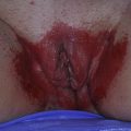
VVLP may present in several forms, and patients may exhibit more than one form of LP concurrently or sequentially. In the largest published study on women with erosive LP (n=114), Cooper and Wojnarowska reported that pain (80%), pruritus (65%), dyspareunia (61%), and irritation (48%) were the most frequent presenting symptoms. In a survey of current practices by 4 vulvar disease experts, published in 2008, on 145 patients, symptoms of soreness were reported in 72% followed by burning (66%) and pruritus (63%), and the most frequent vulvar signs were erosions (74%), red and/or purple coloration (66%), scarring (60%), and apareunia (35%).
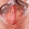
Typical papulosquamous LP of the glabrous skin is infrequently seen on the vulva ( Fig. 1 ). Patients more commonly present with erythema and erosions, but peripheral reticulations may also be noted. Atrophic or erosive LP of the mucous membranes presents with erythema, tender denuded epithelium, and desquamative vulvitis or vaginitis ( Figs. 2–4 ). A similar clinical presentation is noted on the gingivae ( Figs. 5 and 6 ). Cooper and Wojnarowska reported that most patients presented with erosions (90%) and/or white reticulations (82%). Isolated Wickham striae may be found on the labia minora and the medial aspect of the labia majora and may be asymptomatic ( Figs. 7–9 ). Hypertrophic LP occurs in 20% to 25% of patients and presents with extensive, white, thickened, and hyperkeratotic plaques of the mucous membranes ( Fig. 10 ).