Key Points
- •
Intraoperative mapping of the right gastroepiploic lymphosome is valuable to identify critical flap components.
- •
No-touch flap elevation will prevent damage to the flap lymphatics and microvasculature.
- •
Harvesting additional capillary networks can minimize venous congestion.
- •
Additional vascular anastomoses may be required to mitigate venous congestion.
Introduction
Although lymphatic surgery has been practiced for over 100 years, advances in lymphatic comprehension, instrumentation, and imaging have resulted in a rebirth of this field. Similarly, advances in techniques and anatomic knowledge have resulted in the renewed interest in the omental flap for reconstruction and lymphatic surgery.
The omentum has been called the “policeman of the abdomen” due to its immunogenic and angiogenic properties. It contains high concentrations of critical factors, including immunoglobulin M and vascular endothelial growth factor protein. Vascularized omental lymphatic transfer not only provides gastroepiploic lymph nodes but also contains critical lymphatic structures, including the omentum-associated lymphoid tissue (OALT) (“milky spots”), which initiate absorption from the peritoneum. With these characteristics, the omentum has become a versatile flap option for reconstructive and lymphatic surgery.
Initial applications of the omentum for the treatment of lymphedema did not gain wide acceptance. Limitations of early approaches included the need for a celiotomy and its use as a pedicled flap, incurring the risk for hernia, bowel infarction, flap ischemia, and loss of the critical flap structures along the arc of rotation. The application of free tissue transplantation and minimally invasive flap harvest reduces donor site morbidity and pedicled flap complications. This chapter will detail the surgical considerations to improve successful outcomes with the vascularized omental lymphatic transfer.
Concepts
This flap has been described as a large, flattened-out lymph node. With meticulous techniques and comprehension of the flap anatomy, the omental lymphatic flap can remain well vascularized and retain its physiologic lymphatic architecture.
Regional Anatomy
Understanding the flap anatomy and extensive variability is critical to successful flap harvest and outcomes. The arterial blood supply to the omentum can come from two dominant pedicles, the right and left gastroepiploic arteries, which run along the greater curvature of the stomach. The right gastroepiploic artery is the largest branch of the gastroduodenal artery off of the celiac trunk. Although this usually arises near the pylorus, the origin may emanate anywhere along the greater curvature of the stomach to the mid-body.
At their origins, the right gastroepiploic artery diameter is usually greater than 2.5 mm, with the vein usually being larger than 3 mm. Although these relatively larger diameters compared to other lymph node transfers may be appealing, it is important to remember that the numerous small branches must be controlled to avoid postoperative complications, such as hematoma. Additionally, like other intestinal flaps, these vessels are superficial, have thinner walls, and are more fragile than non-intestinal flaps, making them more susceptible to devascularization, kinks, twists, and compression, especially as the flap does not have the benefit of a skin paddle to support its inset.
The branching and connections of the omentum vary significantly. The anastomosis between the left and right gastroepiploic vessels is weak in 60% and absent in over 20% of patients. Identifying this relationship is essential when harvesting a larger flap or splitting a flap. Several branches will perfuse into the omentum, including the right, middle, and left omental branches. If a trans-omental arch is present and contiguous with the major omental branches, including it within the flap is valuable to improve the capillary network.
The omental lymphatic flap includes a complete organ of lymphatic structures within its lymphosome. The commonly named gastroepiploic lymph nodes include lymph node stations 6 (subpyloric) and 4d (right gastroepiploic). These lymph nodes can vary in size and may not be present or visible on radiologic imaging. Although utilizing the gastroepiploic lymph nodes alone may be performed, incorporating the entire lymphosome does not require a significant amount of extra work and can help the patient only by providing additional lymphatic tissue and capillary beds to reduce venous congestion. The OALTs are essentially lymph nodes without capsules. They initiate absorption into the peritoneal cavity and may be more effective than the gastroepiploic lymph nodes. These are generally smaller than the gastric lymph nodes and present variably along the apron of the omentum, making the inclusion of these structures in intraoperative localizing imaging important. The efferent lymphatics from the OALTs can be easily damaged as they are friable and superficially coalesce toward large efferent lymphatics alongside the right gastroepiploic vessels.
Patient Selection
As with any lymphatic surgical procedure, patient selection is paramount to successful outcomes. All patients must be optimized with complete decongestive therapy prior to surgical consideration. Inadequate optimization will increase the potential for postoperative seromas, cellulitis, ineffective flap function, and flap loss. This flap is generally offered to patients with a history of excellent therapy compliance, moderate to more advanced lymphedema, severe fibrosis on examination, a history of recurrent cellulitis, insufficient control with other lymphatic surgeries, and contraindications to other vascularized lymph node transfer options. Morbidly obese patients may be better candidates for a minimally invasive vascularized omental lymphatic transfer over alternative lymph node transfers given their increased risk for donor site lymphedema or delayed wound healing.
Setting patient expectations is also critical for this flap. As the flap does not include a skin paddle, some patients may require a monitoring window covered by a skin graft, which is not visually appealing. Since the flap does not carry intrinsic structural support, patients may require unique positioning and activity restrictions during the postoperative state. Patients must also understand that, although it is a powerful flap option, the vascularized omental lymphatic transfer is not a cure for their lymphedema.
Preoperative Considerations
Preoperative imaging of the recipient site is important for all lymph node transfers. Preoperative imaging of the abdominal donor site for the vascularized omental lymphatic transfer will not significantly alter management. There is no risk for donor site lymphedema, and intraoperative mapping is more important for the delineation of critical flap structures. If the patient had previous imaging studies, these should be reviewed as they may reveal anatomic variants to expect during the flap dissection. Although this flap is effective for patients with a history of cellulitis episodes, the flap is very susceptible to infection in the perioperative state. Patients with recent or recurrent infections should be aggressively treated with antibiotics preoperatively to address the chronic infectious biofilm.
Patient comorbidities may alter the approach to this flap. Severe respiratory or cardiac disease may preclude major abdominal entry surgery. Alternative flaps should be utilized in those with a history of mantle field radiation to the abdomen. A history of previous abdominal surgery or abdominal infections is not a contraindication to the use of this flap but may affect the expected outcomes due to preexisting intrinsic flap fibrosis. Significant fibrosis of the omentum or a previous omentectomy history, with imaging confirming a preserved pedicle, may require conversion to a gastroepiploic lymph node–only flap.
Surgical Techniques
Flap Design
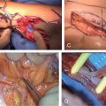
After either open or minimally invasive entry into the abdomen, critical flap structures are identified. The unidirectional right gastroepiploic lymphosome can be mapped via a dual injection technique. Utilizing an equal mixture of indocyanine green, isosulfan blue, technetium-99m, and albumin, the right apron is injected with a small-caliber needle to avoid spillage. This will also assist with the delineation of the residual left gastroepiploic lymphosome if a split flap is planned. The gastroepiploic lymph nodes are then mapped by submucosal gastric dye injection to determine the amount of pedicle harvested.
If a minimally invasive flap harvest is performed, injection of the apron can be performed percutaneously with a spinal needle. Gastroepiploic lymph nodes are best visualized via intraluminal stomach injection performed through an upper endoscopy. Laparoscopic fluorescence imaging and radioactive tracer probes are required to identify the OALTs to be included within the flap.
Once the flap boundaries and volume are determined by this mapping, the recipient pocket can be tailored to the anticipated flap volume to prevent over dissection and dead space. Omental thickness can be variable, independent of body habitus, and should be visualized prior to creating the recipient pocket. The recipient pocket should be debulked of the fibrotic lymphedematous soft tissue and deep fascia, preserving superficial veins and cutaneous nerves, for a recipient bed that is conducive to lymphangiogenesis.
Flap Harvest
Abdominal exploration is grossly performed to rule out comorbid pathology. Omental adhesions are released to allow for flap dissection. Visualization of the omentum is utilized to identify the anatomic variability of the pedicle origin, pedicle continuity, omental vessel branches, and trans-omental arch presence.
Entry into the lesser sac can be performed by separating the transverse colon from the omentum. This plane can oftentimes be fused either anatomically or due to a previous infection or surgery. The lesser sac is usually preserved at its lateral-most extent just medial to the splenocolic ligament and is a reliable entry point into the lesser sac. In the event of significant fibrosis, retrograde flap elevation may be performed by separating the short gastrics. Division of the short gastrics should be performed from lateral to medial due to potential anatomic variability of the pedicle. The left gastroepiploic can be divided early to minimize damage by flap manipulation, and the right gastroepiploic origin should be dissected proximal enough to incorporate the subpyloric lymph nodes within the flap. Given the fragile structure of the flap, a no-touch flap elevation utilizing patient positioning, retraction of adjacent structures, and minimal flap handling during the dissection is preferred to prevent damage to the flap lymphatics and microvasculature.
Open flap harvest can be performed through various incisions. It is important to have an incision of adequate length for appropriate exposure to minimize flap trauma. Short gastrics can be controlled with vascular clips to minimize the risk of thermal spread to the pedicle or gastric ileus. Because the epigastric incision has the highest risk for incisional hernia, it is important to be aware of any preexisting diastasis and incorporate the pillars of the rectus complexes within the fascial closure.
Minimally invasive flap harvest may decrease abdominal morbidity. Due to the fragile flap structure, laparoscopy may be preferred over robotic or even open approaches for improved haptic feedback and for minimization of flap traction or manipulation. A port is placed for camera visualization and flap extraction. An additional two to three working ports are required for dissection. Atraumatic instruments with no-touch flap harvesting should be used to minimize lymphatic disruption. Ultrasonic energy devices with the least amount of thermal spread should be used. When the short gastrics are not long enough for energy devices, vascular clips or erring toward the thick-walled stomach should be performed to minimize pedicle damage and potential thrombosis. Excess thermal injury or manipulation of the stomach may require gastric decompression postoperatively. Flap extraction is performed under direct vision through the camera port, which should be extended adequately to minimize flap trauma, usually to 2–3 cm.
Versatility is an attractive feature of this flap. The flap can be split into two flaps for dual flap inset or for bilateral cases, but splitting the omentum may result in an insufficient second flap. Alternatively, this author prefers to utilize the shared donor site morbidity and perform a minimally invasive mesenteric lymph node transfer concurrently for dual-inset patients. This flap can also be harvested concurrently with a DIEP flap for breast reconstruction if a simultaneous groin lymph node transfer is contraindicated. If the DIEP pedicle is harvested in an open fashion, the same paramedian defect through the posterior rectus fascia is the preferred intra-abdominal access to avoid further damage to the abdominal wall. If the DIEP flap pedicle is harvested in a minimally invasive fashion, the DIEP pedicle can be harvested first through the properitoneal space, then transitioned intracorporeally for the omental lymphatic flap.
Flap Inset
Flap revascularization is performed depending on the patient’s anatomy and chosen recipient site. As the gastroepiploic vein does not contain valves, venous anastomosis distal to a recipient valve is preferred. The efferent right gastroepiploic lymphatic can also be anastomosed to a recipient venule for an anatomic and physiologic reconstruction.
Venous congestion is a problematic outcome encountered with this flap. Its incidence can be reduced significantly by harvesting more capillary networks of the omental flap, including the trans-omental arch if present, instead of essentially utilizing the pedicle alone as in the gastroepiploic flap. If an intact trans-omental arch is present, no further venous anastomoses are usually required. Performing both left and right gastroepiploic venous anastomoses, preferably to the superficial and deep venous systems, for dual physiologic venous outflow can reduce postoperative flap swelling. In the event of recipient lymphatic venous hypertension, especially of the lower extremity, or significant fibrosis of the flap from previous surgery impairing the capillary beds, an arteriovenous fistula of the left gastroepiploic vessels can be performed. The addition of a fistula will engorge the flap early postoperatively, but this will normalize and become supple over time. Alternatively, a flow-through anastomosis of the left gastroepiploic artery may autoregulate the arterial inflow to reduce venous congestion. Listening to the venous outflow with a pencil or implantable Doppler after implementing these options can help guide the surgeon as to which of these options is preferred.
A meticulous flap inset must be performed to prevent folding of the flap and to optimize the surface area of contact with the recipient bed. As the flap does not have intrinsic structural integrity, the flap pocket must be fashioned to account for postoperative swelling without leaving too much space, to prevent traction on the anastomoses and pedicle. A monitoring skin graft is usually harvested from the recipient site or from the flap extraction incisions and allows for additional compliance. The abdomen is re-inspected at the end of the procedure to verify hemostasis.
Postoperative Care
Experience in flap monitoring is paramount for this procedure as it is not a common flap and does not have a skin paddle. The flap appearance can be dynamic with examination findings, which may result in re-exploration on postoperative day 1 but to be expected on postoperative day 5. Expectations of flap evolution, including changes in Doppler signals or flap appearance, should be relayed to the healthcare provider team daily. If thermal spread was close to the stomach, nasogastric tube decompression for 24 hours to avoid gastroparesis and distension is considered. Lower extremities with an arteriovenous fistula can begin an early dependent protocol. Complete decongestive therapy is resumed at 3 months postoperatively. It is prudent to delay removal of the monitoring window to over 12 months postoperatively after the flap has improved the lymphatic fibrosis of the extremity and when concurrent debulking can be performed if necessary.
Outcomes
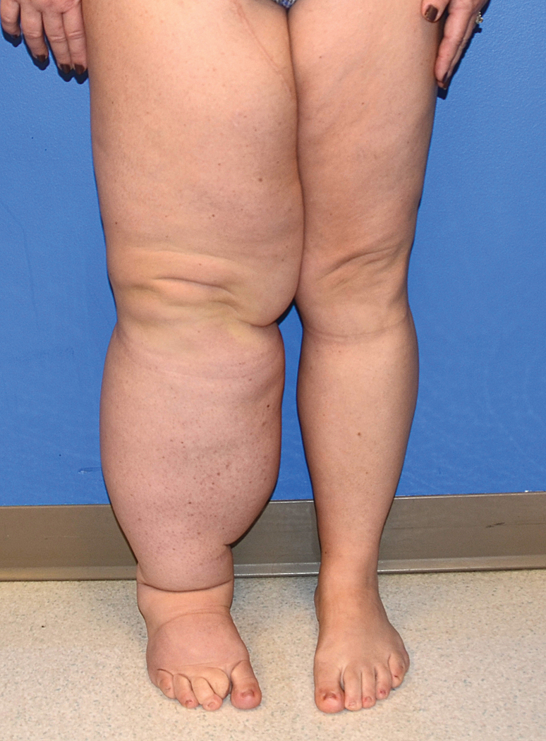
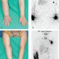
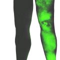
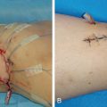
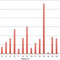
The author has performed over 100 vascularized omental lymphatic transfers, with similar successful outcomes to those previously published, including 83% symptomatic improvement and 22% volume improvement ( Figs. 17.1–17.5 ). Improvements in fibrosis and cellulitis episodes are critically important outcomes when utilizing this flap. Potential donor site complications include pancreatitis, ileus, hernia, and bowel injury. Recipient site complications include hematomas, seromas, cellulitis, and flap loss.
Related posts:
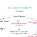
 Lymphoscintigraphy Interpretation, Staging, and Lymphedema Grading
 Anatomy and Structural Physiology of the Lymphatic System
 Vascularized Lymph Node Transfer from the Groin
 Private: The Campisi Approach for Lymphatic Surgery
 Private: Tracking Outcomes Following Lymphedema Treatments
 Private: Microsurgical Procedures: Vascularized Lymph Node Transfer from the Thoracodorsal Axis
Stay updated, free articles. Join our Telegram channel

Full access? Get Clinical Tree


