Transcutaneous lower blepharoplasty with orbitomalar suspension and fat redraping


Table 10.1

Indications for surgery


Cosmetically displeasing lower eyelid fat prolapse with dermatochalasis
Functionally, when glasses rest upon excessive lower eyelid fat prolapse and causes lower eyelid ectropion


Table 10.2

Preoperative evaluation


Prior facial surgery or trauma
Dry eye symptoms
Prior refractive surgery
Degree of dermatochalasis and fat prolapse
History of prior fillers to lower eyelid
Presence of midfacial ptosis
Presence of double convex deformity of lower eyelid
Presence of lower eyelid laxity or other eyelid malpositions


Introduction


Transcutaneous lower blepharoplasty may be considered when addressing cosmetic lower eyelid fat prolapse with associated lower eyelid rhytids. When lower eyelid fat prolapse is the primary concern, the transconjunctival approach, as discussed in Chapter 9 , is a more appropriate choice.


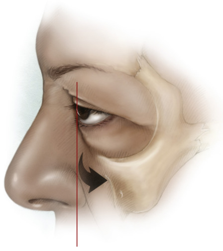
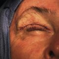
The preoperative evaluation should focus on aesthetic changes due to fat prolapse and skin redundancy as well as the contour changes of the lower eyelid and midfacial junction. Functional changes such as lower eyelid laxity, eyelid retraction, lagophthalmos and blink dynamics should be evaluated and addressed at the time of surgery. Patients with a prior history of blepharoplasty should be evaluated for eyelid retraction and eyelid closure and treatment should be directed at correction of these malpositions prior to blepharoplasty ( Chapter 34 , Chapter 35 , Chapter 36 ). Dry eye and tear film status should be determined and treated prior to surgery as well. Patients who have had keratorefractive surgery should have a careful evaluation for dry eye as this may worsen after blepharoplasty. Patients with a negative vector in which maxillary hypoplasia and a prominent globe are present are at high risk of lower eyelid retraction with transcutaneous lower blepharoplasty ( Figure 10.1 ) and are more appropriate for the transconjunctival approach ( Chapter 9 ). Photo documentation is essential and should be performed in the frontal plane, three-quarter, and side views to demonstrate changes after surgery.


Figures 10.1


Negative vector

The negative vector is present when the globe projects anterior to the inferior orbital rim. This may be due to several factors including thyroid-related orbitopathy, myopia and maxillary retrusion. The risk of eyelid malposition is high in this group of patients. Midfacial elevation such as orbitomalar suspension or an inferior orbital rim implant may be beneficial.


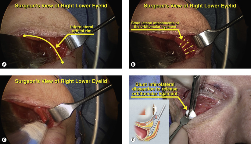
Several adjunctive procedures can be performed during trans­cutaneous lower blepharoplasty. Redraping of fat pedicles can address the double convex deformity of the lower eyelid ( Chapter 9 ). Judicious lower eyelid tightening, conservative skin removal and suborbicularis oculi fat lifting (orbitomalar suspension) at the time of surgery can minimize the risk of lower eyelid retraction and lagophthalmos. We routinely perform orbitomalar suspension on all cases of transcutaneous lower blepharoplasty to lessen the risk of lower eyelid malpositions as well as for the aesthetic benefits to the midface.
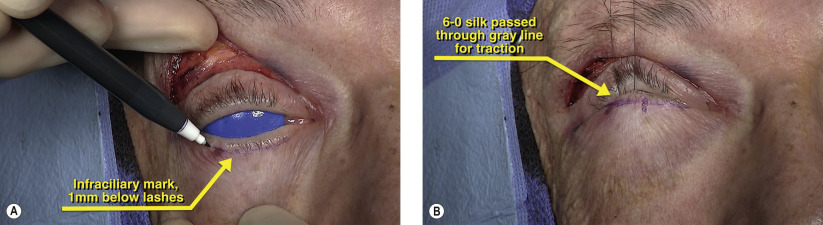
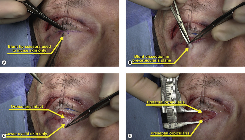
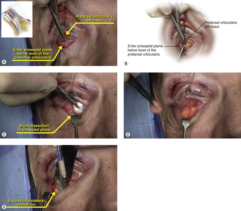


Surgical Technique


Figures 10.2A and 10.2B


Incision planning

Transcutaneous access to the lower eyelid is performed through an infraciliary incision within 1 mm of the eyelashes ( Figure 10.2A ). The incision should be marked before the infiltration of local anesthetic to minimize distortion. The marking should initially extend from the punctum to the lateral canthal angle. Local anesthetic containing 1 : 200,000 epinephrine is given both through the conjunctival approach as well as along the transcutaneous marking to minimize orbicularis bleeding. When injecting transcutaneously, the needle is injected in a pre-orbicularis plane and used to hydrodissect the skin from the underlying muscle. A 6-0 silk suture is then passed through the gray line and placed on superior traction ( Figure 10.2B ). Prior to the start of surgery, a corneal shield is put in place.


Figures 10.3A–D


Infraciliary incision

With superior traction on the eyelid, the infraciliary incision is made with blunt tip Westcott scissors ( Figure 10.3A ). The incision should be kept very close to the eyelashes to maximally conceal the scar. When making this incision, the inferior edge of the skin is tented up with 0.5 forceps to dissect the skin away from the underlying orbicularis. With adequate delivery of the local anesthetic in the pre-orbicularis plane, the skin should easily dissect free from the muscle. Minimizing trauma to the pretarsal orbicularis will lessen the chance of developing postoperative ectropion from an atonic muscle. Using the Westcott scissors, dissection is continued inferiorly with the blunt tips of the scissors directed towards the skin in the pre-orbicularis plane ( Figure 10.3B ). Care is also taken when handling the thinned lower eyelid skin as this is reflected inferiorly ( Figure 10.3C ). The dissection is continued inferiorly for at least 4 mm below the eyelid margin to expose the preseptal orbicularis ( Figure 10.3D ).


Figures 10.4A–D


Preseptal dissection

Once the dissection in the pre-orbicularis plane is complete, the anterior orbit is entered. Sharp dissection is performed at the level of the preseptal orbicularis at a level below the pretarsal orbicularis ( Figures 10.4A and 10.4B ). This preseptal orbicularis window is extended medially and laterally with sharp dissection. Once the preseptal plane has been exposed, blunt dissection with a peanut sponge or cotton-tip applicator is performed with inferior countertraction using a Senn retractor ( Figure 10.4C ). Further blunt dissection in this avascular plane exposes all three lower eyelid fat pads and is continued to the inferior orbital rim ( Figures 10.4D and 10.4E ). Any fine bleeders are coagulated with bipolar cautery.


Figures 10.5A–D


Release of orbitomalar ligament

The orbitomalar ligament is the primary retaining ligament of the midface which anchors the suborbicularis oculi fat (SOOF) to the lateral orbital rim. Release of the orbitomalar ligament and superior suspension of the SOOF provides aesthetic elevation of the midface and protection against lower eyelid retraction. The orbitomalar ligament has broad midfacial attachments and the stoutest attachments are to the inferolateral orbital rim, as shown in Figures 10.5A and 10.5B . A malleable retractor is placed along the inferolateral orbital rim with a Senn retractor providing countertraction. Cutting cautery is used in the dissection along the inferolateral orbital rim to lyse attachments of the orbitomalar ligament ( Figure 10.5C ). Once the orbitomalar ligament has been released, a peanut sponge is used to bluntly dissect any residual attachments to the lateral orbital rim ( Figure 10.5D ). Care is taken during the inferolateral dissection along the orbital rim as the zygomaticofacial artery and nerve exit via its foramen. If bleeding occurs from this foramen, gentle bipolar cautery should be applied to prevent significant midfacial hemorrhage. Neurosensory loss is minimal and recovery is rapid and uneventful.

Only gold members can continue reading. Log In or Register to continue

Stay updated, free articles. Join our Telegram channel

May 16, 2019 | Posted by in Reconstructive surgery | Comments Off on Transcutaneous lower blepharoplasty with orbitomalar suspension and fat redraping

Full access? Get Clinical Tree

Get Clinical Tree app for offline access