Transconjunctival lower blepharoplasty with fat redraping


Table 9.1

Indications for surgery


Cosmetically displeasing lower eyelid fat prolapse without significant dermatochalasis
Functionally, when glasses rest upon prolapsed lower eyelid fat and causes lower eyelid ectropion


Table 9.2

Preoperative evaluation


Prior facial surgery or trauma (including symblepharon)
Dry eye symptoms
Prior refractive surgery
Degree of dermatochalasis and fat prolapse
History of prior fillers to lower eyelid
Presence of midfacial ptosis
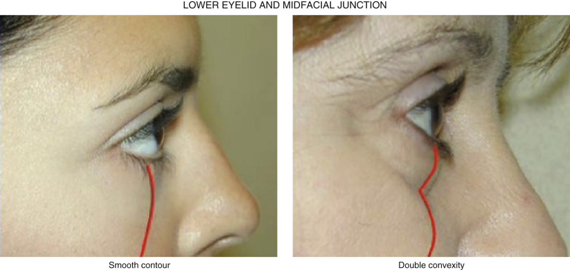
Presence of double convex deformity of lower eyelid
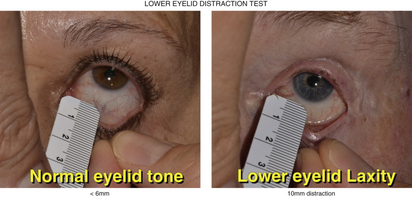
Presence of lower eyelid laxity or other eyelid malpositions


Introduction


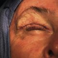
Transconjunctival lower blepharoplasty is well suited for patients with lower eyelid fat prolapse and minimal skin redundancy ( Figure 9.1 ). This approach is appealing to many patients as a skin incision is avoided and there is no orbicularis incision and potential for denervation. The drawbacks of this procedure compared to the transcutaneous approach ( Chapter 10 ) are the difficulty with a smaller incision, poorer exposure, potential for conjunctival chemosis and posterior lamellar shortening. Several adjunctive procedures can be performed with transconjunctival blepharoplasty including: skin pinch, chemical peel/laser resurfacing, lateral canthoplasty and fat redraping. With all of these adjuncts, the orbicularis is left intact, minimizing but not eliminating the risk of lower eyelid retraction and ectropion.


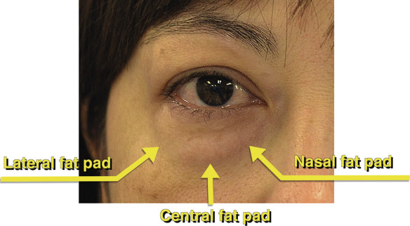
The preoperative evaluation should focus on ruling out co-exist eyelid malpositions such as ectropion, entropion, lower eyelid retraction and lagophthalmos which may predispose to dry eye. A lower eyelid distraction test is performed to assess laxity ( Figure 9.2 ). Normal eyelid tone is 6 mm or less and any laxity greater than 6 mm should be addressed with a lid tightening procedure such as a canthoplasty. Post-LASIK patients should wait at least 3 months after refractive surgery before considering lower blepharoplasty. The conjunctiva should be inspected for any signs of cicatricial changes. Minimal skin redundancy should be noted and if present, a decision to perform concurrent skin pinch or chemical or laser resurfacing should be considered at the time of surgery or postoperatively. Photographs in the frontal, profile, and side planes should be taken before and after surgery.


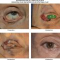
In youth, there is a smooth transition from the lower eyelid to the midface, but with aging, there is unmasking of the inferior orbital rim and generation of the double convex deformity ( Figure 9.3 ). Conservative blepharoplasty with fat redraping should be discussed with younger patients as aged-related fat atrophy may result in a sunken appearance with senescence if subtractive blepharoplasty is performed. Fat redraping can blunt the double convexity and restore a natural eyelid and midfacial transition.


Preoperative Evaluation
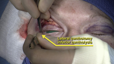
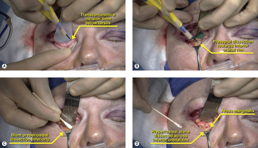
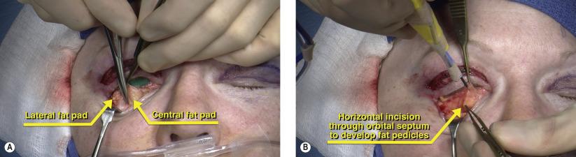
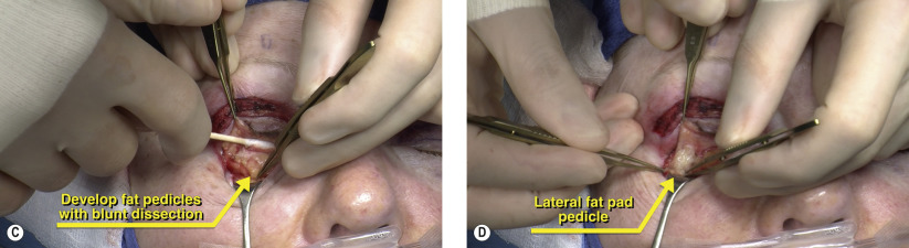


Figure 9.1


Evaluation of lower eyelid contour

The lower eyelid fat pads are evaluated and documented with natural lighting. Prolapse of all three lower eyelid fat pads can be seen in this case with minimal skin redundancy. In the preoperative holding area, the lower eyelid fat pads are marked while the patient is sitting up and prior to sedation and infiltration of local anesthetic.


Figure 9.2


Lower eyelid distraction test

The lower eyelid is pulled away from the globe. With normal eyelid tone, the distance from the sclera to the eyelid should be 6 mm or less. In the right panel, there is lower eyelid laxity of 10 mm. During blepharoplasty, lower eyelid tightening should be performed to minimize the risk of postoperative eyelid malposition ( Chapter 25 , Chapter 29 ).


Figure 9.3


Lower eyelid and midfacial junction

On the left panel, there is a harmonious transition from the lower eyelid to the cheek in the youthful eyelid. With aging, there is unmasking of the orbital rim leading to the double convex deformity. These changes may be due to loss of orbital and cheek fat, attenuation of the orbitomalar ligament and maxillary retrusion.


Surgical Technique


Figure 9.4


Lateral canthotomy and inferior cantholysis

In patients with normal lower eyelid tone, access to the lower eyelid fat may be difficult. In such cases, a lateral canthotomy and inferior cantholysis may be necessary to achieve suitable access to the lower eyelid. The canthotomy is kept small at 1 mm and the cantholysis is restricted to the inferior crus of the lateral canthal tendon ( Figure 9.4 ). When performing fat redraping, access to the inferior orbital rim is enhanced with canthal release.


Figures 9.5A–D


Transconjunctival incision

Using a protective eyelid plate and corneal protector, a transconjunctival incision is made approximately 4 mm below the inferior tarsal border with cutting cautery ( Figure 9.5A ). The cauterization is performed cautiously near the medial eyelid where iatrogenic punctal injury may occur. Once the conjunctiva has been incised, a preseptal dissection is performed towards the inferior orbital rim ( Figure 9.5B ). Blunt dissection with a cotton tip applicator can be used to gently dissect in this avascular plane to expose each of the lower eyelid fat pads ( Figure 9.5C ). If fat redraping will be performed, then a preperiosteal dissection is performed along the arcus marginalis to expose the inferior orbital rim ( Figure 9.5D ). The periosteum along the inferior orbital rim must be kept intact so that the fat can be redraped to the periosteum. Alternatively, fat pedicles can be redraped in a subperiosteal fashion depending on surgeon preference.

May 16, 2019 | Posted by in Reconstructive surgery | Comments Off on Transconjunctival lower blepharoplasty with fat redraping

Full access? Get Clinical Tree

Get Clinical Tree app for offline access