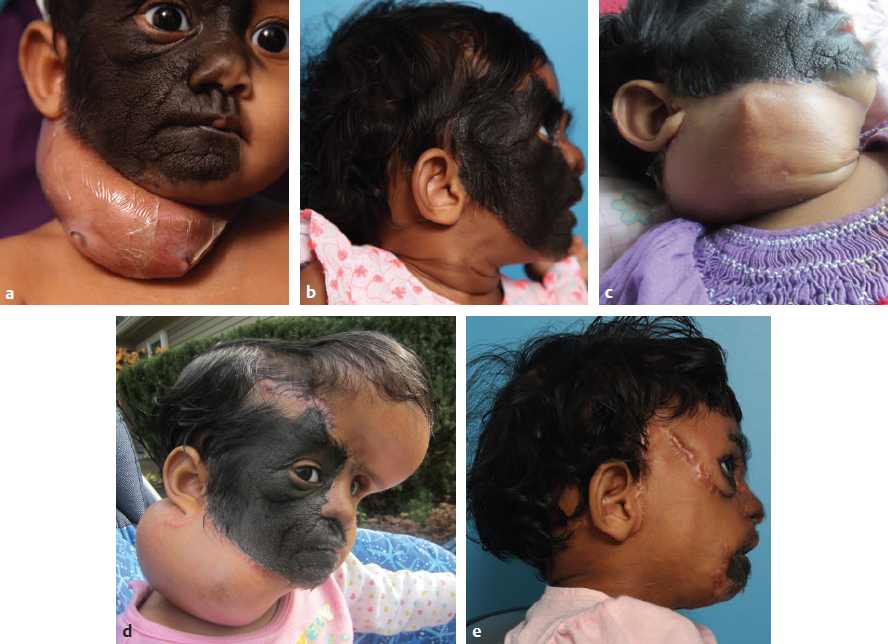
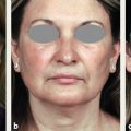
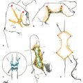
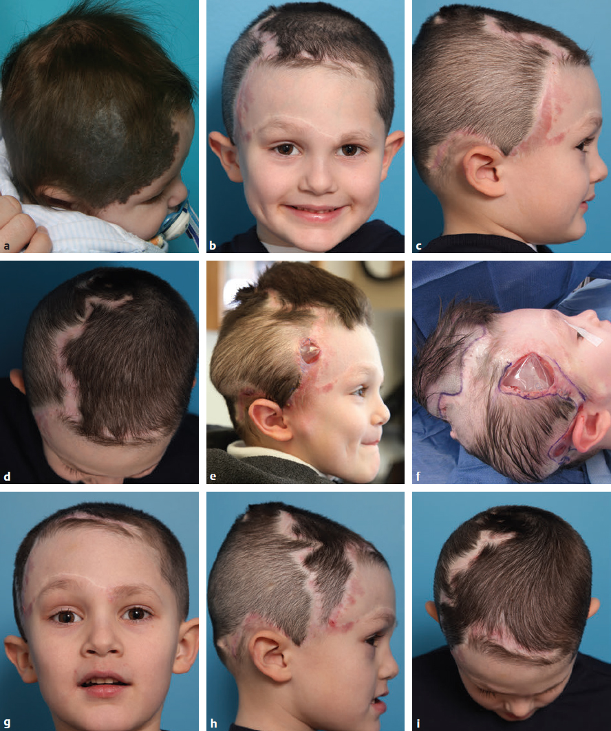
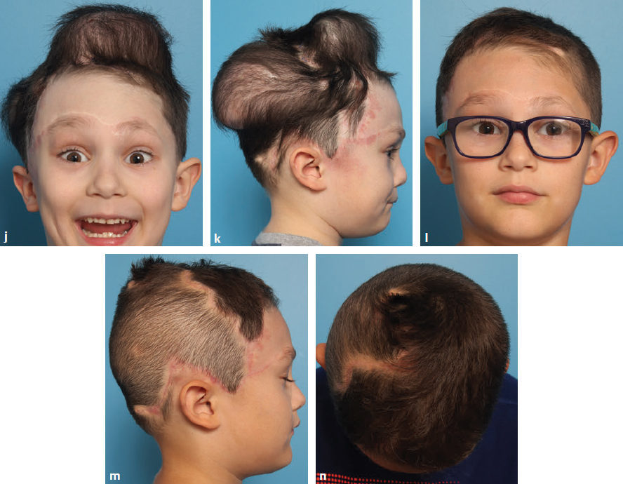
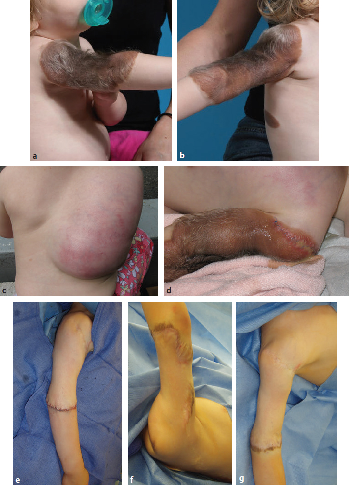
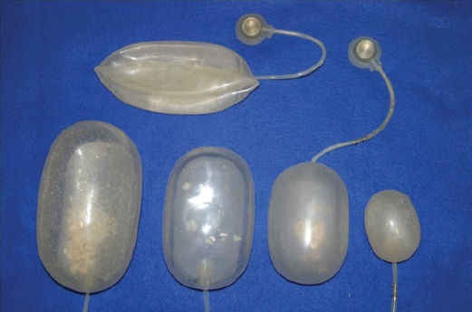
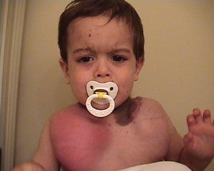
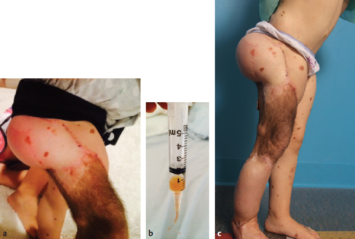
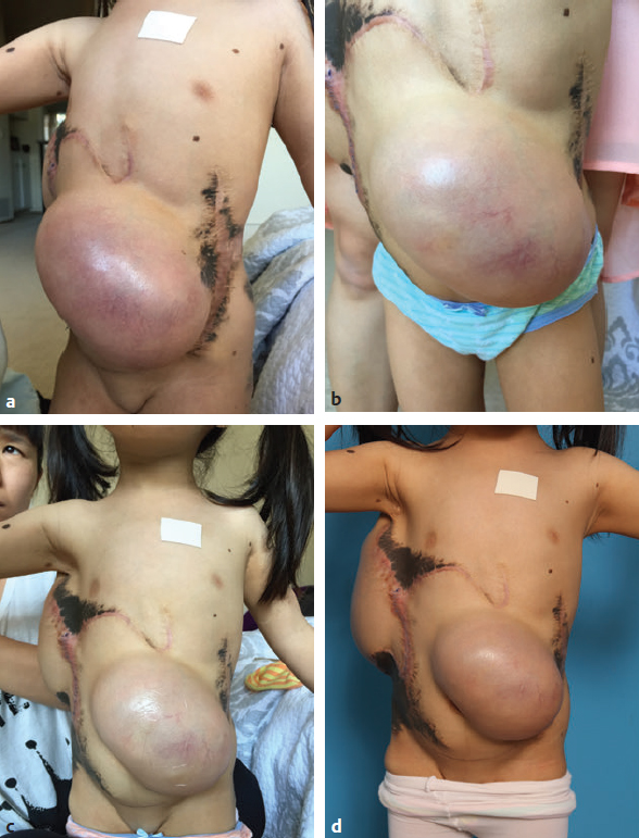
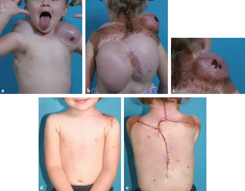
CHAPTER Large and giant congenital melanocytic nevi present a unique reconstructive challenge. Nevomelanocytes are known to penetrate through the full thickness of skin and into the subcutaneous fat. Often they invade nerve and blood vessels. On rare occasions they may penetrate fascia, muscle, and internal organs.1,2 Thus excision of the full thickness of nevus tissue is required to remove most of the nevus cells and minimize subsequent reappearance of nevus cells with possible long-term sequelae. The principle of reconstruction is to replace like with like. Serial excision can often debulk these massive lesions but rarely removes them completely. Excision and split-thickness skin graft, if the excision is carried out to the fascial level, can result in both poor aesthetic outcome and risk of functional disturbance, except for select situations.3 Partial-thickness removal such as dermabrasion, curettage, chemical peel, and laser treatment all have problems with recurrence, because these modalities only eliminate the superficial portion of the nevus. Later degeneration of the remaining nevus has been reported.4 When direct excision and primary closure are not a possibility, tissue expansion is the workhorse treatment modality for melanocytic lesions, for large scars, for vascular lesions, and as an adjunct in reconstruction of some craniofacial deformities. Since 1980, I (B.B.) have dedicated my practice to refining the techniques used in the treatment of nevi and other skin and soft tissue defects using tissue expansion.5–7 This vast experience has garnered a host of successes, as well as some expected and unex pected complications, for which effective management strategies have been developed. This chapter relays this expertise so the reader can avoid some common mistakes and successfully manage a variety of complications involving tissue expansion. Summary Box Avoiding Unfavorable Results and Complications During Expansion Complication Major Infection Flap ischemia Implant exposure or extrusion Device failure Minor Seroma Wound dehiscence Pain Flipped or inaccessible port Postexpansion Major Flap ischemia Infection Wound dehiscence Minor Delayed wound healing Scar widening Flap congestion Seroma Most patients with melanocytic nevi treated with tissue expansion are children, although we have gained some important insights from a small group of adult patients. Juvenile skin, although elastic, does not have the laxity of adult skin, and the local flaps used in adults are often difficult in children. The decision to excise large and giant nevi in children is sometimes controversial.8–10 In our practice the ideal patient is one with a supportive family structure that understands the involvement required to achieve successful outcomes using expansion and can take an active role in the process. Preferably, the patient does not have symptomatic neurocutaneous melanosis,11 a developmental disability, or comorbid conditions that may outweigh the benefits of excision and reconstruction. The patient must also have enough normal skin with which to expand. This does not require a certain percentage but needs to be sufficient to undergo serial expansion to reduce the nevus size so that clothing can cover what remains and provide some social benefit. There is no ideal age to begin expansion, but in our practice we have noted that younger patients often tolerate expansion very well and have little or no memory of the process when they are older. Younger patients are often easier to control and to limit their activities around the time of surgery to optimize healing. We place expanders in children starting at 6 months of age when the anesthetic risk is low.12,13 Many of our patients approach us via the Internet or email and the initial consultation is done via pictures or web-based video conference. For any assessment of a new patient regarding an expander the surgeon must evaluate the extent of the defect or lesion to be treated and the available donor tissue. With experience it becomes easier to estimate how much can be accomplished by a single round of expansion and what defects will require more. Further assessment of the patient focuses on the donor skin surrounding the lesion. The surgeon must ask: What is the tissue quality? Are there preexisting scars? Are there satellite lesions? Is there infection or another wound? What structures will limit the placement or the amount of expansion that can be achieved? The answers to these questions will help delineate the number and the size of the expanders to be used and the ideal placement locations and will identify potential pitfalls. Cases can be matched against patients with similar nevus distribution as a guide to the number of expanders and rounds of expansion that might be required to complete the excision and reconstruction. Once the size and location of the expanders has been chosen, the next step is to determine the length of time that will be required to expand the tissue. We anticipate that with weekly expander injections, most expanders can be filled to the desired volume in 10 to 12 weeks. The volumes vary with the expander size and area to be expanded, and specific volumes are not set but are gauged by capillary refill and the feel of the expander. Therefore for nearly all our patients we plan the removal and reconstruction for 11 to 12 weeks after the expander placement (expansion over 7 to 8 weeks for smaller facial nevi). For smaller expanders or for smaller defects for which we are using a larger expander but do not require the full volume of expansion, we shorten the time as needed. Whether the families decide to come weekly to the clinic for injections or, more commonly, expand at home after careful instructions, the parents and doctor must agree to an open and regular dialog (email, digital images, or phone communication when needed). In this manner, complications can often be avoided or treated soon enough to avoid compromising the expander or flap. Many of our patients perform home expansion. Therefore they need to be prepared to maintain an open line of communication with physicians and nurses regarding the appearance of skin flaps, any fevers, red skin, incision drainage, or other issues. We also require photographic documentation to be provided so that an ongoing assessment of the progress can be made. For patients who elect to undergo office-based expansion, they need to commit to visit the office on a weekly basis. Patients who are willing to meet the aforementioned criteria are considered good candidates for tissue expansion. It is vital that patients be made aware of what a fully inflated expander will look like in a given location. It might be shocking on first impression, and the management of these expectations preoperatively is vital. We provide multiple photographs depicting a variety of cases for patients to visually gain this understanding. Adult patients who have employers or professions that would not tolerate the visual appearance of an expander are encouraged to arrange time off or reassignment during their expansion. This will ensure the least disruption to their livelihood, and they can be more comfortable, improving the chances at a successful outcome. Some parents prefer to have their children undergo the expansion process during the summer months to avoid the social stigma in some scholastic settings. Others choose the opposite approach so as not to miss any summer activities such as swimming or camp. High-risk tissue expansion patients are typically those who have undergone multiple expansions in the past (Fig. 46.1). Resulting scars and thinner flaps increase the risk of exposure of the implant. Patients who are prone to infections, such as those with recurrent otitis media, are at increased risk of expander infection. In these cases we recommend myringotomy tubes before the start of expansion to reduce the risk of bacterial seeding via the bloodstream once the expander has been placed. Other patients may be prescribed oral antibiotics for the course of expansion if they have a history of expander infection. Nevus quality also affects the chances for success. Nevi that have fine villous hair are found to be at higher risk for infection and delayed healing at incision sites (even with optimal wound care the fine hairs seem to incite inflammation and secondary infection) (Fig. 46.2). For these lesions and for scar reconstruction, incisions are typically placed outside of the lesion to prevent these complications. We always stress trimming or cutting hair that is near incisions to avoid the buildup of crust, scab, or other irritation that can lead to local infection. Fig. 46.1 A high-risk patient for expanders. (a) He had a large neural nevus of forehead and scalp. (b–d) After multiple rounds of expansion and initial reconstruction, he presented with residual nevus of the temporal forehead and scar alopecia. (e,f) Two expanders were placed in the frontal and temporal scalp. The expander on the right temple was exposed through the thin scar and residual nevus. (g–i) Tissue loss from implant exposure required reconstruction using the expanded frontal scalp, resulting in unfavorable scalp scars and scar alopecia, and did not achieve sufficient tissue gain to fully excise the residual nevus. (j,k) Repeat expansion with a parietal expander placed in a more posterior position, remote from the scars. (l–n) At 6 months postoperatively, after complete excision of the nevus and revision and reorientation of the scalp scars in a more favorable position. Finally, the most successful outcomes arise from cooperative and collaborative doctor–patient relationships. There is much that the physician cannot control during the time the expander is in place. Although it is easy to provide guidance and reassurance, the parent or adult patient is respon sible for the care, making and keeping appointments, and maintaining an open dialog about early signs of infection or wound problems. The patient or caregiver possesses equally half of the responsibility to achieve a good outcome. It is worth noting that many of the mentioned approaches to incipient infection, or antibiotic regimens in serial expansion cases, have arisen through careful day-to-day (sometimes hour-by-hour) observation by the parents. Tissue expansion can be considered as a continuous procedure that lasts 3 months rather than simply as a two-stage reconstructive surgery. Patients or parents who do not grasp this concept of expansion, fail to appreciate the time commitment, wish to rush the process, or present a controlling attitude toward the surgical plan are often poor candidates. Patients or parents who appear overly concerned with their appearance during expansion or refuse to accept scars or any variation in scarring offer red flags to the surgeon. Even the most straightforward tissue expander reconstruction can fail because of a noncompliant or overly controlling patient personality. By the same token, physicians who do not appreciate the importance of the preoperative plan and cannot commit the necessary time to the ongoing needs of patients through the expansion process are bound to fail. Fig. 46.2 A high-risk patient for an expander. (a,b) She had a large hairy nevus of the arm; villous hair texture increases the risk of infection. An expander was placed through the border of the nevus at the shoulder into the right upper back to expand the parascapular flap. (c) A red expander developed midway through expansion. (d) An incision drained frank pus. Hair was trimmed from around the incision, and it was allowed to continue draining. Oral antibiotics were given for the remainder of expansion. (e–g) Expansion was completed and reconstruction was successful. Fig. 46.3 The ideal expanders are smooth rectangular expanders with remote ports. Silicone backing prevents folds and migration of the expander. Sizes vary depending on the location and tissue requirement. The ideal goal of tissue expansion is to achieve adequate tissue coverage with the fewest rounds of expansion. Scars should be well concealed, avoid contracture or tethering, and have as little asymmetry as possible. Therefore the design of an expanded flap is of major importance and is impacted directly by the orientation of the expander. Although the early dogma of tissue expansion emphasized designing advancement flaps only, experience over more than three decades has demonstrated that expanded transposition and rotation flaps are commonly preferable because they provide greater versatility in flap design and range.7,11,14 In addition, the incision for expander placement was originally designed at a right angle to the direction of expansion, but this approach can greatly limit flap design in serial expansion cases. Several types of tissue expanders exist based on the shape, size, and type of filling valve. We most commonly use rectangular or loaf-shaped expanders to treat congenital nevi (as well as virtually all expansion cases). Expander volumes have a wide range and vary according to the anatomical site. Standard sizes include 70 mL, 250 mL, 350 mL, 500 mL, 750 mL, 1000 mL, and 1200 mL (Fig. 46.3). Each of these types can safely hold twice the listed volume, and overexpansion is common. Although custom expanders may be required in the rare expansion of nasal skin, the concept of shaping an expander to only fit within an area of normal skin adjacent to the lesion will limit the later size of the flap. We prefer to use the largest expander that can safely be placed, because it is better to work with more tissue than less. Although placing a larger expander through an incision further within the border of a nevus will expand the nevus as well as normal skin, the result will be to gain more expansion of the normal skin than can be obtained with a smaller expander. The expander preferably has a reinforced backing, which creates some directional expansion but also allows the initial excess expander envelope to be folded beneath the backing, which creates a low profile at the time of placement. The silicone quality should be soft and pliable to avoid unnecessarily sharp folds that can compromise the flap. Saline is delivered in a controlled fashion via the valve port, which is placed at some distance from the expander and overlying firm tissue. Although integrated ports have been used by some surgeons, we use remote ports in all cases, with none externalized, despite the fact that the parents typically do the expander injections. Because the skin overlying the port can be readily anesthetized with a topical anesthetic, we see no benefit in externalizing the ports. Regarding the donor site, the surgeon must match the color, texture, and contour of the recipient site to maximize the aesthetic and functional outcome. The donor site tissue must be free of infection, or have stable scars, to minimize the risk of expander failure or extrusion. Careful selection of expander size is imperative in areas with thin donor skin to avoid expander folds or prominence, which can create areas of excessive pressure and skin compromise. In most cases, the expanders are placed through an incision within the border of the lesion. In cases where expansion has been used repeatedly and scars are present both at the border of the remaining nevus and at junctions of prior flaps, the new expanders should be placed through those scars that are least likely to be stressed by the weight or pull of the new expander (i.e., away from the most dependent points). In other cases, like unstable scar, nevus with fine villous hair, vascular tumor, and craniofacial deformities, the incisions are planned outside the border of the defect or on occasion at a distant site. A pocket is dissected to allow placement of the expander, with positioning of the port in a separate pocket over a region with firm skeletal support for ease of outpatient filling. Flap dissection needs to be done with as little trauma or stretch to the overlying skin as possible. We prefer to use Bovie cautery judiciously and rely mainly on sharp dissection with scissors and blunt finger dissection of the pocket with precision bipolar cautery. Most flaps are raised just above the typically avascular fascial plane. The flap pocket should not be larger than the footprint of the expander (plus a few centimeters to allow for the previously mentioned excess width of the unexpanded envelope) to avoid expander migration. However, it must be large enough to prevent folds in the expander from pressing into the flap, which can compromise the blood supply. Once the expander is placed fully within the pocket, the flap is examined to ensure there is no tethering tissue still present. If there is, the expander is removed and further dissection is performed. Very limited dissection is done for the placement of the port so that it does not migrate toward or under the expander in the postoperative period. The port should be placed a good distance away from the expander over firm skeletal tissue. To achieve this we sometimes use a separate counterincision through which to place the port. This may be particularly important in later stages of serial expansion when the site for ideal port placement is shorter than ideal. Placing the port in a separate pocket and tunneling the tubing through a tunnel no wider than the width of the tubing can prevent migration of the port. Extension tubing is used if the distance is greater than the manufactured tube length. The surgeon should keep in mind that when overexpansion is expected, the expander may encroach on the injection port toward the later stage of expansion if the port is placed too close to the expander initially. Partial fill of the expander (10–15% of the listed volume) ensures the expander is properly positioned without surface folds that can cause pressure against the skin flap to be expanded. It is important to check the patency of the port before waking the patient. If saline cannot be drawn out or injected in, the tubing may be kinked or the port flipped or placed too deeply in the tissue. These problems need to be addressed before completion of surgery to avoid reoperation. Closed suction drains are placed for 3 to 10 days to control the potential dead space from wide undermining and to remove serosanguinous fluid. Our preference is to use 19-gauge butterfly intravenous (IV) lines, reversing the tubing and cutting holes in the last 4 to 5 cm. The butterfly needle is inserted in a vacutainer tube for suction. Serial injections are started 7 to 10 days after insertion if the skin flaps are in excellent condition and continue on a weekly basis for 8 to 12 weeks. Most pediatric patients follow a home expansion protocol, with injections performed by the parents under the direction of our nursing staff and surgeons. A broad-spectrum antibiotic is started at the time of surgery and is continued until the drains are removed. By maintaining a low threshold for reinstating antibiotic therapy in the presence of suspected infection, most infections can be controlled before potential loss of the expander. Fig. 46.4 An infected expander. During the expansion process, which lasts 8 to 12 weeks depending on the size of the expander and amount of expansion required, close communication with the patient or family is critical. Although infection of a tissue expander occurs in 6 to 9% of cases,15–18 it is rarely an indication to remove a tissue expander. Early identification of symptoms that indicate a problem often prevents expander loss. Patients may report fever, pain, drainage, or red skin over the expander, but equally important is a report of early upper respiratory infection or temperature elevation in the absence of any changes in the area of the expander (Fig. 46.4). In these cases we start antibiotics and observe the patient closely. If fever or redness persists, we attempt fluid aspiration from the expander pocket. This is done by placing a needle into the skin above the remote port and pulling back slightly so the tip of the needle is still under the skin, but out of the port. Aspiration of as little as 1 mL of fluid will often allow antibiotic levels to reach affective levels quickly enough to control the infection and save the expander (Fig. 46.5). This fluid can also be sent for culture to properly taper antibiotic coverage. Finally, in rare instances, persistent fever or a red expander requires intramuscular or IV antibiotics to achieve improvement and salvage the expander. When patients have an active fever or have any suspected infection, we hold all expander fills. If and when the patient improves and fever resolves, we resume expansion 2 to 7 days later. If expander infection persists and is not responsive to IV antibiotics, the expander is removed. Often, some amount of reconstruction can still be performed with whatever amount of expansion had been gained before the illness. The surgeon must consider that the flap may be somewhat compromised by the infectious process, and it is best to use direct advancement in these cases and not to attempt transposition of the flap. Fig. 46.5 (a) This patient developed a red expander with fever partway through expansion. (b) The patient was prescribed oral antibiotics, and fluid was drawn from around the port. The fever abated and expansion continued. (c) The patient is shown at the completion of expansion before reconstruction. Flap ischemia is also rare, but is noted when the flap has areas of pink or purple discoloration. If this occurs immediately after surgery, expansion is delayed until the flap recovers. Later in expansion, some color change is normal as the vascularity of the skin flap is augmented. Late flap ischemia is typically a result of overzealous expansion. In these cases, removal of fluid will improve the skin (Fig. 46.6). When patients or parents are expanding at home, it is very important to instruct them on proper flap assessment such as skin quality, tension, and capillary refill. At other times ischemia may be related to the flap design itself or an overly thin flap. In these cases we place a large, clear, semiocclusive dressing (such as Tegaderm or Opsite) over the flap in the area of compromise (see Fig 46.6b) This is left in place continuously and only replaced if it becomes dislodged. This extra layer helps to support and protect the skin. Often after a few days to a week, the vascularity will improve to the point that expansion can resume. If the skin continues to be compromised, to thin, or even to expose the implant, the dressing can help salvage the remainder of the flap. If other expanders are in place, the extra support and protection provided by the dressing allows completion of expansion by the other expanders, avoiding an additional trip to the operating room (Fig. 46.7). Fig. 46.6 A young girl with a trunk nevus, after multiple stages of tissue expansion and reconstruction. (a) A nearly fully expanded 500-mL expander, with the skin flap overlying the abdominal expander appearing stressed. (b) A parent removed 10 mL of saline and applied a clear semiocclusive dressing. (c) The patient is shown 1 week after the removal of the saline. (d) The flap is shown immediately before reconstruction without additional fill. Fig. 46.7 A young patient with a cape nevus. (a–c) Exposure of the expander occurred in two locations. The areas were treated with a clear semiocclusive dressing to support the remaining flap. Her back expanders continued expansion until complete. (d,e) After reconstruction. If the ischemia or incipient flap compromise is related to a point or a fold in the expander we place one or two layers of foam tape (such as Microfoam tape, 3M) over the area and leave it in place. Expansion is continued on a regular schedule. This has the effect of increasing resistance of the skin in the area of prominence, in effect distributing the pressure over a wider area and lessening the pinpoint stress on the flap. With the added support of the tape, the fold will eventually flatten with ongoing expansion and the skin will often recover, allowing the foam tape to be removed (Fig. 46.8).
46
Tissue Expansion for Pigmented Lesions and Other Defects
Avoiding Unfavorable Results and Complications in Tissue Expansion
Preoperative Planning and Patient Selection
Intraoperative Decision-Making
Placement of Expander
Expansion
Plastic Surgery Key
Fastest Plastic Surgery & Dermatology Insight Engine