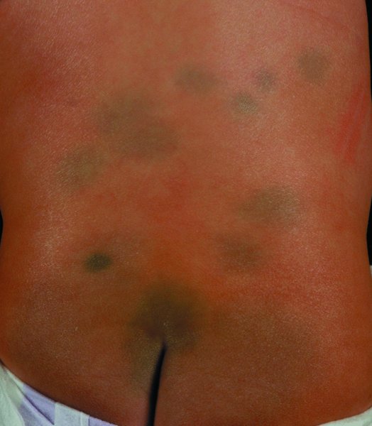
15 Shakespeare knew how the skin changes with age: from the shining morning face of youth to the dry hand, yellow cheek and white beard of old age. In this chapter, we bring together the skin conditions encountered in different age groups, and add two more to Shakespeare’s seven ages – those of the fetus and the pregnant woman. In utero, the skin remains one cell thick until weeks 4–6, when two layers can be seen; by weeks 8–11, there are three layers. Hair follicles start to appear at about 9 weeks, as do nails. Sebaceous glands arrive by 14 weeks, and pigmentation by months 4–6. Free nerve endings begin to develop in the skin at about 7 weeks but the central connections required for a fetus to appreciate pain are not complete until weeks 23–25. Some families are at high risk of having a child with an intolerable genetic skin disorder. Prenatal diagnosis coupled with genetic counselling is then required, as early as possible so that selective termination is easy and safe. In the past, a fetal skin biopsy was sometimes taken under ultrasound guidance to help such families. However, this technique cannot be undertaken before 15 weeks’ gestation and has gradually been superseded by DNA-based diagnostic screening of amniotic fluid cells (at 12–15 weeks) or chorionic villi samples (at 10–12 weeks). This type of testing has been used for conditions such as epidermolysis bullosa (p. 122; see Figure 9.13), severe ichthyoses including harlequin fetus (p. 47) and oculocutaneous albinism (p. 270). Pre-implantation diagnosis is gradually replacing prenatal diagnosis for couples known to be at high risk of severe Mendelian disorders, or structural chromosomal abnormalities. In vitro fertilization is carried out, and then the embryo biopsied and examined for the abnormality in question by fluorescence in situ hybridization, or by polymerase chain reaction (PCR). An embryo free of disease can then be re-implanted, removing the need for selective termination. At birth, the skin is covered, wholly or partly, with a whitish slimy layer, the vernix caseosa. This comes off over the first few days, although some think that cradle cap (scaling of the scalp during the first weeks of life) is a result of its localized persistence. An infant’s skin is often mottled at birth (cutis marmorata) and peripheral cyanosis is common, as is a generalized erythema lasting for a day or two. The term harlequin skin change refers to an appearance seen in a few babies when they lie on their side; the uppermost part of their body becomes pale and is sharply demarcated from a redder lower half. Preterm infants may be covered with fine lanugo hairs. In full-term babies there is usually some loss of scalp hair over the first few weeks. Multiple milia (tiny white epidermal cysts) are seen in about half of all babies. The tiny yellowish papules on the face of many babies are sebaceous glands that have hypertrophied under the influence of maternal androgens that have passed via the placenta. Neonatal acne (p. 158) may have the same trigger. Rarely, maternal autoantibodies pass through the placenta. The child of a mother with subacute lupus erythematosus (p. 129) and anti Ro (SS-A) antibodies, for example, may develop an elevated, often annular, erythema (neonatal lupus erythematosus). This clears over the first few months, but may be associated with congenital heart block. Other important changes seen at birth include those caused by underlying genetic disorders. A collodion baby (p. 47), for example, whose shiny smooth skin looks almost as though it has been painted with collodion, may have an underlying non-bullous ichthyosiform erythroderma (see Figure 9.13 or lamellar ichthyosis (p. 47). Incontinentia pigmenti (p. 348) is in its linear vesicular stage at birth. In the severe junctional type of epidermolysis bullosa (p. 124), the newborn child has a mixture of large raw areas and flaccid blisters. In contrast, in tuberous sclerosis (p. 345), tiny white patches may be the only manifestation at birth. Common birthmarks include congenital melanocytic naevi (p. 282), Mongolian spots (Figure 15.1) and haemangiomas (portwine stains, p. 302; salmon patches, p. 302). Capillary cavernous haemangiomas (strawberry naevi) appear within a few weeks of birth and then tend to regress slowly over the next few years (p. 303). Figure 15.1 Mongolian spot. The stratum corneum is fully formed at birth, and so barrier function is normal except in premature babies. Nevertheless, newborn skin tolerates irritants poorly. Primary irritant reactions are common after prolonged contact with faeces and urine in the napkin and peri-anal areas, although severe napkin (diaper) dermatitis has become much less common since the introduction of disposable napkins (p. 83). True allergic contact eczema is rare in infancy. Atopic eczema (p. 87) commonly starts before the age of 6 months, often appearing on the face with a patchy non-specific distribution elsewhere. There is large surface area to body weight ratio in babies and the risk of side effects from absorption of topically applied medications (e.g. topical corticosteroids and scabicides) is increased. Sadly, practitioners may see signs suggestive of physical abuse. Contusions, abrasions, lacerations, burns and scalds, associated with broken bones or other evidence of trauma are classic features of the battered baby, but the signs may be more subtle. In any event, disorders responsible for the blisters and raw areas, such as epidermolysis bullosa (p. 122) or for bruising (Table 11.7), such as a coagulation or platelet defect, must be excluded. Parents soon learn that nurseries and schools are mixed blessings for health. Snuffles, coughs and colds are usually considered an inevitable part of the growing-up process and essential for the development of an effective immune system. But exposure and close contact at school also bring a host of skin infections and infestations; for example impetigo (p. 215), warts (p. 227), molluscum contagiosum (p. 234), head lice (p. 249) and scabies (p. 253). Such unwelcome guests are seldom accepted passively by mothers who require full explanations to lessen blame.
The Skin at Different Ages
Fetal skin
Infancy (the first year of life)
Childhood
Related posts:


Stay updated, free articles. Join our Telegram channel

Full access? Get Clinical Tree


