(1)
Plastic Aesthetic and Laser Surgery, Hygeia hospital, Athens, Greece
Keywords
CanthopexySingle-suture traction techniqueCanthal ligamentInferior lateral retinaculumSkin traction versus skin contractionCanthopexy is not a novelty. It refers to a less invasive procedure compared to that of canthoplasty, and it is designed to reinforce the existing canthal tendon and the surrounding structures by surgical suturing without removing it from its normal attachment. It has been used widely for both reconstructive and aesthetic reasons. Access to the canthal ligament can be achieved through the lower eyelid, the upper eyelid skin incision, or a small separate incision [1–3].
In our series, no extra incision was performed other than the upper eyelid incision, and the scope added to the previously published studies was the suspension of the lower eyelid in different vectors, so that this traction in combination with OOM contraction would enhance the appearance and skin texture of the lower eyelid (Fig. 5.1).
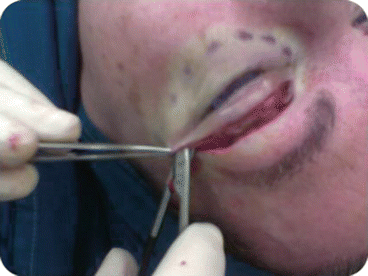
Fig. 5.1
The upper blepharoplasty incision is used for the SSTT procedure. Blunt dissection is performed in suborbicularis plane, toward the attachment of the inferior crux of the ligament to the tarsal plate
Because dissection of tissue is minimal, local hemorrhage, ecchymosis, and edema are avoided. No lysis of the lateral retinaculum or other procedures in the orbital rim area are necessary. A permanent suture is used to secure the canthal ligament to the periosteum of the lateral orbital rim. This superolateral movement of the lateral canthal ligament tightens the lower eyelid skin (skin traction) in a different and superior manner than that of laser skin resurfacing (skin contraction).
5.1 Anatomy of the Lateral Canthal Ligament
Also called the lateral canthal tendon, the lateral canthal ligament is comprised of a superior crux from the superior tarsus and an inferior crux from the inferior tarsus (Fig. 5.2). The superior and inferior crux of the lateral canthal tendons fuse at the lateral border of the tarsal plates to join the lateral retinaculum, a condensation of several anatomical structures that inserts onto the lateral orbital tubercle of Whitnall. This tubercle is located 2–4 mm posterior to the lateral orbital rim, 10–12 mm inferior to the frontozygomatic suture, and at the level of the lateral commissure. The lateral retinaculum consists of fibers from the lateral horn of the levator aponeurosis, Lockwood’s ligament, check ligaments of the lateral rectus muscle, fibers of the suspensory ligaments of the lacrimal gland, and some deep fibers of the pretarsal orbicularis oculi muscle. In the space between the lateral retinaculum and the more anteriorally placed orbital septum is sometimes found a small fat pad, Eisler’s pocket [4].
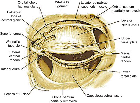
Fig. 5.2
Anatomy of the lateral canthal tendon and related structures
The lateral canthus is positioned approximately 2 mm higher than the medial canthus.
The lateral canthal ligament is a clinically important anatomical structure. The inferior crux of the canthal ligament is used for the canthopexy techniques, by plication and anchoring on the periosteum of the orbital rim.
5.2 The Technique
The single-suture traction technique is performed as the final step of the operation when the patient undergoes upper and lower blepharoplasty. In many cases, this technique is applied also in patients who only require upper or lower blepharoplasty alone. In upper blepharoplasty patients, the existing blepharoplasty incision serves as the approach point for the identification of the inferior crux of the lateral canthal tendon. In lower blepharoplasty patients who also require this maneuver, an extra small incision at the lateral part of the upper eyelid crease above the lateral canthus is needed. Through this incision, and in submuscular plane, a tunneling toward the inferior crux of the lateral canthal ligament will offer identification of this anatomical element. Incision is not necessary to exceed 1 cm in length, and is carefully placed in the upper eyelid crease to avoid visible postoperative scar (Fig. 5.3).
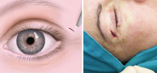
Fig. 5.3
A short incision of 1 cm in length is placed in the lateral part of the upper eyelid crease above the canthus for the SSTT to be executed in patients who only require lower eyelid transconjunctival blepharoplasty. (L) Incision in graphic, (R) incision intraoperatively with 6/0 nonabsorbable suture already passed through the inferior crux of the lateral canthal tendon
The SSTT can also be applied during open (transcutaneous) blepharoplasty of the lower eyelid with the same procedure as in the transconjunctival technique (Fig. 5.4).
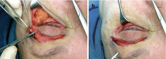
Fig. 5.4
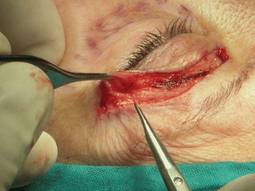
Lower transcutaneous blepharoplasty. Intraoperative view of SSTT applied via the upper blepharoplasty incision. (a) Lower limb ( inferior crux ) of canthal tendon identified. 6/0 prolene suture placed through. (b) Suture anchored on the periosteum and knot tightened
The procedure starts with blunt scissors dissection and tunneling under the OOM segment, which is present at the temporal part of the upper blepharoplasty incision. Care should be taken to avoid bleeding of the lateral palpebral artery and the vessels of the proximal part of its vascular arcade which are located at this point. Although difficult to avoid puncturing branches of this vascular component, upward direction of the scissors when the muscle is perforated minimizes this risk. In case of bleeding, blunt coagulation of this area is enough to stop it (Fig. 5.5).
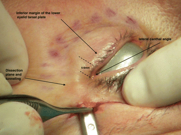
Fig. 5.5
Scissors directed from the muscle entry point toward the canthal angle inferior to the inferior margin of the tarsal plate 5–6 mm toward the eyelid, to complete the tunneling through which the inferior crux of the canthal tendon will be plicated
The scissors are then directed in a vector connecting the initial point where the muscle is perforated to the canthal angle at the fusion of the upper and lower eyelid points and slightly inferior to the inferior margin of the lower eyelid tarsal plate, the dissection extending 5–6 mm parallel to it under the lower eyelid skin (Figs. 5.1 and 5.6).