(1)
Plastic Aesthetic and Laser Surgery, Hygeia hospital, Athens, Greece
Keywords
Orbital bony structurePalpebral anatomyOrbicularis oculi muscleEyelid vascular supply and innervationAging anatomy of the orbitLinesGroovesFolds1.1 The Bony Structure of the Orbit
The orbit is the cavity or socket of the skull in which the eye and its appendages are situated. “Orbit” can refer to the bony socket [1], or it can also be used to imply the contents [2].
The bony margins of the orbital canal in humans do not derive from a single bone, but a mosaic of seven embryologically distinct structures: the zygomatic bone laterally, the sphenoid bone, with its lesser wing forming the optic canal and its greater wing forming the lateral posterior portion of the bony orbital process, the maxillary bone inferiorly and medially, which, along with the lacrimal and ethmoid bones, forms the medial wall of the orbital canal.
The roof (superior wall) is formed primarily by the orbital plate frontal bone and also the lesser wing of sphenoid near the apex of the orbit. The orbital surface presents medially by trochlear fovea and laterally by lacrimal fossa.
The floor (inferior wall) is formed by the orbital surface of maxilla, the orbital surface of zygomatic bone, and the minute orbital process of palatine bone. Medially, near the orbital margin, is located the groove for nasolacrimal duct. Near the middle of the floor is located the infraorbital groove which leads to the infraorbital foramen. The floor is separated from the lateral wall by inferior orbital fissure, which connects the orbit to pterygopalatine and infratemporal fossa.
The medial wall is formed primarily by the orbital plate of ethmoid, as well as contributions from the frontal process of maxilla, the lacrimal bone, and a small part of the body of the sphenoid. It is the thinnest wall of the orbit, evidenced by pneumatized ethmoidal cells.
The lateral wall is formed by the frontal process of zygomatic and more posteriorly by the orbital plate of the greater wing of sphenoid. The bones meet at the zygomaticosphenoid suture.
The base, which opens in the face, has four borders. The following bones take part in their formation (Fig.1.1):
- 1.
Superior margin: frontal bone and sphenoid
- 2.
Inferior margin: maxilla, palatine, and zygomatic
- 3.
Medial margin: ethmoid, lacrimal bone, and maxillary bone
- 4.
Lateral margin: zygomatic and sphenoid
Tip
The upper lateral part of the orbital rim consisted from the zygomatic bone is involved in the several canthopexy techniques as the point of the periosteal suture suspension, for the support of the lower eyelid (see arrow in Fig. 1.1)
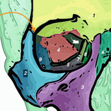
Fig. 1.1
1 Frontal bone, 2 Zygomatic bone, 3 Maxilla, 4 Sphenoid bone, 5 Ethmoid bone, 6 Nasal bone (not part of orbit), 7 Palatine bone, 8 Lacrimal bone
1.2 The Muscular Extraocular and Palpebral Anatomy
1.2.1 The Extraocular Muscles
The extraocular muscles are the six muscles that control movement of the eye and one muscle that controls eyelid elevation (levator palpebrae) (Table 1.1) . The extraocular muscles are supplied mainly by the branches of the ophthalmic artery. In the table below, the innervation, insertion, and action in neutral position of each extraocular muscle are described [3].
Table 1.1
Extra ocular muscles
Muscle | Innervation | Insertion | Neutral position |
|---|---|---|---|
Superior rectus | Oculomotor nerve (superior branch) | Eye (anterior, superior surface) | Elevation Incyclotorsion Adduction |
Inferior rectus | Oculomotor nerve (inferior branch) | Eye (anterior, inferior surface) | Depression Extorsion Adduction |
Lateral rectus | Abducens nerve | Eye (anterior, lateral surface) | Abduction |
Medial rectus | Oculomotor nerve (inferior branch) | Eye (anterior, medial surface) | Adduction |
Superior oblique | Trochlear nerve | Eye (posterior, superior, lateral surface) | Intorsion Depression Abduction |
Inferior oblique | Oculomotor nerve (inferior branch) | Eye (posterior, inferior, lateral surface) | Extorsion Elevation Abduction |
Levator palpebrae superioris | Oculomotor nerve | Tarsal plate of upper eyelid | Retracts and elevates eyelid |
Tip
In upper blepharoplasty procedures, attention should be focused in avoiding damage of the levator muscle aponeurosis, which anatomically is located partially behind the septum and the retroseptal fat and partially under the superior part of the pretarsal portion of the orbicularis oculi muscle. Damage to this muscle, or its innervation, can cause ptosis, the drooping of the eyelid.
In lower transconjunctival blepharoplasty, attention has to be focused in the posterior lamella at the inferior border of the capsulopalpebral fascia and the retractor system, in an effort to avoid damage of the inferior oblique muscle and scarring formation of the tissues.
1.2.2 The Orbicularis Oculi Muscle (OOM)
The orbicularis oculi muscle lies directly underneath the surface of the skin, around the eyes. Its function is to close the eyelid and to help in the passing and draining of tears through the punctum, canaliculi, and lacrimal sac, all parts of the tear drainage system.
The orbicularis oculi muscle is composed of three parts: the orbital portion, the palpebral portion, and the lacrimal portion (Fig. 1.2). The orbital portion overlies the orbital rim, closes the eyelids firmly, and is controlled by voluntary action. The palpebral portion overlies the eyelids, closes the eyelids gently in involuntary or reflex blinking. The palpebral portion is divided into three parts: the pretarsal portion, the preseptal portion, and the ciliary portion. The lacrimal portion compresses the lacrimal sac, which receives tears from the lacrimal ducts and conveys them into the nasolacrimal duct.
The OOM is innervated by the temporal and zygomatic branches of the facial nerve (cranial nerve VII). Its blood supply comes from the branches of the ophthalmic artery
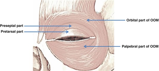
Fig. 1.2
Orbital and palpebral parts of orbicularis oculi muscle. Palpebral part is subdivided into preseptal and pretarsal parts
Tip
The OOM is greatly involved in the blepharoplasty procedures. Its firm attachment with the overlying eyelid skin reflects any procedure undertaken on the muscle (i.e. contraction, dissection, etc.) to the skin, and thus to the texture and aesthetic improvement of the periorbital region.
1.3 The Vascular Palpebral Anatomy
Branches of the internal and external carotid arteries supply the eyelids. The ophthalmic artery branches off the internal carotid artery and supplies different parts of the eyelid. At the inner part of the upper eyelid, the ophthalmic artery splits into two and traverses outward to supply both the upper and the lower eyelids. The branch that supplies the lower eyelid is in fact a branch that arises from the superior marginal vessel (that supplies the upper eyelid). The superior and inferior marginal vessels that arise from the ophthalmic artery together form the marginal arcade. This arcade is prone to injury and bleeding during blepharoplasty.
Related posts:
Stay updated, free articles. Join our Telegram channel

Full access? Get Clinical Tree


