Purpura is extravasation of red blood cells into the skin or mucous membrane. For this reason, purpuric lesions do not blanch on diascopy (pressing on the lesion with a glass slide or finger). The differential diagnosis for purpura is broad, but it can be quickly narrowed by classifying the lesions based on their morphology, as well as other clinical and laboratory findings.1,2 The clinical descriptive terms for purpura are listed below, and their respective tables describing the differential diagnosis are referenced.
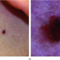
Petechiae: Flat lesions, macules ≤4 mm (Figure 25-1), typically initially bright red and then fade to a rust color (Tables 25-1 and 25-2).
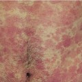
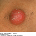
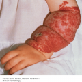
Ecchymosis: Flat lesions, macules and/or patches, >5 mm (Figures 25-2 and 25-3), typically initially red or purple, but may fade to yellow, brown, or green (Table 25-3).
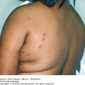
Palpable purpura: Elevated, round or oval, red or purple papules and/or plaques (Figure 25-4), sometimes barely palpable (Table 25-4).
Retiform purpura: Stellate or branching lesions, with angular or geometric borders (Figure 25-5). These are often palpable plaques, but can present as nonpalpable patches as well (Tables 25-5 and 25-6).
| Disease/Disorder | Clinical Features and Associations | Initial Diagnostic Evaluation |
|---|---|---|
| Low platelets (<150,000/μL) | Mucocutaneous bleeding (eg, epistaxis, increased bleeding with menses or minor cuts) Splenomegaly and lymphadenopathy may be present | CBC with differential and peripheral smear; If anemic, check reticulocyte count, LDH, haptoglobin, and bilirubin If hemolysis is present, check PTT, PT/INR, fibrinogen, D-dimer, Coombs, ANA. If inconclusive, consider bone marrow biopsy |
| Immune thrombocytopenia (ITP)3 | F > M Primary: Insidious onset Secondary: Associated with underlying disease, for example, viral infections | Platelets <100, 000 Diagnosis of exclusion Primary: Isolated decreased platelets Secondary: May relate to viral process (can obtain serology for HIV, HCV, HBV, EBV, or PCR for parvovirus and CMV), H. pylori, ANA, pregnancy, APLA, or TSH |
| Thrombotic thrombocytopenic purpura (TTP)1 | Pentad: thrombocytopenia, hemolytic anemia, changes in mental status, fever, and renal dysfunction. Do not need all 5 for diagnosis It may be idiopathic, familial, drug-induced, from pregnancy, HIV, autoimmune disease, or hematopoietic stem cell transplant HUS: Thrombocytopenia, MAHA, and renal dysfunction; usually in children with prodrome of bloody diarrhea | Labs that can be observed: ↓ Platelets and MAHA, schistocytes on p.smear, ↑reticulocyte count, positive hemolysis labs (↑LDH, ↑indirect bilirubin, ↓haptoglobin), nlPTT & INR, ↑creatinine, ↓ADAMSTS13 (in idiopathic TTP) |
| Disseminated intravascular coagulation (DIC)4 | Spectrum in hematologic abnormalities can have bleeding and/or thrombosis. May also have soft-tissue bleeding in muscle and joints May be related to trauma, shock, infection, malignancy, and obstetric complication | ↑PT/INR, ↑PTT, ↓ fibrinogen, positive fibrin degradation product/D-dimer, and hemolysis labs |
| Drug-induced1 | Petechiae, purpura due to drugs such as quinine, bactrim | Generally, isolated thrombocytopenia |
| Bone marrow suppression1 | Associated with aplastic anemia, MDS, drugs (eg, thiazides and antibiotics), alcohol, cirrhosis, myelofibrosis, granulomas from infections, hematologic, or solid malignancies | Peripheral smear may show pancytopenia, blasts, hypersegmented PMNs, leukoerythroblastic changes (teardrop cells, nucleated RBCs, immature WBCs) |
| Disease/Disorder | Clinical Features and Associations | Initial Diagnostic Evaluation |
|---|---|---|
| Platelet function disorder5,6 | Mucocutaneous bleeding | Look for splenomegaly and LAD Labs: CBC with differential, peripheral smear, BUN, creatinine, LFTs, and platelet aggregation tests. If suspect vWD or dysproteinemia, obtain vWF studies or SPEP/UPEP, respectively |
| Congenital or inherited5 | vWD: usually AD | vWD: ↓ vW factor, ↓ vWF activity (measured by ristocetin co-factor assay) ↓ factor VIII, confirm with vWF multimer analysis |
| Acquired (eg, drug-induced, liver disease, uremia, dysproteinemia, acquired vWD)5 | Associated medications include: ASA, NSAIDS, clopidogrel, ticlopidine Acquired vWD may be associated with malignancy, autoimmune disease, hypothyroidism, or drugs | Liver disease may show abnormal LFTs or ↑PT/INR, ↑PTT Uremia will show ↑ BUN Dysproteinemias will show abnormalities on SPEP/UPEP |
| Thrombocytosis secondary to myelofibrosis6 | Myelofibrosis maybe primary or secondary to other malignancy Massive splenomegaly, ± fatigue, weight loss, and fever | Peripheral smear shows leukoerythroblastic changes and bone marrow biopsy shows a “dry tap” with severe fibrosis |
| No platelet abnormality | ||
| Pigmented purpuric eruptions, capillaritis | Clustered petechial hemorrhage, often with a background of yellow brown discoloration often on lower extremities. Most common in middle aged to older men For example, Schamberg’s disease, “cayenne pepper” appearance | No systemic findings A biopsy maybe needed to differentiate lesions from vasculitis |
| Hypergammaglobulinemic purpura of Waldenstrom | Often in women, with recurrent crops of petechiae/purpura on lower extremities that burn/sting Primary or secondary to autoimmune disease (eg, Sjogren’s or SLE), RA, less likely hematologic malignancy | Hypergammaglobulinemia on SPEP ↑ titer of IgG or IgA RF (standard RF tests only detect IgM RF) ↑ESR ± positive ANA, anti-Ro, anti-La |
| Intravascular, local pressure, or trauma | Can be caused by valsava (eg, from vomiting or constipation) or blood pressure cuff | Can occur without an underlying lab abnormality |
| Disease/Disorder | Clinical Features and Associations | Initial Diagnostic Evaluation |
|---|---|---|
| Procoagulant defects1 | Susceptible to having deeper bleeding, into soft tissue | ↑PT/INR, ±↑PTT |
| Anticoagulant | For example, warfarin | ↑PT/INR |
| Liver disease | History of alcohol abuse, hepatitis, and primary liver disease | ↑PT/INR, ↑PTT, abnormal LFTs |
| Vitamin K deficiency | State of malnutrition (eg, alcoholic), malabsorption (eg, from antibiotics), and liver disease | ↑PT/INR |
| DIC | See Table 25-1 | |
| Platelet disorders | See Table 25-1 | |
| Decreased dermal support of vessels and minor trauma1 | ||
| Solar or senile purpura | Older age and sun damaged skin usually on forearms | None |
| Corticosteroid therapy (systemic and topical) | Cushing’s syndrome stigmata (eg, central obesity, dorsocervical fat pad, moon facies) Skin may reveal atrophy and telangiectasia | None |
| Scurvy (vitamin C deficiency) | Diet lacking in vitamin C Perifollicular hemorrhage, corkscrew hairs, gingival bleeding, and tooth loss | ↓Serum ascorbic acid deficiency and possibly other vitamins (eg, B12 and folate) |
| Systemic amyloidosis | All have hepatosplenomegaly, GI involvement (diarrhea, protein loss), and macroglossia May also have cardiac, renal, and liver involvement. Periorbital waxy ecchymotic papules, purpura with minor trauma, and “pinch-purpura” | SPEP/UPEP and free light chain, ± bone marrow bx CBC with differential, LFTs, BUN, creatinine, urinalysis, and EKG Biopsy of abdominal subcutis fat pad reveals apple-green birefringence on Congo red stain Consider genetic testing for hereditary form |
| Genetic disease with collagen or elastin defects | Ehler–Danlos Syndrome: stretchable skin and flexible joints, usually AD inheritance Pseudoxanthoma elasticum: appearance of “plucked chicken skin” on neck | Genetic testing for specific mutations depending on the suspected condition |
| Disease/Disorder | Clinical Features and Associations | Initial Diagnostic Evaluation |
|---|---|---|
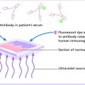
| Leukocytoclastic vasculitis, small and small-medium vessel vasculitis7–9 | Purpuric papules or plaques typically on distal legs or dependent areas | Skin biopsy for routine histology and another for DIF |
| Idiopathic (IgG, IgM, or IgA), secondary to drugs or infection | IgA lesions can appear targetoid | In addition to skin biopsies, careful review of medications, and recent/current infections |
| Henoch–Schönlein purpura | M > F. Usually age <20 years Tetrad: purpura (universal), arthritis (82%), nephritis (40%), abdominal pain (63%), or gastrointestinal hemorrhage (33%) May appear targetoid, on extensor areas and buttock Extension to trunk/upper extremities can indicate renal involvement | Two skin biopsies should be done, one for routine histology and one for DIF Obtain BUN/Cr, urinalysis |
| Urticarial vasculitis | Persistence of hive-like lesions >24 hours, often burn > itch. Can have concomitant systemic symptoms (eg, LAD, arthralgia, angioedema, and fever). Can be caused by drug (eg, ACE inhibitors, penicillin, and sulfonamides), autoimmune disease, hematologic or other malignancies, and infections | In addition to skin biopsies, the following lab findings may be helpful: ↓Complement (eg, CH50, C4, C3, Clq) may relate to a variety of systemic processes, for example, SLE, malignancy, and infection Creatinine and UA. For suspected infection screen for HBV, HCV, and heterophile serology For suspected autoimmune condition screen with autoantibody test |
| Mixed cryoglobulinemia (Type II and III) | Usually acral distribution Weakness, livedo reticularis, leg ulcers, HSM, and Raynaud’s phenomenon Can also have glomerulonephritis and peripheral neuropathy | In addition to skin biopsies, the following labs may be helpful: Cryoglobulins (proteins that precipitate from serum or plasma when cooled) Type II: Sjogren’s, SLE evaluation. Type III: Hepatitis C RNA, ESR, and C4, RF |
| Rheumatic vasculitis (eg, RA, SLE, Sjogren’s syndrome) | Digital ischemia, livedo reticularis, pericarditis, bowel ischemia, peripheral neuropathy | In addition to skin biopsies, the following lab findings may be helpful: + ANA and ↓complement (SLE) + RF, anti-CCP (RA) + ANA, RF, anti-Ro or La (Sjogren’s) |
| Anti-neutrophilic cytoplasmic antibody (ANCA) associated | ||
| Wegener’s granulomatosis | Usually young and middle-aged adults Can have a nasal or oral (eg, sinusitis, saddle-nose deformity), pulmonary (eg, infiltrate, nodule, and cavity), or renal involvement (hematuria, RBC casts) | In addition to skin biopsies, +ANCA (90%), c-ANCA (anti-PR3) |
| Churg–Strauss | Usually age 30-40 years, with HLA-DRB4 Can also involve lungs (eg, asthma), peripheral nerves, heart, and kidneys | In addition to skin biopsies, CBC with differential, showing eosinophilia +ANCA (50%), p-ANCA (anti-MPO), or c-ANCA (anti-PR3) |
| Microscopic polyangiitis | Can involve the kidney and lungs | In addition to skin biopsies, +ANCA (70%), p-ANCA (anti-MPO) |
Related posts:
Stay updated, free articles. Join our Telegram channel

Full access? Get Clinical Tree


